
Article Figures & Data
Figures
- fig 1.
Calcification in aspergillosis.
A, Coronal CT scan shows irregular nodular (short arrow) and fine punctate (long arrow) calcification within the inflammatory tissue at the center of the opacified left maxillary sinus.
B, Photomicrograph of surgical specimen shows mottled calcifications (arrow) embedded within the fungal mycelium (hematoxylin-eosin, original magnification ×100).
C, Darkly stained fungal hyphae (short arrows) are seen around the nodular calcification (long arrow) (Gomori's methenamine silver stain, original magnification ×100).
- fig 2.
Calcification in aspergillosis. Coronal CT scan shows nodular (short arrow) and linear (long arrow) calcifications located centrally in the right maxillary sinus.
fig 3. Calcification in nonfungal sinusitis.
A, Smoothly marginated linear calcification (large arrow) is seen near the floor of the left maxillary sinus. The calcification is located at the periphery of the sinus and there is a thin layer of soft-tissue density (small arrows) separating the calcification from the sinus wall.
B, Photomicrograph of surgical specimen shows calcification (short arrow) within the thickened fibrotic submucosal layer of the maxillary sinus. Vascular congestion with dilated capillaries (long arrows) is seen in the edematous superficial submucosal layer (hematoxylin-eosin, original magnification ×100).
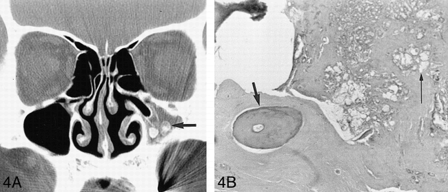
- fig 4.
Ossification in nonfungal sinusitis.
A, CT scan shows round and eggshell lesions (arrow) in the left maxillary sinus. The calcific density lesions are located near the inferior wall of the sinus and are separated from the bony wall.
B, Photomicrograph of surgical specimen shows well-marginated round woven bone (short arrow) embedded within the fibrotic submucosal layer of the maxillary sinus. There is glandular hyperplasia (long arrow) caused by inflammation in the submucosa of the maxillary sinus (hematoxylin-eosin, original magnification ×100).


Tables
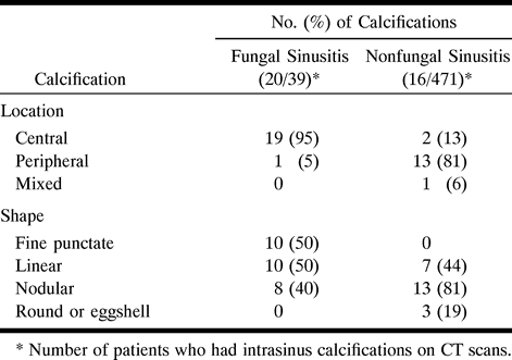
 TABLE 1:
CT findings of calcification in 510 patients with either fungal or nonfungal sinusitis


