Advertisement

Main menu

  • Home
  • Content
    • Current Issue
    • Accepted Manuscripts
    • Article Preview
    • Past Issue Archive
    • Video Articles
    • AJNR Case Collection
    • Case of the Week Archive
    • Case of the Month Archive
    • Classic Case Archive
  • Special Collections
    • AJNR Awards
    • Low-Field MRI
    • Alzheimer Disease
    • ASNR Foundation Special Collection
    • Photon-Counting CT
    • View All
  • Multimedia
    • AJNR Podcasts
    • AJNR SCANtastic
    • Trainee Corner
    • MRI Safety Corner
    • Imaging Protocols
  • For Authors
    • Submit a Manuscript
    • Submit a Video Article
    • Submit an eLetter to the Editor/Response
    • Manuscript Submission Guidelines
    • Statistical Tips
    • Fast Publishing of Accepted Manuscripts
    • Graphical Abstract Preparation
    • Imaging Protocol Submission
    • Author Policies
  • About Us
    • About AJNR
    • Editorial Board
    • Editorial Board Alumni
  • More
    • Become a Reviewer/Academy of Reviewers
    • Subscribers
    • Permissions
    • Alerts
    • Feedback
    • Advertisers
    • ASNR Home

User menu

  • Alerts
  • Log in

Search

  • Advanced search
American Journal of Neuroradiology

American Journal of Neuroradiology

ASHNR American Society of Functional Neuroradiology ASHNR American Society of Pediatric Neuroradiology ASSR
  • Alerts
  • Log in

Advanced Search

  • Home
  • Content
    • Current Issue
    • Accepted Manuscripts
    • Article Preview
    • Past Issue Archive
    • Video Articles
    • AJNR Case Collection
    • Case of the Week Archive
    • Case of the Month Archive
    • Classic Case Archive
  • Special Collections
    • AJNR Awards
    • Low-Field MRI
    • Alzheimer Disease
    • ASNR Foundation Special Collection
    • Photon-Counting CT
    • View All
  • Multimedia
    • AJNR Podcasts
    • AJNR SCANtastic
    • Trainee Corner
    • MRI Safety Corner
    • Imaging Protocols
  • For Authors
    • Submit a Manuscript
    • Submit a Video Article
    • Submit an eLetter to the Editor/Response
    • Manuscript Submission Guidelines
    • Statistical Tips
    • Fast Publishing of Accepted Manuscripts
    • Graphical Abstract Preparation
    • Imaging Protocol Submission
    • Author Policies
  • About Us
    • About AJNR
    • Editorial Board
    • Editorial Board Alumni
  • More
    • Become a Reviewer/Academy of Reviewers
    • Subscribers
    • Permissions
    • Alerts
    • Feedback
    • Advertisers
    • ASNR Home
  • Follow AJNR on Twitter
  • Visit AJNR on Facebook
  • Follow AJNR on Instagram
  • Join AJNR on LinkedIn
  • RSS Feeds

AJNR Awards, New Junior Editors, and more. Read the latest AJNR updates

Research ArticleBRAIN

Successful Radiosurgical Treatment of Arteriovenous Malformation Accompanied by Venous Malformation

Hiroki Kurita, Tomio Sasaki, Masao Tago, Yuu Kaneko and Takaaki Kirino
American Journal of Neuroradiology March 1999, 20 (3) 482-485;
Hiroki Kurita
  • Find this author on Google Scholar
  • Find this author on PubMed
  • Search for this author on this site
Tomio Sasaki
  • Find this author on Google Scholar
  • Find this author on PubMed
  • Search for this author on this site
Masao Tago
  • Find this author on Google Scholar
  • Find this author on PubMed
  • Search for this author on this site
Yuu Kaneko
  • Find this author on Google Scholar
  • Find this author on PubMed
  • Search for this author on this site
Takaaki Kirino
  • Find this author on Google Scholar
  • Find this author on PubMed
  • Search for this author on this site
  • Article
  • Figures & Data
  • Info & Metrics
  • Responses
  • References
  • PDF
Loading

Abstract

Summary: We present a patient with a rare cerebrovascular malformation consisting of a typical arteriovenous malformation (AVM) with a nidus and a venous malformation (VM) in a single lesion. The AVM component was successfully obliterated by radiosurgery, whereas the VM was completely preserved. Radiosurgery can be an effective treatment technique for treating this type of malformation because it allows targeted obliteration of the AVM yet carries a low risk of damaging the venous drainage toward and away from the VM.

Various associations of two discrete vascular malformations of the brain are known, and each combination is recognized as a distinct clinicopathologic entity (1, 2). Several authors have described venous malformation (VM) with arteriovenous shunting that can be considered an immature form of arteriovenous malformation (AVM). Nonetheless, coexistence of an AVM and a VM within a single lesion is extremely rare. We report a case of true AVM-VM coexistence that was successfully treated by gamma-knife radiosurgery. Our experience adds to the knowledge concerning the clinical features of and therapeutic strategies for this peculiar cerebrovascular malformation.

Case Report

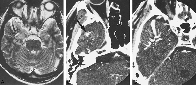
A 39-year-old man noticed transient dysarthria in June 1996, and underwent neuroradiologic examination at a local hospital. MR imaging showed an infarction affecting the left corona radiata, and an unruptured vascular malformation in the right temporal lobe (Fig 1A). He was treated conservatively for his ischemic event, and then was referred to us for further evaluation.

fig 1.
  • Download figure
  • Open in new tab
  • Download powerpoint
fig 1.

T2-weighted MR image (3500/96/1) shows unruptured VM of the right antero-temporal lobe (A), and thin-slice dynamic CT shows disappearance of nidus enhancement (arrow) and large draining vein (B and C).

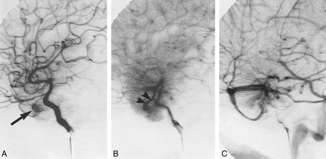
Physical and neurologic examinations were normal. Thin-slice dynamic CT revealed small nidus enhancement and a large draining vein (Fig 1B and C). Cerebral angiography showed a right temporal AVM that was fed by a branch of the middle cerebral artery. After forming a distinct nidus, the draining vein of this AVM was connected to an abnormal vein, into which many other finer veins also drained to form a typical “caput medusae” appearance of a venous malformation. The abnormal central vein drained into the right sphenopetrosal vein. Dysmorphism of the sylvian veins was also evident (Fig 2).

fig 2.
  • Download figure
  • Open in new tab
  • Download powerpoint
fig 2.

Serial right internal carotid angiogram reveals the AVM-VM malformation.

A, Arterial phase shows a small AVM nidus fed by the branch of the middle cerebral artery (arrow).

B, Later arterial phase shows a single AVM drainer (arrow heads) joining collecting veins along with converging small medullary veins.

C, Venous phase shows the typical “caput medusae” appearance of a VM. Note the dysmorphism of the sylvian vein.

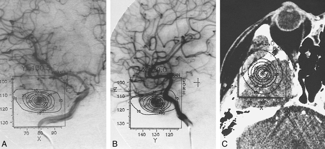
The patient underwent gamma-knife radiosurgery in October 1996. The treatment strategy was to irradiate only the nidus of the AVM to prevent obstructing the regional venous outflow through the VM. Isodose planning was performed on the basis of stereotactic cerebral angiography and CT. Radiation (25 Gy) was delivered to the periphery of the nidus in a single dose using one isocenter (Fig 3).

fig 3.
  • Download figure
  • Open in new tab
  • Download powerpoint
fig 3.

Anteroposterior (A) and lateral (B) views on a stereotactic right internal carotid angiogram, and stereotactic CT (C) show AVM component of lesion. At radiosurgery, 25 Gy is delivered to 50% isodose line using 1 isocenter (maximum dose 50 Gy).

The postradiosurgical course was uneventful. At the patient's 1-year follow-up, MR imaging revealed no parenchymal radiation injury, hemorrhage, or ischemia (Fig 4A). CT showed disappearance of nidus enhancement (Fig B and C). Cerebral angiography showed total obliteration of the AVM and preservation of the VM (Fig 5).

fig 4.
  • Download figure
  • Open in new tab
  • Download powerpoint
fig 4.

T2-weighted MR image obtained 1 year after radiosurgery shows no hemorrhagic or ischemic complications (A). Thin-slice dynamic CT shows absence of nidus enhancement (B and C).

fig 5.
  • Download figure
  • Open in new tab
  • Download powerpoint
fig 5.

Serial right internal carotid angiogram obtained 1 year after radiosurgery shows total obliteration of the AVM and preservation of the VM during the arterial (A), late arterial (B), and venous (C) phases

Discussion

There have been several reports describing a hybrid malformation consisting of an AVM and VM as a rare subset of mixed cerebrovascular malformations (1, 3–8). The majority of the reported cases, however, are in fact a combination of a typical VM with its classic triad (regional venous dysmorphism, convergence of deep collecting veins, and a deep central draining vein) and additional arteriovenous shunting (1, 3–5, 8). Such anomalies, named “arterialized VMs” by some authors (1, 7), do not have a discrete nidus but a single or multiple arterial fistulization, and therefore are considered to be a transitional form between an AVM and a VM. A hybrid malformation consisting of a typical AVM with a nidus and a classic VM in a single lesion is extremely rare. To our knowledge, the present case is only the third instance of this type of malformation (6, 8).

It is now accepted that VMs are a congenital anatomic variant of normal venous drainage that generally follow a benign clinical course (9–12). Hemorrhagic complications are rare, and many of the hemorrhagic cases are ascribed to other coexisting VMs (1). The risk of cerebral venous infarction occurring after excision or obliteration of a VM is significant and well known (1, 6, 9, 12). Consequently, when an AVM and VM coexist, it is advisable to treat the AVM while preserving the VM, which should serve as an important channel of the venous drainage from the regional brain (1, 6, 8). Limited surgical excision of the AVM component would be a valid option (1, 6) if the nidus is well separated from the major portion of the associated VM. Nonetheless, there remains a risk of unintentional damage to the venous return that could lead to a hemorrhagic or ischemic complication (6). Endovascular embolization can also be considered, but the blood-flow pattern around the lesion can be complicated, and may hinder the total obliteration. Although there are limitations regarding lesion size, radiosurgery would be a treatment of choice when the two lesions are located too closely for a safe surgical excision or embolization of the AVM.

Several authors have discussed the use of radiosurgery for the management of AVM-VM coexistence. In their radiosurgical series of VMs, Lindquist et al (5) reported two arterialized VM cases in which the component detected in the arterial phase was irradiated, resulting in disappearance of the arteriovenous shunt. On the contrary, Mullan et al (7) mentioned similar transitional forms of AVMs and VMs that persisted despite radiosurgical treatment. These reports indicate that radiosurgery effectively occludes the arteriovenous fistula in selected patients with arterialized VM. The results remain controversial, however, because it is sometimes difficult to target small or multiple arteriovenous fistulas for obliteration by radiation.

Conclusion

We report a case of an AVM-VM coexistence that was successfully treated by radiosurgery. Although rare, the case presented here represents another good indication for radiosurgical treatment.

Acknowledgments

We are grateful to Bernhard Meyer of the Department of Neurosurgery, University of Bonn, and to Keisuke Ueki of the Department of Neurosurgery, University of Tokyo, for their helpful comments and suggestions.

Footnotes

  • ↵1 Address reprint requests to Hiroki Kurita, M.D., Ph.D., Neurochirurgische Universitätklinik, Albert-Ludwings-Universität Freiburg, Breisacher Straβe 64, D-79106, Freiburg, Germany.

References

  1. ↵
    Awad IA, Robinson JR Jr, Mohanty S, Estes ML. Mixed vascular malformations of the brain: clinical and pathogenetic considerations. Neurosurgery 1993;33:179-188
    PubMed
  2. Robinson JR Jr, Award IA, Masaryk TJ, Estes ML. Pathological heterogeneity of angiographically occult vascular malformations of the brain. Neurosurgery 1993;33:547-555
    PubMed
  3. Fierstien SB, Pribram HW, Hieshima G. Angiography and computed tomography in the evaluation of cerebral venous malformations. Neuroradiol 1979;17:137-148
    PubMed
  4. Hirata Y, Matsukado Y, Nagahiro S, Kuratsu J. Intracerebral venous angioma with arterial blood supply: a mixed angioma. Surg Neurol 1986;25:227-232
    PubMed
  5. ↵
    Lindquist C, Guo WY, Karlsson B, Steiner L. Radiosurgery for venous angiomas. J Neurosurg 1993;78:531-536
    PubMed
  6. ↵
    Meyer B, Stangl AP, Schramm J. Association of venous and true arteriovenous malformation: a rare entity among mixed vascular malformation of the brain. J Neurosurg 1995;83:141-144
    PubMed
  7. ↵
    Mullan S, Mojtahedi S, Johnson DL, MacDonald RL. Cerebral venous malformation-arteriovenous malformation transition forms. J Neurosurg 1996;85:9-13
    PubMed
  8. Itoyama Y, Kitano I, Fujioka S, et al. [Mixed angioma of the brain: report of two cases]. No Shinkei Geka 1994;22:465-469 (Jpn)
    PubMed
  9. ↵
    Garner TB, Curling OD, Kelly DL Jr, Laster DW. The natural history of intracranial venous angiomas. J Neurosurg 1991;75:715-755
    PubMed
  10. Kondziolka D, Dempsey PK, Lunsford LD. The case for conservative management of venous angiomas. Can J Neurol Sci 1991;18:295-299
    PubMed
  11. Rigamonti D, Spetzler RF, Medina M, Rigamonti K, Geckle DS, Pappas C. Cerebral venous malformations. J Neurosurg 1990;73:560-564
    PubMed
  12. Lasjaunias P, Burrows P, Planet C. Developmental venous anomalies (DVA): the so-called venous angioma. Neurosurg Rev 1986;9:233-242
    CrossRefPubMed
  • Received May 28, 1998.
  • Accepted after revision September 8, 1998.
  • Copyright © American Society of Neuroradiology
View Abstract
PreviousNext
Back to top

In this issue

American Journal of Neuroradiology
Vol. 20, Issue 3
1 Mar 1999
  • Table of Contents
  • Index by author
Advertisement
Print
Download PDF
Email Article

Thank you for your interest in spreading the word on American Journal of Neuroradiology.

NOTE: We only request your email address so that the person you are recommending the page to knows that you wanted them to see it, and that it is not junk mail. We do not capture any email address.

Enter multiple addresses on separate lines or separate them with commas.
Successful Radiosurgical Treatment of Arteriovenous Malformation Accompanied by Venous Malformation
(Your Name) has sent you a message from American Journal of Neuroradiology
(Your Name) thought you would like to see the American Journal of Neuroradiology web site.
CAPTCHA
This question is for testing whether or not you are a human visitor and to prevent automated spam submissions.
Cite this article
Hiroki Kurita, Tomio Sasaki, Masao Tago, Yuu Kaneko, Takaaki Kirino
Successful Radiosurgical Treatment of Arteriovenous Malformation Accompanied by Venous Malformation
American Journal of Neuroradiology Mar 1999, 20 (3) 482-485;

Citation Manager Formats

  • BibTeX
  • Bookends
  • EasyBib
  • EndNote (tagged)
  • EndNote 8 (xml)
  • Medlars
  • Mendeley
  • Papers
  • RefWorks Tagged
  • Ref Manager
  • RIS
  • Zotero
0 Responses
Respond to this article
Share
Bookmark this article
Successful Radiosurgical Treatment of Arteriovenous Malformation Accompanied by Venous Malformation
Hiroki Kurita, Tomio Sasaki, Masao Tago, Yuu Kaneko, Takaaki Kirino
American Journal of Neuroradiology Mar 1999, 20 (3) 482-485;
del.icio.us logo Twitter logo Facebook logo Mendeley logo
  • Tweet Widget
  • Facebook Like
  • Google Plus One

Jump to section

  • Article
    • Abstract
    • Case Report
    • Discussion
    • Conclusion
    • Acknowledgments
    • Footnotes
    • References
  • Figures & Data
  • Info & Metrics
  • Responses
  • References
  • PDF

Related Articles

  • No related articles found.
  • PubMed
  • Google Scholar

Cited By...

  • Pathomechanisms of Symptomatic Developmental Venous Anomalies
  • Crossref
  • Google Scholar

This article has not yet been cited by articles in journals that are participating in Crossref Cited-by Linking.

More in this TOC Section

  • Multimodal CT Provides Improved Performance for Lacunar Infarct Detection
  • Optimal MRI Sequence for Identifying Occlusion Location in Acute Stroke: Which Value of Time-Resolved Contrast-Enhanced MRA?
  • Evaluating the Effects of White Matter Multiple Sclerosis Lesions on the Volume Estimation of 6 Brain Tissue Segmentation Methods
Show more BRAIN

Similar Articles

Advertisement

Indexed Content

  • Current Issue
  • Accepted Manuscripts
  • Article Preview
  • Past Issues
  • Editorials
  • Editor's Choice
  • Fellows' Journal Club
  • Letters to the Editor
  • Video Articles

Cases

  • Case Collection
  • Archive - Case of the Week
  • Archive - Case of the Month
  • Archive - Classic Case

More from AJNR

  • Trainee Corner
  • Imaging Protocols
  • MRI Safety Corner

Multimedia

  • AJNR Podcasts
  • AJNR Scantastics

Resources

  • Turnaround Time
  • Submit a Manuscript
  • Submit a Video Article
  • Submit an eLetter to the Editor/Response
  • Manuscript Submission Guidelines
  • Statistical Tips
  • Fast Publishing of Accepted Manuscripts
  • Graphical Abstract Preparation
  • Imaging Protocol Submission
  • Evidence-Based Medicine Level Guide
  • Publishing Checklists
  • Author Policies
  • Become a Reviewer/Academy of Reviewers
  • News and Updates

About Us

  • About AJNR
  • Editorial Board
  • Editorial Board Alumni
  • Alerts
  • Permissions
  • Not an AJNR Subscriber? Join Now
  • Advertise with Us
  • Librarian Resources
  • Feedback
  • Terms and Conditions
  • AJNR Editorial Board Alumni

American Society of Neuroradiology

  • Not an ASNR Member? Join Now

© 2025 by the American Society of Neuroradiology All rights, including for text and data mining, AI training, and similar technologies, are reserved.
Print ISSN: 0195-6108 Online ISSN: 1936-959X

Powered by HighWire