
Article Figures & Data
Figures
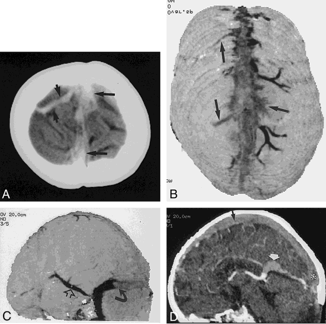
- fig 1.
4-year-old boy on L-asparaginase therapy.
A and B, Integral rendered CTCVI scans show detailed cortical venous anatomy, including the anastomotic veins of Trolard (small closed arrows), the superior sagittal sinus (large straight arrow), and the transverse (open arrows) and occipital (curved arrow) sinus.
C, The same data can be segmented to isolate the circle of Willis.
D, Integral rendering with sagittal cut planes reveals the patent sylvian aqueduct (arrow) and corpus callosum (asterisk), again from the same 60-second scan. Note the deep cerebral artery and venous detail.
- fig 2.
17-day-old boy who was clinically dehydrated with increased head circumference after resection of a medulloblastoma 2 days before these images were obtained.
A, Noncontrast vertex CT section shows high attenuation within the sagittal sinus and multiple cortical veins (arrows).
B, Integral rendered vertex view of a CTCVI scan clearly shows the lack of enhancement in the sagittal sinus and in several cortical veins (arrows). CTCVI confirms that the increased density on the noncontrast CT scan was related to thrombosis rather than to hemoconcentration.
C, Filling defect (curved arrow) in the transverse sinus is clearly seen in this posterolateral integral rendered image. Note the enlarged Labbé vein, which reconstitutes the sigmoid and jugular veins (open arrow).
D, Sagittal cut-plane reformatted image clearly shows the thrombosis of the superior sagittal sinus (black arrow), vein of Galen (white arrow), and torcular Herophili (asterisk).
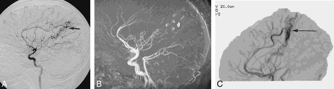
- fig 3.
9-year-old girl with a history of suspected AVM.
A, Lateral DSA image from a right internal carotid injection shows an AVM supplied by the pericallosal artery (arrow).
B, The AVM is seen less well on this lateral MIP image from a 3D time-of-flight MR angiogram (arrow), most likely owing to a combination of turbulence and in-plane flow artifacts.
C, The lateral MIP image of the CTCVI study is not susceptible to flow-related artifacts and clearly shows the malformation with its pericallosal supply (arrow).
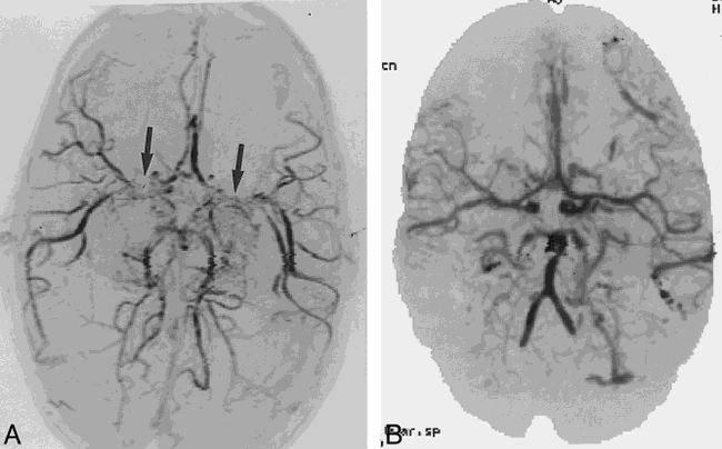
- fig 4.
4-year-old boy examined for increased head circumference and engorgement of superficial veins of the scalp.
A, 3D time-of-flight MR angiogram suggests moya-moya disease with bilateral supraclinoid carotid stenosis (arrows). The good flow distally in the middle cerebral arteries was puzzling.
B, CTCVI scan reveals patent proximal middle cerebral arteries as well as distal internal carotid arteries. Multiple small arteriovenous fistulas were also seen and confirmed at angiography. The MR signal loss was most likely related to the extremely high flow.


Tables

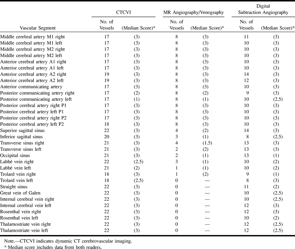
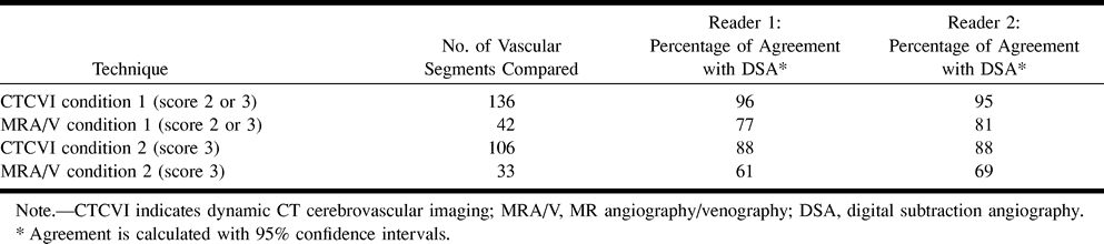
- TABLE 2:
Comparison of CTCVI with MR angiography/venography and digital subtraction angiography


