Advertisement

Main menu

  • Home
  • Content
    • Current Issue
    • Accepted Manuscripts
    • Article Preview
    • Past Issue Archive
    • Video Articles
    • AJNR Case Collection
    • Case of the Week Archive
    • Case of the Month Archive
    • Classic Case Archive
  • Special Collections
    • AJNR Awards
    • Low-Field MRI
    • Alzheimer Disease
    • ASNR Foundation Special Collection
    • Photon-Counting CT
    • View All
  • Multimedia
    • AJNR Podcasts
    • AJNR SCANtastic
    • Trainee Corner
    • MRI Safety Corner
    • Imaging Protocols
  • For Authors
    • Submit a Manuscript
    • Submit a Video Article
    • Submit an eLetter to the Editor/Response
    • Manuscript Submission Guidelines
    • Statistical Tips
    • Fast Publishing of Accepted Manuscripts
    • Graphical Abstract Preparation
    • Imaging Protocol Submission
    • Author Policies
  • About Us
    • About AJNR
    • Editorial Board
    • Editorial Board Alumni
  • More
    • Become a Reviewer/Academy of Reviewers
    • Subscribers
    • Permissions
    • Alerts
    • Feedback
    • Advertisers
    • ASNR Home

User menu

  • Alerts
  • Log in

Search

  • Advanced search
American Journal of Neuroradiology

American Journal of Neuroradiology

ASHNR American Society of Functional Neuroradiology ASHNR American Society of Pediatric Neuroradiology ASSR
  • Alerts
  • Log in

Advanced Search

  • Home
  • Content
    • Current Issue
    • Accepted Manuscripts
    • Article Preview
    • Past Issue Archive
    • Video Articles
    • AJNR Case Collection
    • Case of the Week Archive
    • Case of the Month Archive
    • Classic Case Archive
  • Special Collections
    • AJNR Awards
    • Low-Field MRI
    • Alzheimer Disease
    • ASNR Foundation Special Collection
    • Photon-Counting CT
    • View All
  • Multimedia
    • AJNR Podcasts
    • AJNR SCANtastic
    • Trainee Corner
    • MRI Safety Corner
    • Imaging Protocols
  • For Authors
    • Submit a Manuscript
    • Submit a Video Article
    • Submit an eLetter to the Editor/Response
    • Manuscript Submission Guidelines
    • Statistical Tips
    • Fast Publishing of Accepted Manuscripts
    • Graphical Abstract Preparation
    • Imaging Protocol Submission
    • Author Policies
  • About Us
    • About AJNR
    • Editorial Board
    • Editorial Board Alumni
  • More
    • Become a Reviewer/Academy of Reviewers
    • Subscribers
    • Permissions
    • Alerts
    • Feedback
    • Advertisers
    • ASNR Home
  • Follow AJNR on Twitter
  • Visit AJNR on Facebook
  • Follow AJNR on Instagram
  • Join AJNR on LinkedIn
  • RSS Feeds

AJNR Awards, New Junior Editors, and more. Read the latest AJNR updates

Research ArticleHead and Neck Imaging

Mucocele Involving the Anterior Clinoid Process: MR and CT Findings

C. C. Tchoyoson Lim, William P. Dillon and Michael W. McDermott
American Journal of Neuroradiology February 1999, 20 (2) 287-290;
C. C. Tchoyoson Lim
  • Find this author on Google Scholar
  • Find this author on PubMed
  • Search for this author on this site
William P. Dillon
  • Find this author on Google Scholar
  • Find this author on PubMed
  • Search for this author on this site
Michael W. McDermott
  • Find this author on Google Scholar
  • Find this author on PubMed
  • Search for this author on this site
  • Article
  • Figures & Data
  • Info & Metrics
  • Responses
  • References
  • PDF
Loading

Article Figures & Data

Figures

  • fig 1.
    • Download figure
    • Open in new tab
    • Download powerpoint
    fig 1.

    Case 1: 32-year-old woman with retroorbital headache and a long history of sinus disease.

    A, Schematic representation of a sagittal section through the sphenoidal sinus (S) shows the relationship between Onodi cell (arrows), optic nerve (O), and pituitary gland (P). (Reprinted with permission from [7].)

    B, T1-weighted (516/14/2 [TR/TE/excitations]) conventional spin-echo MR image shows an area of increased signal intensity (arrow) anterior and superior to the sphenoidal sinus (S), corresponding to the Onodi cell seen in A. Note that this is part of the posterior ethmoidal air cells (arrowhead).

    C, Axial T1-weighted image (600/14/2) shows the mucus-filled Onodi cell extending into the left anterior clinoid process (arrow) and inferior to the optic nerve (arrowheads). Note that on axial images the optic nerve appears to course through the mucocele.

    D, Coronal T1-weighted (550/14/2) (top) and T2-weighted (3700/105/2) (bottom) fast spin-echo MR images show that the mass (arrow) underlying the optic nerve (arrowhead) is of high signal intensity on the T1-weighted sequence and of intermediate intensity on the T2-weighted sequence.

  • fig 2.
    • Download figure
    • Open in new tab
    • Download powerpoint
    fig 2.

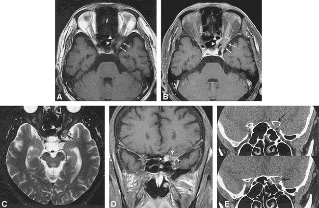
    Case 2: 61-year-old man with sudden-onset diplopia and forehead numbness. Fourth cranial nerve palsy as well as V1 sensory loss was found on physical examination.

    A, Axial T1-weighted image (600/14/2) shows a lesion of intermediate signal intensity extending laterally (arrows) as well as medially (arrowheads) around the left optic nerve. Note that, as in fig 1C, the optic nerve appears to course through the lesion, a clue to the sinus origin of the mass.

    B, Contrast-enhanced T1-weighted (500/20/2) image with fat saturation reveals mild peripheral enhancement (arrows, arrowheads).

    C, Axial T2-weighted (4000/105/2) fast spin-echo MR image shows the mass is isointense (arrow) to slightly hyperintense (arrowhead) relative to brain parenchyma. Mild inflammatory sinus disease is present in the ethmoidal air cells bilaterally.

    D, Coronal contrast-enhanced T1-weighted (583/14/2) conventional spin-echo sequence with fat saturation shows the enhancing mass (arrow) involving the left anterior clinoid process (arrowheads) extending into the orbital apex.

    E, Contiguous 1-mm high-resolution coronal CT scans show a soft-tissue attenuation lesion of the left anterior clinoid process (arrow) extending into the orbital apex. The cortex of the clinoid process is focally expanded and destroyed. A small polypoid soft-tissue mass (arrowhead) is also noted along the lateral surface of the left sphenoidal air cells.

PreviousNext
Back to top

In this issue

American Journal of Neuroradiology
Vol. 20, Issue 2
1 Feb 1999
  • Table of Contents
  • Index by author
Advertisement
Print
Download PDF
Email Article

Thank you for your interest in spreading the word on American Journal of Neuroradiology.

NOTE: We only request your email address so that the person you are recommending the page to knows that you wanted them to see it, and that it is not junk mail. We do not capture any email address.

Enter multiple addresses on separate lines or separate them with commas.
Mucocele Involving the Anterior Clinoid Process: MR and CT Findings
(Your Name) has sent you a message from American Journal of Neuroradiology
(Your Name) thought you would like to see the American Journal of Neuroradiology web site.
CAPTCHA
This question is for testing whether or not you are a human visitor and to prevent automated spam submissions.
Cite this article
C. C. Tchoyoson Lim, William P. Dillon, Michael W. McDermott
Mucocele Involving the Anterior Clinoid Process: MR and CT Findings
American Journal of Neuroradiology Feb 1999, 20 (2) 287-290;

Citation Manager Formats

  • BibTeX
  • Bookends
  • EasyBib
  • EndNote (tagged)
  • EndNote 8 (xml)
  • Medlars
  • Mendeley
  • Papers
  • RefWorks Tagged
  • Ref Manager
  • RIS
  • Zotero
0 Responses
Respond to this article
Share
Bookmark this article
Mucocele Involving the Anterior Clinoid Process: MR and CT Findings
C. C. Tchoyoson Lim, William P. Dillon, Michael W. McDermott
American Journal of Neuroradiology Feb 1999, 20 (2) 287-290;
del.icio.us logo Twitter logo Facebook logo Mendeley logo
  • Tweet Widget
  • Facebook Like
  • Google Plus One

Jump to section

  • Article
    • Abstract
    • Case Reports
    • Discussion
    • Conclusion
    • Acknowledgments
    • Footnotes
    • References
  • Figures & Data
  • Info & Metrics
  • Responses
  • References
  • PDF

Related Articles

  • No related articles found.
  • PubMed
  • Google Scholar

Cited By...

  • Sphenoid sinus mucocele causing third nerve paralysis: CT and MR findings
  • Acute visual loss by an Onodi cell
  • Crossref
  • Google Scholar

This article has not yet been cited by articles in journals that are participating in Crossref Cited-by Linking.

More in this TOC Section

  • ASL Sensitivity for Head and Neck Paraganglioma
  • Post SRS Peritumoral Hyperintense Signal of VSs
  • Contrast Enhanced Pituitary CISS/FIESTA
Show more Head and Neck Imaging

Similar Articles

Advertisement

Indexed Content

  • Current Issue
  • Accepted Manuscripts
  • Article Preview
  • Past Issues
  • Editorials
  • Editor's Choice
  • Fellows' Journal Club
  • Letters to the Editor
  • Video Articles

Cases

  • Case Collection
  • Archive - Case of the Week
  • Archive - Case of the Month
  • Archive - Classic Case

More from AJNR

  • Trainee Corner
  • Imaging Protocols
  • MRI Safety Corner
  • Book Reviews

Multimedia

  • AJNR Podcasts
  • AJNR Scantastics

Resources

  • Turnaround Time
  • Submit a Manuscript
  • Submit a Video Article
  • Submit an eLetter to the Editor/Response
  • Manuscript Submission Guidelines
  • Statistical Tips
  • Fast Publishing of Accepted Manuscripts
  • Graphical Abstract Preparation
  • Imaging Protocol Submission
  • Evidence-Based Medicine Level Guide
  • Publishing Checklists
  • Author Policies
  • Become a Reviewer/Academy of Reviewers
  • News and Updates

About Us

  • About AJNR
  • Editorial Board
  • Editorial Board Alumni
  • Alerts
  • Permissions
  • Not an AJNR Subscriber? Join Now
  • Advertise with Us
  • Librarian Resources
  • Feedback
  • Terms and Conditions
  • AJNR Editorial Board Alumni

American Society of Neuroradiology

  • Not an ASNR Member? Join Now

© 2025 by the American Society of Neuroradiology All rights, including for text and data mining, AI training, and similar technologies, are reserved.
Print ISSN: 0195-6108 Online ISSN: 1936-959X

Powered by HighWire