Advertisement

Main menu

  • Home
  • Content
    • Current Issue
    • Accepted Manuscripts
    • Article Preview
    • Past Issue Archive
    • Video Articles
    • AJNR Case Collection
    • Case of the Week Archive
    • Case of the Month Archive
    • Classic Case Archive
  • Special Collections
    • AJNR Awards
    • Low-Field MRI
    • Alzheimer Disease
    • ASNR Foundation Special Collection
    • Photon-Counting CT
    • View All
  • Multimedia
    • AJNR Podcasts
    • AJNR SCANtastic
    • Trainee Corner
    • MRI Safety Corner
    • Imaging Protocols
  • For Authors
    • Submit a Manuscript
    • Submit a Video Article
    • Submit an eLetter to the Editor/Response
    • Manuscript Submission Guidelines
    • Statistical Tips
    • Fast Publishing of Accepted Manuscripts
    • Graphical Abstract Preparation
    • Imaging Protocol Submission
    • Author Policies
  • About Us
    • About AJNR
    • Editorial Board
    • Editorial Board Alumni
  • More
    • Become a Reviewer/Academy of Reviewers
    • Subscribers
    • Permissions
    • Alerts
    • Feedback
    • Advertisers
    • ASNR Home

User menu

  • Alerts
  • Log in

Search

  • Advanced search
American Journal of Neuroradiology

American Journal of Neuroradiology

ASHNR American Society of Functional Neuroradiology ASHNR American Society of Pediatric Neuroradiology ASSR
  • Alerts
  • Log in

Advanced Search

  • Home
  • Content
    • Current Issue
    • Accepted Manuscripts
    • Article Preview
    • Past Issue Archive
    • Video Articles
    • AJNR Case Collection
    • Case of the Week Archive
    • Case of the Month Archive
    • Classic Case Archive
  • Special Collections
    • AJNR Awards
    • Low-Field MRI
    • Alzheimer Disease
    • ASNR Foundation Special Collection
    • Photon-Counting CT
    • View All
  • Multimedia
    • AJNR Podcasts
    • AJNR SCANtastic
    • Trainee Corner
    • MRI Safety Corner
    • Imaging Protocols
  • For Authors
    • Submit a Manuscript
    • Submit a Video Article
    • Submit an eLetter to the Editor/Response
    • Manuscript Submission Guidelines
    • Statistical Tips
    • Fast Publishing of Accepted Manuscripts
    • Graphical Abstract Preparation
    • Imaging Protocol Submission
    • Author Policies
  • About Us
    • About AJNR
    • Editorial Board
    • Editorial Board Alumni
  • More
    • Become a Reviewer/Academy of Reviewers
    • Subscribers
    • Permissions
    • Alerts
    • Feedback
    • Advertisers
    • ASNR Home
  • Follow AJNR on Twitter
  • Visit AJNR on Facebook
  • Follow AJNR on Instagram
  • Join AJNR on LinkedIn
  • RSS Feeds

AJNR Awards, New Junior Editors, and more. Read the latest AJNR updates

Research ArticleHead and Neck Imaging

Vasa Vasorum: Another Cause of the Carotid String Sign

Monty A. Martin and Tom R. Marotta
American Journal of Neuroradiology February 1999, 20 (2) 259-262;
Monty A. Martin
  • Find this author on Google Scholar
  • Find this author on PubMed
  • Search for this author on this site
Tom R. Marotta
  • Find this author on Google Scholar
  • Find this author on PubMed
  • Search for this author on this site
  • Article
  • Figures & Data
  • Info & Metrics
  • Responses
  • References
  • PDF
Loading

Article Figures & Data

Figures

  • fig 1.
    • Download figure
    • Open in new tab
    • Download powerpoint
    fig 1.

    Lateral left carotid angiograms in 60-year-old man with two prior CVAs.

    A, Early phase shows multiple serpiginous vessels projected over expected course of ICA (arrows).

    B, Later phase of A.
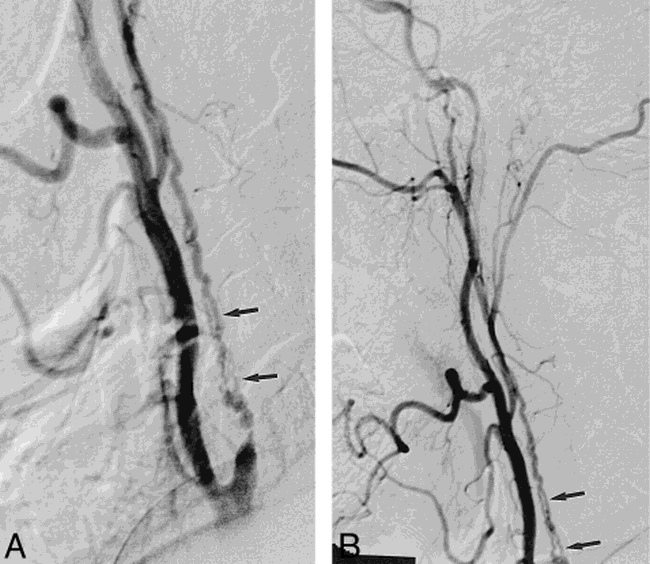
  • fig 2.
    • Download figure
    • Open in new tab
    • Download powerpoint
    fig 2.

    Lateral right carotid angiograms in 64-year-old woman with one prior CVA.

    A, Early phase shows internal carotid stump (curved arrow) and collateral vessel originating from below ICA takeoff (straight arrows).

    B and C, Later phases show continuation of collateral vessel to opacify distal ICA.

  • fig 3.
    • Download figure
    • Open in new tab
    • Download powerpoint
    fig 3.

    Lateral right carotid angiograms in 66-year-old man with multiple TIAs and one CVA.

    A and B, Multiple serpiginous vessels project over expected course of ICA (arrows) with anterograde filling of the distal ICA.
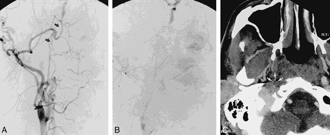
  • fig 4.
    • Download figure
    • Open in new tab
    • Download powerpoint
    fig 4.

    Frontal right carotid angiograms in 39-year-old man approximately 5 months after right carotid dissection with occlusion.

    A and B, Short-segment serpiginous network of vessels bridge narrowed cervical ICA to petrous portion of ICA (arrows).

    C, CT angiogram, transverse view, shows multiple small vessels (arrows) at perimeter of reduced-caliber thrombosed ICA.

PreviousNext
Back to top

In this issue

American Journal of Neuroradiology
Vol. 20, Issue 2
1 Feb 1999
  • Table of Contents
  • Index by author
Advertisement
Print
Download PDF
Email Article

Thank you for your interest in spreading the word on American Journal of Neuroradiology.

NOTE: We only request your email address so that the person you are recommending the page to knows that you wanted them to see it, and that it is not junk mail. We do not capture any email address.

Enter multiple addresses on separate lines or separate them with commas.
Vasa Vasorum: Another Cause of the Carotid String Sign
(Your Name) has sent you a message from American Journal of Neuroradiology
(Your Name) thought you would like to see the American Journal of Neuroradiology web site.
CAPTCHA
This question is for testing whether or not you are a human visitor and to prevent automated spam submissions.
Cite this article
Monty A. Martin, Tom R. Marotta
Vasa Vasorum: Another Cause of the Carotid String Sign
American Journal of Neuroradiology Feb 1999, 20 (2) 259-262;

Citation Manager Formats

  • BibTeX
  • Bookends
  • EasyBib
  • EndNote (tagged)
  • EndNote 8 (xml)
  • Medlars
  • Mendeley
  • Papers
  • RefWorks Tagged
  • Ref Manager
  • RIS
  • Zotero
0 Responses
Respond to this article
Share
Bookmark this article
Vasa Vasorum: Another Cause of the Carotid String Sign
Monty A. Martin, Tom R. Marotta
American Journal of Neuroradiology Feb 1999, 20 (2) 259-262;
del.icio.us logo Twitter logo Facebook logo Mendeley logo
  • Tweet Widget
  • Facebook Like
  • Google Plus One

Jump to section

  • Article
    • Abstract
    • Methods
    • Results
    • Discussion
    • Conclusion
    • Footnotes
    • References
  • Figures & Data
  • Info & Metrics
  • Responses
  • References
  • PDF

Related Articles

  • No related articles found.
  • PubMed
  • Google Scholar

Cited By...

  • Development of Collateral Vessels after Anterior Circulation Large Vessel Occlusion in Pediatric Arterial Ischemic Stroke Relates to Stroke Etiology: A Longitudinal Study
  • Republished: Pipeline embolization device thrombosis induced peri-construct collateral channels
  • Carotid Near-Occlusion: A Comprehensive Review, Part 1--Definition, Terminology, and Diagnosis
  • Pipeline embolization device thrombosis induced peri-construct collateral channels
  • Vasa Vasorum Enhancement on Computerized Tomographic Angiography Correlates With Symptomatic Patients With 50% to 70% Carotid Artery Stenosis
  • Recanalisation of the internal carotid artery via the vasa vasorum after coil occlusion
  • Arterial Wall Enhancement Overlying Carotid Plaque on CT Angiography Correlates With Symptoms in Patients With High Grade Stenosis
  • Carotid Pseudostring Sign From Vasa Vasorum Collaterals
  • Total Occlusion versus Hairline Residual Lumen of the Internal Carotid Arteries: Accuracy of Single Section Helical CT Angiography
  • Crossref
  • Google Scholar

This article has not yet been cited by articles in journals that are participating in Crossref Cited-by Linking.

More in this TOC Section

  • FDG Uptake and Thyroid Cancer Post-BRAF Therapy
  • Normal Facial Nerve Enhancement on VIBE Sequence
  • Comprehensive Review of Inner Ear Anatomy on Photon-Counting CT
Show more Head and Neck Imaging

Similar Articles

Advertisement

Indexed Content

  • Current Issue
  • Accepted Manuscripts
  • Article Preview
  • Past Issues
  • Editorials
  • Editor's Choice
  • Fellows' Journal Club
  • Letters to the Editor
  • Video Articles

Cases

  • Case Collection
  • Archive - Case of the Week
  • Archive - Case of the Month
  • Archive - Classic Case

More from AJNR

  • Trainee Corner
  • Imaging Protocols
  • MRI Safety Corner

Multimedia

  • AJNR Podcasts
  • AJNR Scantastics

Resources

  • Turnaround Time
  • Submit a Manuscript
  • Submit a Video Article
  • Submit an eLetter to the Editor/Response
  • Manuscript Submission Guidelines
  • Statistical Tips
  • Fast Publishing of Accepted Manuscripts
  • Graphical Abstract Preparation
  • Imaging Protocol Submission
  • Evidence-Based Medicine Level Guide
  • Publishing Checklists
  • Author Policies
  • Become a Reviewer/Academy of Reviewers
  • News and Updates

About Us

  • About AJNR
  • Editorial Board
  • Editorial Board Alumni
  • Alerts
  • Permissions
  • Not an AJNR Subscriber? Join Now
  • Advertise with Us
  • Librarian Resources
  • Feedback
  • Terms and Conditions
  • AJNR Editorial Board Alumni

American Society of Neuroradiology

  • Not an ASNR Member? Join Now

© 2025 by the American Society of Neuroradiology All rights, including for text and data mining, AI training, and similar technologies, are reserved.
Print ISSN: 0195-6108 Online ISSN: 1936-959X

Powered by HighWire