
Abstract
BACKGROUND AND PURPOSE: Ultrasmall particles of iron oxide (USPIO) constitute a contrast agent that accumulates in cells from the mononuclear phagocytic system. In the CNS they may accumulate in phagocytic cells such as macrophages. The goal of this study was to compare USPIO-enhanced MR images with conventional T2-weighted images and gadolinium-enhanced T1-weighted images in a model of experimental autoimmune encephalomyelitis (EAE).
METHODS: Nine rats with EAE and four control rats were imaged at 4.7 T and 1.5 T with conventional T1- and T2-weighted sequences, gadolinium-enhanced T1-weighted sequences, and T2-weighted sequences obtained 24 hours after intravenous injection of a USPIO contrast agent, AMI-227. Histologic examination was performed with hematoxylin-eosin stain, Perls' stain for iron, and ED1 immunohistochemistry for macrophages.
RESULTS: USPIO-enhanced images showed a high sensitivity (8/9) for detecting EAE lesions, whereas poor sensitivity was obtained with T2-weighted images (1/9) and gadolinium-enhanced T1-weighted images (0/9). All the MR findings in the control rats were negative. Histologic examination revealed the presence of macrophages at the site where abnormalities were seen on USPIO-enhanced images.
CONCLUSION: The high sensitivity of USPIO for macrophage activity relative to other imaging techniques is explained by the histologic findings of numerous perivascular cell infiltrates, including macrophages, in EAE. This work supports the possibility of intracellular USPIO transport to the CNS by monocytes/macrophages, which may have future implications for imaging of human inflammatory diseases.
Ultrasmall particles of iron oxide (USPIO) are dextran-coated iron oxide nanoparticles that are internalized into a variety of cells, among which are cells of the mononuclear phagocytic system (1, 2). Owing to the presence of iron, they produce a susceptibility effect that is observable on T2- and T2*-weighted MR images as low signal intensity (1). They are currently used as a contrast agent for human lymphography in phase III studies. After time, these particles are found inside macrophages in normal lymph nodes (3).
Few experimental studies have applied USPIO-enhanced MR imaging to the CNS (4, 5). USPIO may either accumulate in experimental glioma cells or in macrophages of the CNS in inflammatory diseases, such as experimental autoimmune encephalomyelitis (EAE). EAE is an animal model of multiple sclerosis (MS) characterized by an inflammatory cell reaction, including lymphocyte and macrophage infiltration and activation of microglial cells (6, 7). EAE has been investigated with several MR imaging techniques, such as conventional T2-weighted sequences, magnetization transfer sequences, diffusion-weighted sequences, and gadolinium contrast-enhanced sequences (8–12). The reported sensitivity of such techniques is variable, depending on the species, the nature of the immunogenic compound, and the MR technique applied. Using a guinea pig model of EAE, Karlik et al (11) found a sensitivity of 50% with gadolinium-enhanced imaging, and Grossman et al (8) found a sensitivity of 42% with T2-weighted imaging.
In the present work, we sought to compare USPIO-enhanced imaging, conventional T2-weighted imaging, and gadolinium-enhanced T1-weighted MR imaging in the depiction of EAE lesions.
Methods
Experimental Acute Encephalomyelitis
An EAE model was induced in 14 female Lewis rats using guinea pig CNS together with Freund's complete adjuvant and H37RA mycobacterium tuberculosis. Immunization was performed by intradermal inoculation of 0.1 mL in each hind leg under general anesthesia. A control group of four rats was inoculated with normal saline solution under the same conditions as the experimental group.
All the animals were examined and weighed every other day and kept in individual cages with standardized conditions of light and free access to water and food. We followed the ethical recommendations that are in use at our institution.
We considered as D0 the day of onset of clinical signs of the disease, which were graded according to the following clinical scale: 0 = normal healthy animal, 1 = loss of tail tonus, 2 = hind limb weakness, 3 = complete hind limb paralysis, 4 = complete paralysis, incontinence, and moribund conditions.
To obtain maximum sensitivity, MR studies were performed 2 days after the emergence of clinical signs; that is, at D2 in the rats with EAE and a clinical score of 2 or above.
Contrast Agents
The USPIO contrast agent was AMI-227 (Sinerem, Laboratory Guerbet, Aulnay, France). It is composed of an iron core surrounded by a dextran coat. The mean diameter of this particle (iron + dextran) is 30 nm, and its blood half-life at the dose of 300 μmol Fe/kg is about 5 to 6 hours in Lewis rats. Because of its size, AMI-227 cannot diffuse easily out of the vascular space so it is mainly cleared from blood by macrophages.
For gadolinium-enhanced imaging, we used gadolinium tetraazacyclododecane (Gd-DOTA) at a dose of 0.5 mmol/kg.
The contrast agents were injected intravenously through the tail vein, using a 24-gauge catheter.
Imaging Protocol
MR studies were performed at 4.7 T on a Brucker Biospec imager using a custom-made bird-cage type head coil of 5 cm in diameter and on a 1.5-T Siemens Magnetom SP 63 with a soft flexible coil for imaging small joints. At 4.7 T, the imaging sequences were as follows: a sagittal localizer spin-echo (SE) T1-weighted (500/20 [TR/TE]) sequence; a nine-section coronal SE T1-weighted (509/18) sequence (matrix = 256 × 256, field of view [FOV] = 4.0 cm, section thickness = 3 mm); and a nine-section coronal SE T2-weighted (2030/60) sequence (matrix = 256 × 256, FOV = 4.0 cm, section thickness = 3 mm). At 1.5 T, imaging sequences included a nine-section coronal SE T1-weighted (400/20/3) sequence (matrix = 256 × 256, FOV = 12 cm, section thickness = 3 mm) and a nine-section coronal turbo SE T2-weighted (5000/20) sequence (matrix = 256 × 256, FOV = 12 cm, thickness = 3 mm). Eight rats with EAE and four control rats were imaged at 4.7 T and one EAE rat at 1.5 T at D2 and D3; that is, 24 hours after USPIO injection.
Sacrifice
Seven animals were sacrificed using an intraperitoneal overdose of pentobarbital sodium; 11 rats were sacrificed withintracardiac perfusion of paraformaldehyde.
Histologic Examinations
Histologic examination was performed on all animal brains with hematoxylin-eosin stain to assess inflammatory infiltrates and with Prussian blue or Perls' technique for iron staining.
The nature of the infiltrated cells was determined by immunohistochemistry in five rats with EAE and in two control rats using ED1, a specific marker for cells with macrophage capacity expressing the ED1 lysosomal antigen (macrophages, monocytes, and active microglia) (6). Correlation of areas with positive ED1 antibodies and abnormal signal intensity on MR images was established by comparing histologic sections with each of the nine MR sections of each sequence. The correlations were performed by two readers, who simultaneously compared MR images with histologic sections.
Results
Clinical Findings
Eleven (79%) of 14 rats with EAE showed clinical abnormalities 10 to 12 days after immunization, nine with a clinical score of 2 or greater at D2. None of the control rats had any clinical abnormalities.
MR Findings
The results of MR imaging are detailed in the Table. On T2-weighted images without USPIO, one rat with EAE had a small focus of signal increase at D2 (Fig 1). Findings in all the control rats were normal. On Gd-DOTA--enhanced T1-weighted images, no abnormal enhancement was observed at 4.7 T and 1.5 T, either in the rats with EAE or in the control group, at D2 and D3. On T2-weighted images obtained 2 hours after injection of USPIO, a global signal decrease of the brain parenchyma was observed in all EAE and control rats. On T2-weighted images obtained 24 hours after USPIO injection (D3), multiple focal areas of abnormal low signal intensity were found within the CNS parenchyma, mainly in the periventricular areas of the cerebellum and brain stem in eight rats with EAE imaged at 4.7 T (Fig 2) and at 1.5 T (Fig 3). None of the control rats had abnormalities. Although the spatial resolution was higher at 4.7 T, the imaging findings were similar at 1.5 and 4.7 T.
MR findings in nine rats with experimental autoimmune encephalomyelitis (EAE) and four control animals

Coronal SE T2-weighted (2030/60/4) MR image obtained at 4.7 T in a rat with EAE. The section is at the level of the midbrain (arrowhead indicates the right cerebral hemisphere). A small area of increased signal intensity is present around the sylvian aqueduct (arrow)

A and B, Coronal SE T2-weighted images of the brains of two rats with clinical EAE, imaged 24 hours after administration of USPIO (AMI-227) at 4.7 T. The section is at the level of the posterior fossa (arrowheads indicate the right cerebellar hemisphere; long arrows, the pons). Numerous lesions are seen as low signal intensities related to the magnetic susceptibility effects caused by iron particles within phagocytic cells (small arrows in A).
fig 3. Coronal turbo SE T2-weighted (5000/20/1) MR image of the brain of a rat with EAE, imaged 24 hours after administration of AMI-227 at 1.5 T. Multiple areas of low signal are seen within the posterior fossa (arrows).
Histologic Findings
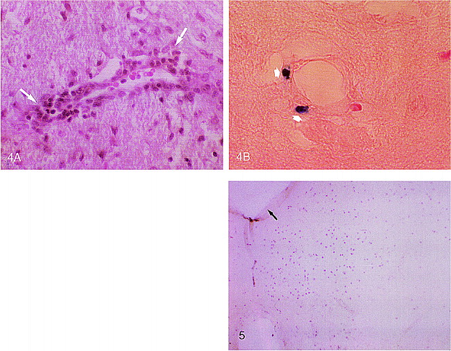
Hematoxylin-eosin stain showed multiple focal areas with perivascular inflammatory infiltrates in the CNS parenchyma of all rats with EAE that were examined (Fig 4A). These infiltrates were located in the same areas in which focal low signal intensities were seen on T2-weighted images 24 hours after injection of AMI-227. There was no sign of edema (enlarged or displaced structures) and no necrosis or hemorrhage.

Histologic sections at a site of low signal on MR images after administration of AMI-227.
A, Hematoxylin-eosin stain shows multiple perivascular inflammatory infiltrates (arrows).
B, Prussian blue stain for iron (Perls' technique) shows staining of perivascular cells (arrows).
fig 5. Histologic section at the level of the posterior fossa (arrow indicates fourth ventricle) shows immunostaining with an ED1 antibody macrophage marker. Macrophage infiltrates correspond to magnetic susceptibility–related low signal intensities on T2-weighted images 24 hours after USPIO injection and to infiltrating cells on hematoxylin-eosin stain.
Prussian blue stain for iron was positive in some areas, showing focal blue intracellular spots in perivascular inflammatory cells (Fig 4B). We did not see iron staining in the intravascular or interstitial spaces in either perfused or nonperfused animals.
With an ED1 antibody macrophage marker, immunostaining was positive in focal areas, mostly around vascular structures (Fig 5). There was good correlation between immunocytochemistry staining, perivascular infiltrating cells, and the presence of low signal intensity on T2-weighted imagesobtained 24 hours after USPIO injection.
Discussion
Our findings demonstrate the capability of USPIO-enhanced MR imaging to show infiltrating cells within the CNS in vivo. USPIO has a long half-life (4 to 5 hours) within the bloodstream before it accumulates within cells of the mononuclear phagocytic system. This explains the presence of global low signal intensity in the brains of both EAE rats and normal control rats 2 hours after USPIO administration. Imaging studies performed 24 hours after administration of USPIO showed the particles to be internalized in the phagocytic cells of EAE animals only. The same time interval between intravenous injection and imaging has been used previously for human lymphography, experimental gliomas, and EAE (4, 5).
In EAE, histologic examinations showed the presence of numerous infiltrating cells in perivascular areas. These cells are attracted from the blood by the secretion of chemoattractant factors from CNS cells (7). The reactive cells usually cross the blood-brain barrier (BBB) by diapedesis (6, 7). Among such cells, monocytes that become macrophages within the CNS are numerous (6). They act as phagocytes and secretory cells, releasing cytokines and free radicals (13). In the brain parenchyma, microglial cells are activated and acquire the same properties as hematogenous macrophages (14). Other pathologic findings, such as edema and demyelination, were not present. This is consistent with the EAE model in Lewis rats injected via whole-brain immunization, which usually produces an acute inflammatory disease with recovery after 5 to 7 days. Hence, imaging and pathologic examination were performed at D2 or D3, during the early process of the disease.
Conventional T2-weighted images without USPIO showed positive findings in only one of nine rats with EAE. In earlier studies, Grossman et al (8) found abnormalities on T2-weighted images in six of 14 EAE guinea pigs studied; Dousset et al (9) observed T2 abnormalities in two of five EAE guinea pigs; and higher sensitivity was found by Stewart et al (10) and Rose et al (15) in monkeys with EAE, in which lesions on T2-weighted images were seen in 100% of clinically affected animals. Signal abnormalities on T2-weighted images depend on the variation of water content in the interstitial space, which in acute EAE is related to the presence of vasogenic edema caused by the inflammatory reaction. Nevertheless, a minimum of water increase is necessary to create contrast, which may not be present in many acute inflammatory lesions. Thus, there is often a discrepancy between histologic findings of disseminated inflammatory cell infiltrates and the number of lesions seen on T2-weighted MR images (10). In the present study, such a discrepancy was observed. Histologically, there was no mass effect or displacement of brain structures that could be related to edema. However, numerous inflammatory cells were seen with hematoxylin-eosin staining.
Gd-DOTA enhancement was not present in this EAE model. One of the reasons that might explain the absence of this enhancement in our work is that we performed some of the studies at 4.7 T. Contrast between enhancing and nonenhancing tissues diminishes with increasing field strength (16). However, to increase the uptake of gadolinium chelates, we used a dose five times higher than the recommended dose for humans. In addition, one animal was imaged at 1.5 T with Gd-DOTA, and no abnormalities were observed, whereas the same animal showed lesions with USPIO enhancement. One previous study established a sensitivity of 47% (nine of 19 clinically affected animals) in guinea pigs with EAE using gadolinium-enhanced imaging (11). This low sensitivity may be explained by a transient or absent BBB disruption (12). Chelates of gadolinium are very small particles as compared with USPIO nanoparticles. Thus, in our study, the discrepancy between negative conventional Gd-DOTA enhancement and positive USPIO uptake suggests that the crossing of free USPIO particles through a ruptured BBB did not occur. Accordingly, two hypotheses may be proposed: 1) the USPIO may have passed through the endothelial cells, been released in the interstitial space, and finally captured by macrophagic cells; and 2) iron particles from the USPIO may have been internalized in hematogenous monocytes/macrophages before the cells reached the inflammatory site. Our work was not designed to discriminate between the two mechanisms. Regardless, it is potentially important to know that in the brain a contrast agent such as USPIO has the potential to delineate the presence of macrophagic cells at a site in which a gadolinium contrast agent may not indicate rupture of the BBB.
Macrophages are considered important effector cells in EAE and MS lesions. In MS, macrophages are involved in many physiopathologic processes, including inflammation and demyelination (17). To increase the ability of MR to depict disease activity, imaging of macrophage activity may be of interest. The use of USPIO might be extended to many CNS diseases in which macrophages play a major role in the pathophysiological process, such as in trauma, infection, and inflammation (18, 19).
Conclusion
MR imaging with USPIO in an EAE model revealed the presence, in vivo, of lesions characterized by perivascular cell infiltrates in sites where conventional T2-weighted images and Gd-DOTA–enhanced T1-weighted images were negative.
Acknowledgments
We are grateful to Valerie Sesay for linguistic assistance and to Bruno Bonnemain, Soraya Benderbous, and Xavier Violas for scientific support.
Footnotes
↵1 Presented at the annual meeting of the American Society of Neuroradiology, Toronto, Canada, May 1997.
↵2 Address reprint requests to Vincent Dousset MD, PhD, Laboratoire de NeuroBiologie et NeuroImagerie Expérimentales, BP 78, Université Victor Segalen Bordeaux2, 144 Rue Léo-Saignat, 33076 Bordeaux, France.
References
- Received April 21, 1998.
- Copyright © American Society of Neuroradiology
In this issue


Jump to section
Related Articles
Cited By...
- Magnetic Resonance Imaging in Multiple Sclerosis
- In vivo nanoparticle imaging of innate immune cells can serve as a marker of disease severity in a model of multiple sclerosis
- Imaging circulating cells and lymphoid tissues with iron oxide nanoparticles
- Imaging Inflammation in Acute Brain Ischemia
- Metalloproteinases Control Brain Inflammation Induced by Pertussis Toxin in Mice Overexpressing the Chemokine CCL2 in the Central Nervous System
- MRI techniques to monitor MS evolution: The present and the future
- Observation on the Integrity of the Blood-Brain Barrier After Microbubble Destruction by Diagnostic Transcranial Color-Coded Sonography