
Article Figures & Data
Figures
- fig 1.
Coronal images through the orbits of a patient with lymphoma of the lacrimal gland. Sequences are T2-weighted SPIR (A), SPIR/FLAIR (B ), T1-weighted SPIR with contrast (C ), and STIR (D).
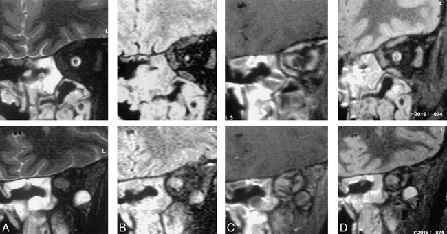
- fig 2.
Coronal images through the orbits of a patient with an orbital apex meningioma. The sequences are T2-weighted SPIR (A), SPIR/FLAIR (B ), T1-weighted SPIR with contrast (C ), and STIR (D).
Images in the mid orbit (top row) show normal appearances on the STIR (top of D) and T2-weighted SPIR (top of A) images. The SPIR/FLAIR image (top of B) clearly shows increased signal in the optic nerve itself. Postcontrast T1-weighted SPIR (top of C) shows thickening of the optic nerve sheath because of meningioma en plaque. Images at the orbital apex (bottom row) demonstrate a mass lesion in the position of the optic nerve–sheath complex. The contrast between the lesion and surrounding tissues is greater on the SPIR/FLAIR image (bottom of B) than on the T2-weighted SPIR (bottom of A) or STIR (bottom of D) images. On the postcontrast T1-weighted SPIR image (bottom of C) the lesion shows inhomogeneous enhancement and is difficult to distinguish from adjacent enhancing extraocular muscles.
- fig 3.
Axial images through the globe in a patient with a small melanotic melanoma with an associated retinal detachment. The SPIR/FLAIR sequence (A) shows the extent of the detachment more clearly than the T2-weighted SPIR (B ) and STIR images (C ), but does not reveal the tumor itself.
- fig 4.
Coronal images through the orbit in a patient with an orbital apex meningioma. The extensive intracranial en plaque spread is shown well on the T1-weighted SPIR sequence with contrast (C ) and also can be appreciated on the T2-weighted SPIR (A) and STIR (D) images it is outlined by high-signal CSF. Although the meningeal thickening can be seen on the SPIR/FLAIR sequences (B ), its presence and extent were not appreciated by either of the radiologists who were reporting on this scan in isolation.
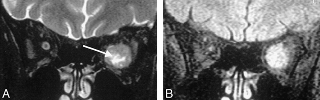
- fig 5.
Coronal images in a patient with an orbital meningioma. A central area of cystic necrosis visible on T2-weighted SPIR images (A, arrow) is not apparent on SPIR/FLAIR images (B).


Tables

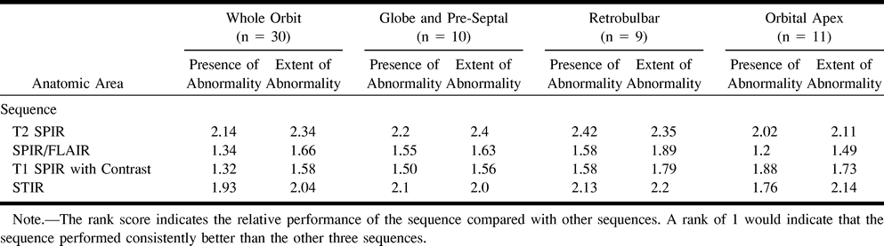
Mean rankings for presence and extent of abnormality for the whole orbit and by orbital region
In this issue


Jump to section
Related Articles
Cited By...
- Enhanced Myocardial Tissue Visualization: A Comparative Cardiovascular Magnetic Resonance Study of Gradient-Spin Echo-STIR and Conventional STIR Imaging
- Choroidal Metastases From Esophageal Adenocarcinoma Responding to Chemotherapy With Cisplatin and Irinotecan
- Intracanalicular Optic Nerve Meningioma: A Serious Diagnostic Pitfall