Advertisement
  • Main menu
  • User menu
  • Search
  • English ▼
    • English
    • Afrikaans
    • Albanian
    • Amharic
    • Arabic
    • Armenian
    • Azerbaijani
    • Basque
    • Belarusian
    • Bengali
    • Bosnian
    • Bulgarian
    • Catalan
    • Cebuano
    • Chichewa
    • Chinese (Simplified)
    • Chinese (Traditional)
    • Corsican
    • Croatian
    • Czech
    • Danish
    • Dutch
    • Esperanto
    • Estonian
    • Filipino
    • Finnish
    • French
    • Frisian
    • Galician
    • Georgian
    • German
    • Greek
    • Gujarati
    • Haitian Creole
    • Hausa
    • Hawaiian
    • Hebrew
    • Hindi
    • Hmong
    • Hungarian
    • Icelandic
    • Igbo
    • Indonesian
    • Irish
    • Italian
    • Japanese
    • Javanese
    • Kannada
    • Kazakh
    • Khmer
    • Korean
    • Kurdish (Kurmanji)
    • Kyrgyz
    • Lao
    • Latin
    • Latvian
    • Lithuanian
    • Luxembourgish
    • Macedonian
    • Malagasy
    • Malay
    • Malayalam
    • Maltese
    • Maori
    • Marathi
    • Mongolian
    • Myanmar (Burmese)
    • Nepali
    • Norwegian
    • Pashto
    • Persian
    • Polish
    • Portuguese
    • Punjabi
    • Romanian
    • Russian
    • Samoan
    • Scottish Gaelic
    • Serbian
    • Sesotho
    • Shona
    • Sindhi
    • Sinhala
    • Slovak
    • Slovenian
    • Somali
    • Spanish
    • Sudanese
    • Swahili
    • Swedish
    • Tajik
    • Tamil
    • Telugu
    • Thai
    • Turkish
    • Ukrainian
    • Urdu
    • Uzbek
    • Vietnamese
    • Welsh
    • Xhosa
    • Yiddish
    • Yoruba
    • Zulu

Main menu

  • Home
  • Content
    • Current Issue
    • Accepted Manuscripts
    • Article Preview
    • Past Issue Archive
    • Video Articles
    • AJNR Case Collection
    • Case of the Week Archive
    • Case of the Month Archive
    • Classic Case Archive
  • Special Collections
    • AJNR Awards
    • Low-Field MRI
    • Alzheimer Disease
    • ASNR Foundation Special Collection
    • Photon-Counting CT
    • View All
  • Multimedia
    • AJNR Podcasts
    • AJNR SCANtastic
    • Trainee Corner
    • MRI Safety Corner
    • Imaging Protocols
  • For Authors
    • Submit a Manuscript
    • Submit a Video Article
    • Submit an eLetter to the Editor/Response
    • Manuscript Submission Guidelines
    • Statistical Tips
    • Fast Publishing of Accepted Manuscripts
    • Graphical Abstract Preparation
    • Imaging Protocol Submission
    • Author Policies
  • About Us
    • About AJNR
    • Editorial Board
    • Editorial Board Alumni
  • More
    • Become a Reviewer/Academy of Reviewers
    • Subscribers
    • Permissions
    • Alerts
    • Feedback
    • Advertisers
    • ASNR Home

User menu

  • Alerts
  • Log in

Search

  • Advanced search
American Journal of Neuroradiology

American Journal of Neuroradiology

ASHNR American Society of Functional Neuroradiology ASHNR American Society of Pediatric Neuroradiology ASSR
  • Alerts
  • Log in

Advanced Search

English ▼
  • English
  • Afrikaans
  • Albanian
  • Amharic
  • Arabic
  • Armenian
  • Azerbaijani
  • Basque
  • Belarusian
  • Bengali
  • Bosnian
  • Bulgarian
  • Catalan
  • Cebuano
  • Chichewa
  • Chinese (Simplified)
  • Chinese (Traditional)
  • Corsican
  • Croatian
  • Czech
  • Danish
  • Dutch
  • Esperanto
  • Estonian
  • Filipino
  • Finnish
  • French
  • Frisian
  • Galician
  • Georgian
  • German
  • Greek
  • Gujarati
  • Haitian Creole
  • Hausa
  • Hawaiian
  • Hebrew
  • Hindi
  • Hmong
  • Hungarian
  • Icelandic
  • Igbo
  • Indonesian
  • Irish
  • Italian
  • Japanese
  • Javanese
  • Kannada
  • Kazakh
  • Khmer
  • Korean
  • Kurdish (Kurmanji)
  • Kyrgyz
  • Lao
  • Latin
  • Latvian
  • Lithuanian
  • Luxembourgish
  • Macedonian
  • Malagasy
  • Malay
  • Malayalam
  • Maltese
  • Maori
  • Marathi
  • Mongolian
  • Myanmar (Burmese)
  • Nepali
  • Norwegian
  • Pashto
  • Persian
  • Polish
  • Portuguese
  • Punjabi
  • Romanian
  • Russian
  • Samoan
  • Scottish Gaelic
  • Serbian
  • Sesotho
  • Shona
  • Sindhi
  • Sinhala
  • Slovak
  • Slovenian
  • Somali
  • Spanish
  • Sudanese
  • Swahili
  • Swedish
  • Tajik
  • Tamil
  • Telugu
  • Thai
  • Turkish
  • Ukrainian
  • Urdu
  • Uzbek
  • Vietnamese
  • Welsh
  • Xhosa
  • Yiddish
  • Yoruba
  • Zulu
  • Home
  • Content
    • Current Issue
    • Accepted Manuscripts
    • Article Preview
    • Past Issue Archive
    • Video Articles
    • AJNR Case Collection
    • Case of the Week Archive
    • Case of the Month Archive
    • Classic Case Archive
  • Special Collections
    • AJNR Awards
    • Low-Field MRI
    • Alzheimer Disease
    • ASNR Foundation Special Collection
    • Photon-Counting CT
    • View All
  • Multimedia
    • AJNR Podcasts
    • AJNR SCANtastic
    • Trainee Corner
    • MRI Safety Corner
    • Imaging Protocols
  • For Authors
    • Submit a Manuscript
    • Submit a Video Article
    • Submit an eLetter to the Editor/Response
    • Manuscript Submission Guidelines
    • Statistical Tips
    • Fast Publishing of Accepted Manuscripts
    • Graphical Abstract Preparation
    • Imaging Protocol Submission
    • Author Policies
  • About Us
    • About AJNR
    • Editorial Board
    • Editorial Board Alumni
  • More
    • Become a Reviewer/Academy of Reviewers
    • Subscribers
    • Permissions
    • Alerts
    • Feedback
    • Advertisers
    • ASNR Home
  • Follow AJNR on Twitter
  • Visit AJNR on Facebook
  • Follow AJNR on Instagram
  • Join AJNR on LinkedIn
  • RSS Feeds

AJNR Awards, New Junior Editors, and more. Read the latest AJNR updates

Research ArticleBrain

The Cerebellum's Role in Reading: A Functional MR Imaging Study

Robert K. Fulbright, Annette R. Jenner, W. Einer Mencl, Kenneth R. Pugh, Bennett A. Shaywitz, Sally E. Shaywitz, Stephen J. Frost, Pawel Skudlarski, R. Todd Constable, Cheryl M. Lacadie, Karen E. Marchione and John C. Gore
American Journal of Neuroradiology November 1999, 20 (10) 1925-1930;
Robert K. Fulbright
  • Find this author on Google Scholar
  • Find this author on PubMed
  • Search for this author on this site
Annette R. Jenner
  • Find this author on Google Scholar
  • Find this author on PubMed
  • Search for this author on this site
W. Einer Mencl
  • Find this author on Google Scholar
  • Find this author on PubMed
  • Search for this author on this site
Kenneth R. Pugh
  • Find this author on Google Scholar
  • Find this author on PubMed
  • Search for this author on this site
Bennett A. Shaywitz
  • Find this author on Google Scholar
  • Find this author on PubMed
  • Search for this author on this site
Sally E. Shaywitz
  • Find this author on Google Scholar
  • Find this author on PubMed
  • Search for this author on this site
Stephen J. Frost
  • Find this author on Google Scholar
  • Find this author on PubMed
  • Search for this author on this site
Pawel Skudlarski
  • Find this author on Google Scholar
  • Find this author on PubMed
  • Search for this author on this site
R. Todd Constable
  • Find this author on Google Scholar
  • Find this author on PubMed
  • Search for this author on this site
Cheryl M. Lacadie
  • Find this author on Google Scholar
  • Find this author on PubMed
  • Search for this author on this site
Karen E. Marchione
  • Find this author on Google Scholar
  • Find this author on PubMed
  • Search for this author on this site
John C. Gore
  • Find this author on Google Scholar
  • Find this author on PubMed
  • Search for this author on this site
  • Article
  • Figures & Data
  • Info & Metrics
  • Responses
  • References
  • PDF
Loading

Article Figures & Data

Figures

  • Tables
  • fig 1.
    • Download figure
    • Open in new tab
    • Download powerpoint
    fig 1.

    Composite maps (first order, see Table) show regional cerebellar activation during the four reading tasks. Column one compares the case task with the line task (C/L), column two compares the word rhyme task with the line task (WR/L), column three compares the non-word rhyme task with the line task (NWR/L), and column four compares the category task with the line task (CT/L). There is a progressive increase in demand on cognitive processing in going from column one to column four. Numbers indicate cerebellar regions that were more active (P = .005, red-yellow scale) in a reading task compared with the line task; letters indicate areas of the cerebellum that were more active (P = .005, blue-purple scale) in the line task compared with a reading task. 1, anterior aspect of the simple lobule; 2, middle and lateral aspects of the posterior superior fissure and adjacent simple lobule and superior semilunar lobule; 3, middle aspect of the horizontal fissure and adjacent superior semilunar lobule and inferior semilunar lobule; 4, middle aspect of the prepyramidal fissure and adjacent inferior semilunar lobule; 5, posterior and lateral aspects of the horizontal fissure and adjacent superior semilunar lobule and inferior semilunar lobule; 6, posterior and lateral aspects of the posterior superior fissure and adjacent simple lobule and superior semilunar lobule; 7, posterior aspect of inferior semilunar lobule; and 8, posterior and medial aspects of the posterior superior fissure and adjacent simple lobule and superior semilunar lobule. a, inferior vermis; b, postpyramidal fissure and medial aspect of the tonsils; c, biventer lobule; d, medial aspect of the biventer lobule; and e, the middle and posterior aspects of the horizontal fissure and adjacent superior semilunar lobule and inferior semilunar lobule. Section locations in each column from superior to inferior correspond to the following approximate y axis positions of the Talairach atlas: −40, −50, −60, −70, −80, and −90.

  • fig 2.
    • Download figure
    • Open in new tab
    • Download powerpoint
    fig 2.

    Composite maps (second order, see Table) contrast cerebellar activation during different reading conditions. The SPMs from the case/line comparison served as a baseline for comparison with the SPMs from other task comparisons to generate the three composite maps: non-word rhyme (non-word rhyme/line versus case/line, column one), word rhyme (word-rhyme/line versus case/line, column two), and category (category/line versus case/line, column three). Numbers indicate cerebellar regions that were more active (P = .005, red-yellow scale) in either category/line, word rhyme/line, or non-word rhyme/line compared with case/line, respectively. Column two (word rhyme) shows no significant difference in activation between word rhyme/line and case/line. In the non-word rhyme condition (column 1, arrow A) , participants performed the same task (judge whether letter strings rhyme) as in the word rhyme condition but on unfamiliar stimuli (non-word letter strings). Activation in the non-word rhyme condition occurred in the medial aspect of posterior superior fissure and adjacent simple lobule and superior semilunar lobule bilaterally (1), the medial and posterior aspects of the superior semilunar lobule bilaterally (3), the posterior aspect of the posterior superior fissure and adjacent simple and superior semilunar lobules bilaterally (4), the posterior and medial aspect of the simple lobule on the right (6), and the posterior and medial aspects of the inferior semilunar lobule on the left (7). In the category condition (column 3, arrow B), participants viewed similar stimuli (word pairs) as in the word-rhyme condition but were required to make a more elaborate semantic analysis (category judgment versus rhyme judgment). Cerebellar activation in the category condition was observed in the right deep nuclear region (2), the middle and posterior aspects of the horizontal fissure and adjacent superior semilunar lobule and inferior semilunar lobule bilaterally (3), the inferior vermis (5), the posterior and medial aspects of the simple lobule bilaterally (6), and the posterior and medial aspects of the inferior semilunar lobule bilaterally (7). There were no areas of the cerebellum that were more active (P = .005, blue-purple scale) in the case/line condition compared with the other reading conditions. Section locations in each column from superior to inferior correspond to the following y axis positions of the Talairach atlas: −40, −50, −60, −70, −80, and −90.

Tables

  • Figures
  • Table1

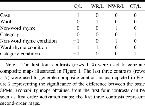
    Coefficients of the linear contrasts used to generate composite activation maps

PreviousNext
Back to top

In this issue

American Journal of Neuroradiology
Vol. 20, Issue 10
1 Nov 1999
  • Table of Contents
  • Index by author
Advertisement
Print
Download PDF
Email Article
Cite this article
0 Responses
Respond to this article
Share
Bookmark this article
The Cerebellum's Role in Reading: A Functional MR Imaging Study
Robert K. Fulbright, Annette R. Jenner, W. Einer Mencl, Kenneth R. Pugh, Bennett A. Shaywitz, Sally E. Shaywitz, Stephen J. Frost, Pawel Skudlarski, R. Todd Constable, Cheryl M. Lacadie, Karen E. Marchione, John C. Gore
American Journal of Neuroradiology Nov 1999, 20 (10) 1925-1930;
del.icio.us logo Twitter logo Facebook logo Mendeley logo
  • Tweet Widget

Jump to section

  • Article
    • Abstract
    • Methods
    • Discussion
    • Conclusion
    • Acknowledgments
    • Footnotes
    • References
  • Figures & Data
  • Info & Metrics
  • Responses
  • References
  • PDF

Related Articles

  • No related articles found.
  • PubMed
  • Google Scholar

Cited By...

  • Consistent cerebellar pathway-cognition associations across pre-adolescents & young adults: a diffusion MRI study of 9000+ participants
  • Preliminary evidence of widespread morphological variations of the brain in dyslexia
  • Crossref
  • Google Scholar

This article has not yet been cited by articles in journals that are participating in Crossref Cited-by Linking.

More in this TOC Section

  • Evaluating the Effects of White Matter Multiple Sclerosis Lesions on the Volume Estimation of 6 Brain Tissue Segmentation Methods
  • Quiet PROPELLER MRI Techniques Match the Quality of Conventional PROPELLER Brain Imaging Techniques
  • Predictors of Reperfusion in Patients with Acute Ischemic Stroke
Show more Brain

Similar Articles

Advertisement

Indexed Content

  • Current Issue
  • Accepted Manuscripts
  • Article Preview
  • Past Issues
  • Editorials
  • Editor's Choice
  • Fellows' Journal Club
  • Letters to the Editor
  • Video Articles

Cases

  • Case Collection
  • Archive - Case of the Week
  • Archive - Case of the Month
  • Archive - Classic Case

More from AJNR

  • Trainee Corner
  • Imaging Protocols
  • MRI Safety Corner
  • Book Reviews

Multimedia

  • AJNR Podcasts
  • AJNR Scantastics

Resources

  • Turnaround Time
  • Submit a Manuscript
  • Submit a Video Article
  • Submit an eLetter to the Editor/Response
  • Manuscript Submission Guidelines
  • Statistical Tips
  • Fast Publishing of Accepted Manuscripts
  • Graphical Abstract Preparation
  • Imaging Protocol Submission
  • Evidence-Based Medicine Level Guide
  • Publishing Checklists
  • Author Policies
  • Become a Reviewer/Academy of Reviewers
  • News and Updates

About Us

  • About AJNR
  • Editorial Board
  • Editorial Board Alumni
  • Alerts
  • Permissions
  • Not an AJNR Subscriber? Join Now
  • Advertise with Us
  • Librarian Resources
  • Feedback
  • Terms and Conditions
  • AJNR Editorial Board Alumni

American Society of Neuroradiology

  • Not an ASNR Member? Join Now

© 2025 by the American Society of Neuroradiology All rights, including for text and data mining, AI training, and similar technologies, are reserved.
Print ISSN: 0195-6108 Online ISSN: 1936-959X

Powered by HighWire
Email this Article

Thank you for your interest in spreading the word on American Journal of Neuroradiology.

NOTE: We only request your email address so that the person you are recommending the page to knows that you wanted them to see it, and that it is not junk mail. We do not capture any email address.

Enter multiple addresses on separate lines or separate them with commas.
The Cerebellum's Role in Reading: A Functional MR Imaging Study
(Your Name) has sent you a message from American Journal of Neuroradiology
(Your Name) thought you would like to see the American Journal of Neuroradiology web site.
CAPTCHA
This question is for testing whether or not you are a human visitor and to prevent automated spam submissions.
Cite this article
Robert K. Fulbright, Annette R. Jenner, W. Einer Mencl, Kenneth R. Pugh, Bennett A. Shaywitz, Sally E. Shaywitz, Stephen J. Frost, Pawel Skudlarski, R. Todd Constable, Cheryl M. Lacadie, Karen E. Marchione, John C. Gore
The Cerebellum's Role in Reading: A Functional MR Imaging Study
American Journal of Neuroradiology Nov 1999, 20 (10) 1925-1930;

Citation Manager Formats

  • BibTeX
  • Bookends
  • EasyBib
  • EndNote (tagged)
  • EndNote 8 (xml)
  • Medlars
  • Mendeley
  • Papers
  • RefWorks Tagged
  • Ref Manager
  • RIS
  • Zotero

We use cookies on this site to enhance your user experience. By clicking any link on this page you are giving your consent for us to set cookies.