Advertisement

Main menu

  • Home
  • Content
    • Current Issue
    • Accepted Manuscripts
    • Article Preview
    • Past Issue Archive
    • Video Articles
    • AJNR Case Collection
    • Case of the Week Archive
    • Case of the Month Archive
    • Classic Case Archive
  • Special Collections
    • AJNR Awards
    • Low-Field MRI
    • Alzheimer Disease
    • ASNR Foundation Special Collection
    • Photon-Counting CT
    • View All
  • Multimedia
    • AJNR Podcasts
    • AJNR SCANtastic
    • Trainee Corner
    • MRI Safety Corner
    • Imaging Protocols
  • For Authors
    • Submit a Manuscript
    • Submit a Video Article
    • Submit an eLetter to the Editor/Response
    • Manuscript Submission Guidelines
    • Statistical Tips
    • Fast Publishing of Accepted Manuscripts
    • Graphical Abstract Preparation
    • Imaging Protocol Submission
    • Author Policies
  • About Us
    • About AJNR
    • Editorial Board
    • Editorial Board Alumni
  • More
    • Become a Reviewer/Academy of Reviewers
    • Subscribers
    • Permissions
    • Alerts
    • Feedback
    • Advertisers
    • ASNR Home

User menu

  • Alerts
  • Log in

Search

  • Advanced search
American Journal of Neuroradiology

American Journal of Neuroradiology

ASHNR American Society of Functional Neuroradiology ASHNR American Society of Pediatric Neuroradiology ASSR
  • Alerts
  • Log in

Advanced Search

  • Home
  • Content
    • Current Issue
    • Accepted Manuscripts
    • Article Preview
    • Past Issue Archive
    • Video Articles
    • AJNR Case Collection
    • Case of the Week Archive
    • Case of the Month Archive
    • Classic Case Archive
  • Special Collections
    • AJNR Awards
    • Low-Field MRI
    • Alzheimer Disease
    • ASNR Foundation Special Collection
    • Photon-Counting CT
    • View All
  • Multimedia
    • AJNR Podcasts
    • AJNR SCANtastic
    • Trainee Corner
    • MRI Safety Corner
    • Imaging Protocols
  • For Authors
    • Submit a Manuscript
    • Submit a Video Article
    • Submit an eLetter to the Editor/Response
    • Manuscript Submission Guidelines
    • Statistical Tips
    • Fast Publishing of Accepted Manuscripts
    • Graphical Abstract Preparation
    • Imaging Protocol Submission
    • Author Policies
  • About Us
    • About AJNR
    • Editorial Board
    • Editorial Board Alumni
  • More
    • Become a Reviewer/Academy of Reviewers
    • Subscribers
    • Permissions
    • Alerts
    • Feedback
    • Advertisers
    • ASNR Home
  • Follow AJNR on Twitter
  • Visit AJNR on Facebook
  • Follow AJNR on Instagram
  • Join AJNR on LinkedIn
  • RSS Feeds

AJNR Awards, New Junior Editors, and more. Read the latest AJNR updates

Research ArticleBrain

MR Blood Oxygenation Level–Dependent Signal Differences in Parenchymal and Large Draining Vessels: Implications for Functional MR Imaging

Timo Krings, Stephan G. Erberich, Florian Roessler, Jürgen Reul and Armin Thron
American Journal of Neuroradiology November 1999, 20 (10) 1907-1914;
Timo Krings
  • Find this author on Google Scholar
  • Find this author on PubMed
  • Search for this author on this site
Stephan G. Erberich
  • Find this author on Google Scholar
  • Find this author on PubMed
  • Search for this author on this site
Florian Roessler
  • Find this author on Google Scholar
  • Find this author on PubMed
  • Search for this author on this site
Jürgen Reul
  • Find this author on Google Scholar
  • Find this author on PubMed
  • Search for this author on this site
Armin Thron
  • Find this author on Google Scholar
  • Find this author on PubMed
  • Search for this author on this site
  • Article
  • Figures & Data
  • Info & Metrics
  • Responses
  • References
  • PDF
Loading

Article Figures & Data

Figures

  • Tables
  • fig 1.
    • Download figure
    • Open in new tab
    • Download powerpoint
    fig 1.

    MR signal time courses for parenchyma, sulcal veins, and bridging veins for single subject in two different functional runs (hand clenching) (S1). Each diamond represents MR signal at one time-point. Temporal resolution of the sequence was 2.2 s/image. Mean percent signal changes increase as a function of vessel size. Note low test-retest variability of percent signal changes within the same subject.

  • fig 2.
    • Download figure
    • Open in new tab
    • Download powerpoint
    fig 2.

    MR signal time course correlation with contrast-enhanced T1-weighted scans and phase-contrast MR angiography (S5). Contrast-enhanced studies reliably show sulcal and superficial veins, whereas the phase-contrast MR angiography with given flow sensitivity demonstrates vessels the size of large bridging veins. Significant change in MR signal (ΔS) is seen in bridging veins even close to superior sagittal sinus. From this data, one can presume that dilution of increased oxyhemoglobin content and therefore decay of ΔS takes place when bridging veins enter large draining sinuses. Arrows point at sulcal veins, arrowheads at large bridging veins.

  • fig 3.
    • Download figure
    • Open in new tab
    • Download powerpoint
    fig 3.

    MR signal time courses for parenchyma, sulcal veins, and bridging veins for subject scanned with higher temporal resolution (556 ms/image) during hand clenching. With a finer temporal resolution, the differences in onset of significant task related MR signal changes between vessels of different diameter can be visualized.

  • fig 4.
    • Download figure
    • Open in new tab
    • Download powerpoint
    fig 4.

    Functional MR and MR signal time course. This patient was evaluated for presurgical planning of a space-occupying lesion near central sulcus (Astrocytoma III) (S7). The first fMR imaging study shows dual activation in slice of interest with parenchymal activation located in close proximity to tumor. A stereotactic biopsy was performed after which the patient suffered from transient mild paresis of the small right hand muscles. A postbiopsy MR image demonstrates that the biopsy involved a previously activated region posterior to the lesion. Functional study again shows dual activation with parenchymal activation close to biopsy tract and lateral venous activity unaffected by biopsy. This case illustrates the importance of differentiating task-related hemodynamic changes of small parenchymal venules and large draining veins.

Tables

  • Figures
  • Table1

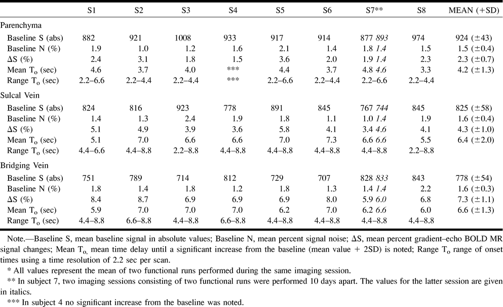
    MR BOLD signal characteristics for veins of different sizes*

PreviousNext
Back to top

In this issue

American Journal of Neuroradiology
Vol. 20, Issue 10
1 Nov 1999
  • Table of Contents
  • Index by author
Advertisement
Print
Download PDF
Email Article

Thank you for your interest in spreading the word on American Journal of Neuroradiology.

NOTE: We only request your email address so that the person you are recommending the page to knows that you wanted them to see it, and that it is not junk mail. We do not capture any email address.

Enter multiple addresses on separate lines or separate them with commas.
MR Blood Oxygenation Level–Dependent Signal Differences in Parenchymal and Large Draining Vessels: Implications for Functional MR Imaging
(Your Name) has sent you a message from American Journal of Neuroradiology
(Your Name) thought you would like to see the American Journal of Neuroradiology web site.
CAPTCHA
This question is for testing whether or not you are a human visitor and to prevent automated spam submissions.
Cite this article
Timo Krings, Stephan G. Erberich, Florian Roessler, Jürgen Reul, Armin Thron
MR Blood Oxygenation Level–Dependent Signal Differences in Parenchymal and Large Draining Vessels: Implications for Functional MR Imaging
American Journal of Neuroradiology Nov 1999, 20 (10) 1907-1914;

Citation Manager Formats

  • BibTeX
  • Bookends
  • EasyBib
  • EndNote (tagged)
  • EndNote 8 (xml)
  • Medlars
  • Mendeley
  • Papers
  • RefWorks Tagged
  • Ref Manager
  • RIS
  • Zotero
0 Responses
Respond to this article
Share
Bookmark this article
MR Blood Oxygenation Level–Dependent Signal Differences in Parenchymal and Large Draining Vessels: Implications for Functional MR Imaging
Timo Krings, Stephan G. Erberich, Florian Roessler, Jürgen Reul, Armin Thron
American Journal of Neuroradiology Nov 1999, 20 (10) 1907-1914;
del.icio.us logo Twitter logo Facebook logo Mendeley logo
  • Tweet Widget
  • Facebook Like
  • Google Plus One

Jump to section

  • Article
    • Abstract
    • Methods
    • Results
    • Discussion
    • Conclusion
    • Footnotes
    • References
  • Figures & Data
  • Info & Metrics
  • Responses
  • References
  • PDF

Related Articles

  • No related articles found.
  • PubMed
  • Google Scholar

Cited By...

  • Multivoxel Pattern of Blood Oxygen Level Dependent Activity can be sensitive to stimulus specific fine scale responses
  • Axial variation of deoxyhemoglobin density as a source of the low-frequency time lag structure in blood oxygenation level-dependent signals
  • Functional Magnetic Resonance Imaging Before and After Aphasia Therapy: Shifts in Hemodynamic Time to Peak During an Overt Language Task
  • Activation in primary and secondary motor areas in patients with CNS neoplasms and weakness
  • Metabolic and electrophysiological validation of functional MRI
  • Functional MRI for presurgical planning: problems, artefacts, and solution strategies
  • Crossref
  • Google Scholar

This article has not yet been cited by articles in journals that are participating in Crossref Cited-by Linking.

More in this TOC Section

  • Multimodal CT Provides Improved Performance for Lacunar Infarct Detection
  • Optimal MRI Sequence for Identifying Occlusion Location in Acute Stroke: Which Value of Time-Resolved Contrast-Enhanced MRA?
  • Evaluating the Effects of White Matter Multiple Sclerosis Lesions on the Volume Estimation of 6 Brain Tissue Segmentation Methods
Show more BRAIN

Similar Articles

Advertisement

Indexed Content

  • Current Issue
  • Accepted Manuscripts
  • Article Preview
  • Past Issues
  • Editorials
  • Editor's Choice
  • Fellows' Journal Club
  • Letters to the Editor
  • Video Articles

Cases

  • Case Collection
  • Archive - Case of the Week
  • Archive - Case of the Month
  • Archive - Classic Case

More from AJNR

  • Trainee Corner
  • Imaging Protocols
  • MRI Safety Corner
  • Book Reviews

Multimedia

  • AJNR Podcasts
  • AJNR Scantastics

Resources

  • Turnaround Time
  • Submit a Manuscript
  • Submit a Video Article
  • Submit an eLetter to the Editor/Response
  • Manuscript Submission Guidelines
  • Statistical Tips
  • Fast Publishing of Accepted Manuscripts
  • Graphical Abstract Preparation
  • Imaging Protocol Submission
  • Evidence-Based Medicine Level Guide
  • Publishing Checklists
  • Author Policies
  • Become a Reviewer/Academy of Reviewers
  • News and Updates

About Us

  • About AJNR
  • Editorial Board
  • Editorial Board Alumni
  • Alerts
  • Permissions
  • Not an AJNR Subscriber? Join Now
  • Advertise with Us
  • Librarian Resources
  • Feedback
  • Terms and Conditions
  • AJNR Editorial Board Alumni

American Society of Neuroradiology

  • Not an ASNR Member? Join Now

© 2025 by the American Society of Neuroradiology All rights, including for text and data mining, AI training, and similar technologies, are reserved.
Print ISSN: 0195-6108 Online ISSN: 1936-959X

Powered by HighWire