Advertisement
  • Main menu
  • User menu
  • Search
  • English ▼
    • English
    • Afrikaans
    • Albanian
    • Amharic
    • Arabic
    • Armenian
    • Azerbaijani
    • Basque
    • Belarusian
    • Bengali
    • Bosnian
    • Bulgarian
    • Catalan
    • Cebuano
    • Chichewa
    • Chinese (Simplified)
    • Chinese (Traditional)
    • Corsican
    • Croatian
    • Czech
    • Danish
    • Dutch
    • Esperanto
    • Estonian
    • Filipino
    • Finnish
    • French
    • Frisian
    • Galician
    • Georgian
    • German
    • Greek
    • Gujarati
    • Haitian Creole
    • Hausa
    • Hawaiian
    • Hebrew
    • Hindi
    • Hmong
    • Hungarian
    • Icelandic
    • Igbo
    • Indonesian
    • Irish
    • Italian
    • Japanese
    • Javanese
    • Kannada
    • Kazakh
    • Khmer
    • Korean
    • Kurdish (Kurmanji)
    • Kyrgyz
    • Lao
    • Latin
    • Latvian
    • Lithuanian
    • Luxembourgish
    • Macedonian
    • Malagasy
    • Malay
    • Malayalam
    • Maltese
    • Maori
    • Marathi
    • Mongolian
    • Myanmar (Burmese)
    • Nepali
    • Norwegian
    • Pashto
    • Persian
    • Polish
    • Portuguese
    • Punjabi
    • Romanian
    • Russian
    • Samoan
    • Scottish Gaelic
    • Serbian
    • Sesotho
    • Shona
    • Sindhi
    • Sinhala
    • Slovak
    • Slovenian
    • Somali
    • Spanish
    • Sudanese
    • Swahili
    • Swedish
    • Tajik
    • Tamil
    • Telugu
    • Thai
    • Turkish
    • Ukrainian
    • Urdu
    • Uzbek
    • Vietnamese
    • Welsh
    • Xhosa
    • Yiddish
    • Yoruba
    • Zulu

Main menu

  • Home
  • Content
    • Current Issue
    • Accepted Manuscripts
    • Article Preview
    • Past Issue Archive
    • Video Articles
    • AJNR Case Collection
    • Case of the Week Archive
    • Case of the Month Archive
    • Classic Case Archive
  • Special Collections
    • Advancing NeuroMRI with High-Relaxivity Contrast Agents
    • AJNR Awards
    • Low-Field MRI
    • Alzheimer Disease
    • ASNR Foundation Special Collection
    • Photon-Counting CT
    • View All
  • Multimedia
    • AJNR Podcasts
    • AJNR SCANtastic
    • Trainee Corner
    • MRI Safety Corner
    • Imaging Protocols
  • For Authors
    • Submit a Manuscript
    • Submit a Video Article
    • Submit an eLetter to the Editor/Response
    • Manuscript Submission Guidelines
    • Statistical Tips
    • Fast Publishing of Accepted Manuscripts
    • Graphical Abstract Preparation
    • Imaging Protocol Submission
    • Author Policies
  • About Us
    • About AJNR
    • Editorial Board
    • Editorial Board Alumni
  • More
    • Become a Reviewer/Academy of Reviewers
    • Subscribers
    • Permissions
    • Alerts
    • Feedback
    • Advertisers
    • ASNR Home

User menu

  • Alerts
  • Log in

Search

  • Advanced search
American Journal of Neuroradiology

American Journal of Neuroradiology

ASHNR American Society of Functional Neuroradiology ASHNR American Society of Pediatric Neuroradiology ASSR
  • Alerts
  • Log in

Advanced Search

English ▼
  • English
  • Afrikaans
  • Albanian
  • Amharic
  • Arabic
  • Armenian
  • Azerbaijani
  • Basque
  • Belarusian
  • Bengali
  • Bosnian
  • Bulgarian
  • Catalan
  • Cebuano
  • Chichewa
  • Chinese (Simplified)
  • Chinese (Traditional)
  • Corsican
  • Croatian
  • Czech
  • Danish
  • Dutch
  • Esperanto
  • Estonian
  • Filipino
  • Finnish
  • French
  • Frisian
  • Galician
  • Georgian
  • German
  • Greek
  • Gujarati
  • Haitian Creole
  • Hausa
  • Hawaiian
  • Hebrew
  • Hindi
  • Hmong
  • Hungarian
  • Icelandic
  • Igbo
  • Indonesian
  • Irish
  • Italian
  • Japanese
  • Javanese
  • Kannada
  • Kazakh
  • Khmer
  • Korean
  • Kurdish (Kurmanji)
  • Kyrgyz
  • Lao
  • Latin
  • Latvian
  • Lithuanian
  • Luxembourgish
  • Macedonian
  • Malagasy
  • Malay
  • Malayalam
  • Maltese
  • Maori
  • Marathi
  • Mongolian
  • Myanmar (Burmese)
  • Nepali
  • Norwegian
  • Pashto
  • Persian
  • Polish
  • Portuguese
  • Punjabi
  • Romanian
  • Russian
  • Samoan
  • Scottish Gaelic
  • Serbian
  • Sesotho
  • Shona
  • Sindhi
  • Sinhala
  • Slovak
  • Slovenian
  • Somali
  • Spanish
  • Sudanese
  • Swahili
  • Swedish
  • Tajik
  • Tamil
  • Telugu
  • Thai
  • Turkish
  • Ukrainian
  • Urdu
  • Uzbek
  • Vietnamese
  • Welsh
  • Xhosa
  • Yiddish
  • Yoruba
  • Zulu
  • Home
  • Content
    • Current Issue
    • Accepted Manuscripts
    • Article Preview
    • Past Issue Archive
    • Video Articles
    • AJNR Case Collection
    • Case of the Week Archive
    • Case of the Month Archive
    • Classic Case Archive
  • Special Collections
    • Advancing NeuroMRI with High-Relaxivity Contrast Agents
    • AJNR Awards
    • Low-Field MRI
    • Alzheimer Disease
    • ASNR Foundation Special Collection
    • Photon-Counting CT
    • View All
  • Multimedia
    • AJNR Podcasts
    • AJNR SCANtastic
    • Trainee Corner
    • MRI Safety Corner
    • Imaging Protocols
  • For Authors
    • Submit a Manuscript
    • Submit a Video Article
    • Submit an eLetter to the Editor/Response
    • Manuscript Submission Guidelines
    • Statistical Tips
    • Fast Publishing of Accepted Manuscripts
    • Graphical Abstract Preparation
    • Imaging Protocol Submission
    • Author Policies
  • About Us
    • About AJNR
    • Editorial Board
    • Editorial Board Alumni
  • More
    • Become a Reviewer/Academy of Reviewers
    • Subscribers
    • Permissions
    • Alerts
    • Feedback
    • Advertisers
    • ASNR Home
  • Follow AJNR on Twitter
  • Visit AJNR on Facebook
  • Follow AJNR on Instagram
  • Join AJNR on LinkedIn
  • RSS Feeds

AJNR Awards, New Junior Editors, and more. Read the latest AJNR updates


Improved Turnaround Times | Median time to first decision: 12 days

Research ArticleSpine

Pseudohemangioma of the Vertebra: An Unusual Radiographic Manifestation of Primary Ewing's Sarcoma

Joshua A. Bemporad, Gordon Sze, John C. Chaloupka and Charles Duncan
American Journal of Neuroradiology November 1999, 20 (10) 1809-1813;
Joshua A. Bemporad
  • Find this author on Google Scholar
  • Find this author on PubMed
  • Search for this author on this site
Gordon Sze
  • Find this author on Google Scholar
  • Find this author on PubMed
  • Search for this author on this site
John C. Chaloupka
  • Find this author on Google Scholar
  • Find this author on PubMed
  • Search for this author on this site
Charles Duncan
  • Find this author on Google Scholar
  • Find this author on PubMed
  • Search for this author on this site
  • Article
  • Figures & Data
  • Info & Metrics
  • Responses
  • References
  • PDF
Loading

Article Figures & Data

Figures

  • fig 1.
    • Download figure
    • Open in new tab
    • Download powerpoint
    fig 1.

    Lateral plain film of lower cervical spine shows compression of C7 vertebra with preservation of C6–7 disk space.

    fig 2. Axial CT scan without contrast (bone window) shows course trabecular pattern with disruption of cortex of vertebral body. Left neural foramen is slightly widened.
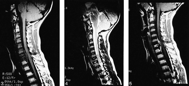
  • fig 3.
    • Download figure
    • Open in new tab
    • Download powerpoint
    fig 3.

    Midline sagittal T1-weighted image of cervical spine shows homogeneous extradural mass compressing spinal cord posteriorly. Mass is low in signal and roughly isointense to gray matter. C7 vertebra is compressed and is decreased in signal with preservation of intervertebral disk spaces.

    fig 4. Sagittal T2-weighted image of cervical spine slightly to left of midline better shows preservation of intervertebral disk spaces. Extradural mass is fairly homogeneous and intermediate-to-high in signal. Compression of C7 vertebra with increase in anteroposterior diameter is obvious.

    fig 5. Midline sagittal T1-weighted image with contrast shows heterogeneous enhancement of extradural mass and C7 vertebral body. There is no abnormal enhancement of the spinal cord.
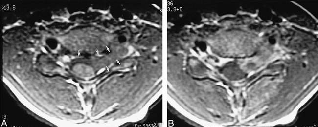
  • fig 6.
    • Download figure
    • Open in new tab
    • Download powerpoint
    fig 6.

    A, T1-weighted axial image through C6–7 intervertebral disk shows extradural mass compressing spinal cord posteriorly. Mass crosses midline and extends out of left neural foramen (arrows).

    B, T1-weighted axial image with contrast shows heterogeneous enhancement of mass.

  • fig 7.
    • Download figure
    • Open in new tab
    • Download powerpoint
    fig 7.

    Superselective angiogram of left costocervical trunk. Late arterial-phase projection shows staining in region of epidural mass.

  • fig 8.
    • Download figure
    • Open in new tab
    • Download powerpoint
    fig 8.

    Photomicrograph reveals that tumor is composed of densely packed small, round-to-oval cells (lower left) with high nuclear-to-cytoplasmic ratio (“small blue cell tumor;” several representative cells are highlighted with small arrowheads). Tumor has infiltrated medullary space of vertebrae. Note bone spicule (letter A) on right of figure. Faint thin-walled vascular channels are present (outlined by large arrowheads) (hematoxylin and eosin, original magnification × 200). Cells were immunopositve for CD99 and immunonegative for LCA and PGP 9.5. (not shown).

  • fig 9.
    • Download figure
    • Open in new tab
    • Download powerpoint
    fig 9.

    A, Proton density–weighted sagittal image of 53-year-old woman with aggressive VH of L3 vertebra. There is decrease of signal in L3 vertebral body. There is a large, homogeneous, low-signal extraosseous mass within spinal canal compressing thecal sac. There is growth of tumor superiorly and inferiorly along posterior aspect of L3 vertebral body. Note that there is preservation of intervertebral disk spaces.

    B, T2-weighted sagittal images (from same patient as 9A) shows that signal in extraosseous component increases on T2-weighted image. T2-weighted image better shows how tumor extends beyond inferior disk space (arrow).

PreviousNext
Back to top

In this issue

American Journal of Neuroradiology
Vol. 20, Issue 10
1 Nov 1999
  • Table of Contents
  • Index by author
Advertisement
Print
Download PDF
Email Article
Cite this article
0 Responses
Respond to this article
Share
Bookmark this article
Pseudohemangioma of the Vertebra: An Unusual Radiographic Manifestation of Primary Ewing's Sarcoma
Joshua A. Bemporad, Gordon Sze, John C. Chaloupka, Charles Duncan
American Journal of Neuroradiology Nov 1999, 20 (10) 1809-1813;
del.icio.us logo Twitter logo Facebook logo Mendeley logo
  • Tweet Widget

Jump to section

  • Article
    • Abstract
    • Case Report
    • Discussion
    • Acknowledgments
    • Footnotes
    • References
  • Figures & Data
  • Info & Metrics
  • Responses
  • References
  • PDF

Related Articles

  • No related articles found.
  • PubMed
  • Google Scholar

Cited By...

  • Spinal Ewing Sarcoma Debuting with Cord Compression: Have We Discovered the Thread of Ariadne?
  • Crossref
  • Google Scholar

This article has not yet been cited by articles in journals that are participating in Crossref Cited-by Linking.

More in this TOC Section

  • CT-Guided C2 Dorsal Root Ganglion Radiofrequency Ablation for the Treatment of Cervicogenic Headache: Case Series and Clinical Outcomes
  • The Evaluation and Prediction of Laminoplasty Surgery Outcome in Patients with Degenerative Cervical Myelopathy Using Diffusion Tensor MRI
  • Subject-Specific Studies of CSF Bulk Flow Patterns in the Spinal Canal: Implications for the Dispersion of Solute Particles in Intrathecal Drug Delivery
Show more Spine

Similar Articles

Advertisement

Indexed Content

  • Current Issue
  • Accepted Manuscripts
  • Article Preview
  • Past Issues
  • Editorials
  • Editor's Choice
  • Fellows' Journal Club
  • Letters to the Editor
  • Video Articles

Cases

  • Case Collection
  • Archive - Case of the Week
  • Archive - Case of the Month
  • Archive - Classic Case

More from AJNR

  • Trainee Corner
  • Imaging Protocols
  • MRI Safety Corner
  • Book Reviews

Multimedia

  • AJNR Podcasts
  • AJNR Scantastics

Resources

  • Turnaround Time
  • Submit a Manuscript
  • Submit a Video Article
  • Submit an eLetter to the Editor/Response
  • Manuscript Submission Guidelines
  • Statistical Tips
  • Fast Publishing of Accepted Manuscripts
  • Graphical Abstract Preparation
  • Imaging Protocol Submission
  • Evidence-Based Medicine Level Guide
  • Publishing Checklists
  • Author Policies
  • Become a Reviewer/Academy of Reviewers
  • News and Updates

About Us

  • About AJNR
  • Editorial Board
  • Editorial Board Alumni
  • Alerts
  • Permissions
  • Not an AJNR Subscriber? Join Now
  • Advertise with Us
  • Librarian Resources
  • Feedback
  • Terms and Conditions
  • AJNR Editorial Board Alumni

American Society of Neuroradiology

  • Not an ASNR Member? Join Now

© 2025 by the American Society of Neuroradiology All rights, including for text and data mining, AI training, and similar technologies, are reserved.
Print ISSN: 0195-6108 Online ISSN: 1936-959X

Powered by HighWire
Email this Article

Thank you for your interest in spreading the word on American Journal of Neuroradiology.

NOTE: We only request your email address so that the person you are recommending the page to knows that you wanted them to see it, and that it is not junk mail. We do not capture any email address.

Enter multiple addresses on separate lines or separate them with commas.
Pseudohemangioma of the Vertebra: An Unusual Radiographic Manifestation of Primary Ewing's Sarcoma
(Your Name) has sent you a message from American Journal of Neuroradiology
(Your Name) thought you would like to see the American Journal of Neuroradiology web site.
CAPTCHA
This question is for testing whether or not you are a human visitor and to prevent automated spam submissions.
Cite this article
Joshua A. Bemporad, Gordon Sze, John C. Chaloupka, Charles Duncan
Pseudohemangioma of the Vertebra: An Unusual Radiographic Manifestation of Primary Ewing's Sarcoma
American Journal of Neuroradiology Nov 1999, 20 (10) 1809-1813;

Citation Manager Formats

  • BibTeX
  • Bookends
  • EasyBib
  • EndNote (tagged)
  • EndNote 8 (xml)
  • Medlars
  • Mendeley
  • Papers
  • RefWorks Tagged
  • Ref Manager
  • RIS
  • Zotero

We use cookies on this site to enhance your user experience. By clicking any link on this page you are giving your consent for us to set cookies.