
Article Figures & Data
Figures
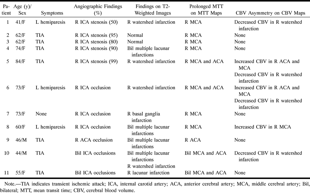
- fig 1.
Patient 2: 62-year-old woman with TIAs.
A, Angiogram shows 95% stenosis of the right ICA (arrow).
B, Findings on T2-weighted MR image are normal.
C, Corresponding MTT map shows extensive high signal intensity in the right MCA distribution, indicating prolonged MTT on the affected side compared with the contralateral side.
D, Corresponding CBV map shows symmetry in signal intensity, indicating normal CBV.
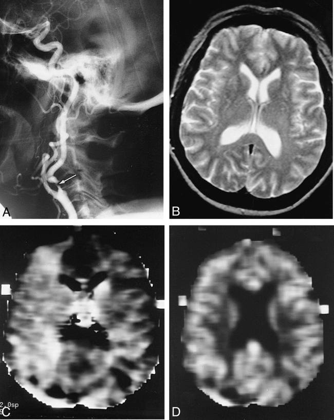
- fig 2.
Patient 3: 62-year-old woman with TIAs.
A, Angiogram shows 80% stenosis of the right ICA.
B, Findings on T2-weighted MR image are normal.
C, Corresponding MTT map shows high signal intensity in the right MCA distribution. The extent of high signal is less than that in patient 2 (fig 1C).
D, Corresponding CBV map shows symmetry in signal intensity.
- fig 3.
Patient 5: 84-year-old woman with TIAs.
A, Angiogram shows 99% stenosis of the right ICA.
B, T2-weighted MR image shows a right posterior watershed infarction (arrowheads).
C, Corresponding MTT map shows extensive high signal intensity in the right MCA and ACA distributions.
D, Corresponding CBV map shows higher signal intensity in the right than in the left ACA distribution (arrows), indicating an elevation of CBV in the right ACA distribution. The higher CBV most likely represents brain parenchyma with maximal vasodilatation, while the lower CBV is most likely caused by neuronal loss (old infarction/gliosis) (arrowheads).
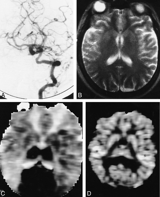
- fig 4.
Patient 11: 55-year-old woman with TIAs.
A, Angiogram shows collateral flow to the cerebral hemispheres from the vertebrobasilar distribution through the posterior communicating arteries. Both ICAs were occluded (not shown).
B, T2-weighted MR image shows no abnormalities except for a lacunar infarction on the right.
C, Corresponding MTT map shows extensive areas with prolonged MTT in the distribution of both ICAs. The only normal blood flow is seen alone at both posterior circulations (dark areas).
D, Corresponding CBV map reveals no evidence of decreased blood volume. Blood volume is increased in the posterior watershed region bilaterally (asterisks).


Tables

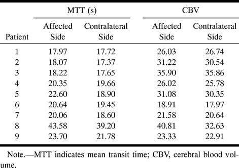
- TABLE 2:
Mean values of MTT and CBV in the affected and contralateral sides


