Advertisement

Main menu

  • Home
  • Content
    • Current Issue
    • Accepted Manuscripts
    • Article Preview
    • Past Issue Archive
    • Video Articles
    • AJNR Case Collection
    • Case of the Week Archive
    • Case of the Month Archive
    • Classic Case Archive
  • Special Collections
    • AJNR Awards
    • Low-Field MRI
    • Alzheimer Disease
    • ASNR Foundation Special Collection
    • Photon-Counting CT
    • View All
  • Multimedia
    • AJNR Podcasts
    • AJNR SCANtastic
    • Trainee Corner
    • MRI Safety Corner
    • Imaging Protocols
  • For Authors
    • Submit a Manuscript
    • Submit a Video Article
    • Submit an eLetter to the Editor/Response
    • Manuscript Submission Guidelines
    • Statistical Tips
    • Fast Publishing of Accepted Manuscripts
    • Graphical Abstract Preparation
    • Imaging Protocol Submission
    • Author Policies
  • About Us
    • About AJNR
    • Editorial Board
    • Editorial Board Alumni
  • More
    • Become a Reviewer/Academy of Reviewers
    • Subscribers
    • Permissions
    • Alerts
    • Feedback
    • Advertisers
    • ASNR Home

User menu

  • Alerts
  • Log in

Search

  • Advanced search
American Journal of Neuroradiology

American Journal of Neuroradiology

ASHNR American Society of Functional Neuroradiology ASHNR American Society of Pediatric Neuroradiology ASSR
  • Alerts
  • Log in

Advanced Search

  • Home
  • Content
    • Current Issue
    • Accepted Manuscripts
    • Article Preview
    • Past Issue Archive
    • Video Articles
    • AJNR Case Collection
    • Case of the Week Archive
    • Case of the Month Archive
    • Classic Case Archive
  • Special Collections
    • AJNR Awards
    • Low-Field MRI
    • Alzheimer Disease
    • ASNR Foundation Special Collection
    • Photon-Counting CT
    • View All
  • Multimedia
    • AJNR Podcasts
    • AJNR SCANtastic
    • Trainee Corner
    • MRI Safety Corner
    • Imaging Protocols
  • For Authors
    • Submit a Manuscript
    • Submit a Video Article
    • Submit an eLetter to the Editor/Response
    • Manuscript Submission Guidelines
    • Statistical Tips
    • Fast Publishing of Accepted Manuscripts
    • Graphical Abstract Preparation
    • Imaging Protocol Submission
    • Author Policies
  • About Us
    • About AJNR
    • Editorial Board
    • Editorial Board Alumni
  • More
    • Become a Reviewer/Academy of Reviewers
    • Subscribers
    • Permissions
    • Alerts
    • Feedback
    • Advertisers
    • ASNR Home
  • Follow AJNR on Twitter
  • Visit AJNR on Facebook
  • Follow AJNR on Instagram
  • Join AJNR on LinkedIn
  • RSS Feeds

AJNR Awards, New Junior Editors, and more. Read the latest AJNR updates

LetterLetter

Aneurysmal Changes at the Basilar Terminus in the Rabbit Elastase Aneurysm Model

H. Meng, S.K. Natarajan, L. Gao, C. Ionita, J. Kolega, A.H. Siddiqui and J. Mocco
American Journal of Neuroradiology March 2010, 31 (3) E35-E36; DOI: https://doi.org/10.3174/ajnr.A2012
H. Meng
  • Find this author on Google Scholar
  • Find this author on PubMed
  • Search for this author on this site
S.K. Natarajan
  • Find this author on Google Scholar
  • Find this author on PubMed
  • Search for this author on this site
L. Gao
  • Find this author on Google Scholar
  • Find this author on PubMed
  • Search for this author on this site
C. Ionita
  • Find this author on Google Scholar
  • Find this author on PubMed
  • Search for this author on this site
J. Kolega
  • Find this author on Google Scholar
  • Find this author on PubMed
  • Search for this author on this site
A.H. Siddiqui
  • Find this author on Google Scholar
  • Find this author on PubMed
  • Search for this author on this site
J. Mocco
  • Find this author on Google Scholar
  • Find this author on PubMed
  • Search for this author on this site
  • Article
  • Figures & Data
  • Info & Metrics
  • Responses
  • References
  • PDF
Loading

We read the interesting report by Dai et al,1 wherein the authors examined histology at the basilar terminus (BT) following right common carotid artery (RCCA) ligation as a part of a procedure to create an elastase aneurysm model in 30 consecutive New Zealand white rabbits at various time points. The authors concluded that in contrast to our previous report,2 there was no bulgelike localized dilation associated with a missing internal elastic lamina (IEL) to suggest microaneurysm or nascent aneurysm formation at the BT, at any time point after RCCA ligation.

We find this report particularly interesting because the first aneurysmal changes at the rabbit BT that we noticed actually occurred in 2 rabbits that had undergone RCCA ligation.3 The rabbits underwent the same surgical procedure as that performed by Dai et al,1 with the same objective of creating the elastase aneurysm model on the RCCA stump for an endovascular device testing.3 We examined whether there were any aneurysmal changes in the basilar bifurcation in these 2 rabbits through histologic staining of contiguous specimens with hematoxylin-eosin (HE), Van Gieson, and trichrome. In the first rabbit, sacrificed 10 weeks after RCCA ligation, we observed loss of IEL, endothelial cells, and smooth muscle cells, a thinned media, and an outward convex bulge at the BT (Fig 1). Fifteen consecutive sections of 15-μm thickness consistently presented this BT bulge with missing IEL and media thinning. We repeated the observation in the second rabbit, which had received incomplete RCCA ligation and was sacrificed 12 weeks later. We saw similar aneurysmal changes, including IEL loss and medial thinning at the BT, albeit with a shallower and longer bulge (Fig 2). These vascular defects were clearly aneurysmal and could not be a staining or sectioning artifact, or misinterpreted branches.

Fig 1.
  • Download figure
  • Open in new tab
  • Download powerpoint
Fig 1.

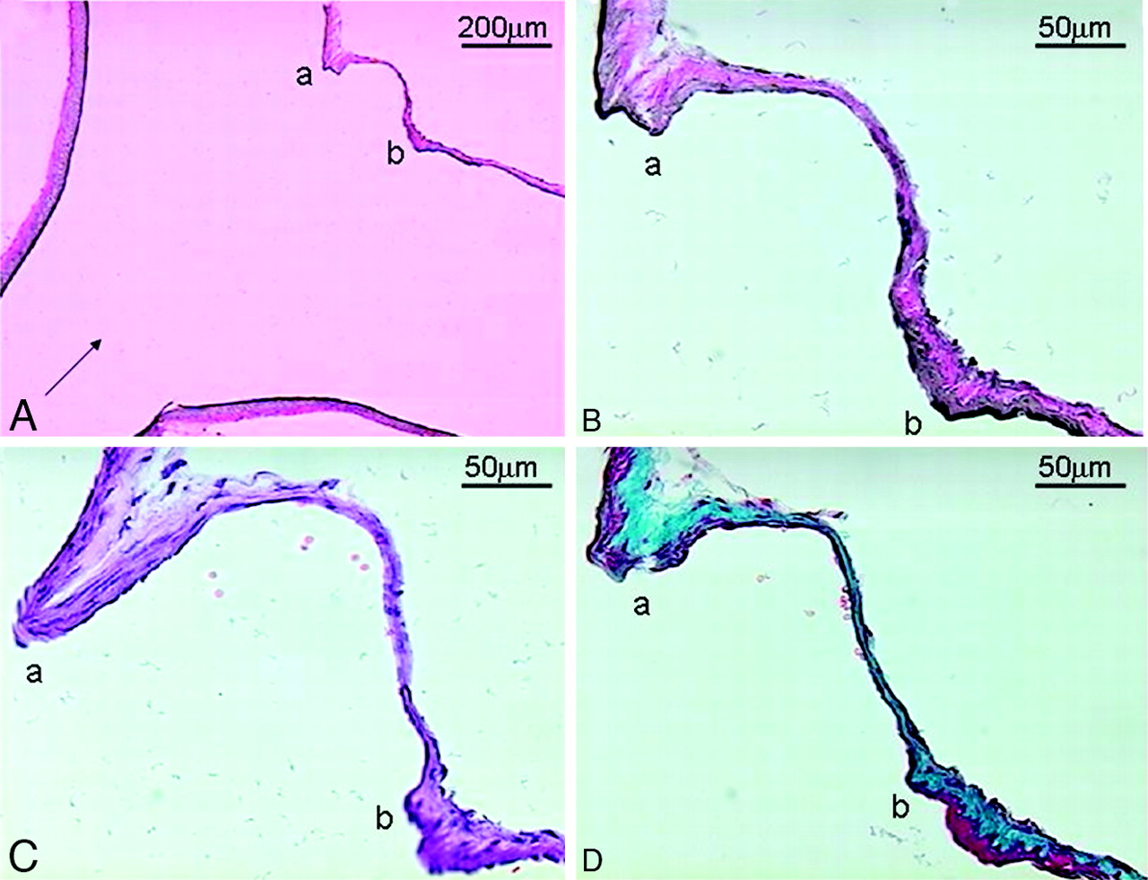
An aneurysm detected at the rabbit BT 10 weeks after RCCA ligation in an elastase aneurysm model. A, Basilar artery−posterior cerebral artery bifurcation shown at a low magnification. Arrow indicates blood flow direction. An aneurysm is observed between a and b, with IEL disruption (a total length of 300 μm) and media thinning occurring in the bulge (Van Gieson, original magnification ×100). B−D, The aneurysm site is shown at a high magnification (original magnification ×400). B, Van Gieson staining shows IEL loss. C, HE staining shows endothelial loss. D, Trichrome staining shows medial thinning.

Fig 2.
  • Download figure
  • Open in new tab
  • Download powerpoint
Fig 2.

Aneurysmal degradation at the rabbit BT 12 weeks after incomplete RCCA ligation in an elastase aneurysm model. A and B, Basilar bifurcation shown at a low magnification (original magnification ×100). Arrow indicates blood flow direction. A shallow bulge with a total disruption length of 600 μm is observed between a and b. A, Van Gieson staining. B, Trichrome staining. C and D, Aneurysmal degradation is shown at a high magnification (original magnification ×400). C, Trichrome staining shows medial loss. D, Van Gieson staining shows IEL disruption.

These 2 cases prompted us to conduct a prospective study of nascent aneurysmal initiation at the BT induced by common carotid artery (CCA) ligation alone, which resulted in the article by Gao et al.2 We performed unilateral or bilateral CCA ligation in otherwise unmanipulated New Zealand white rabbits and prospectively examined aneurysmal changes at the BT 12 weeks later. We observed nascent aneurysm formation and its dose dependence on basilar artery flow increase. Since then, we have shifted our model to perform bilateral CCA ligation only. We have seen prominent IEL loss at the BT in every one of our rabbits, as early as 2 days and 5 days post-CCA ligation. We are in the process of drafting these results.

Our experience with both the elastase model (reported here) and the nascent BT aneurysm model2 obviously differs from that of Dai et al.1 We are concerned that in their study, Van Gieson staining was done after washing off previous HE staining on the same specimen. The reliability of the de-staining and re-staining technique in detecting IEL loss has not been verified. Reports in prostate cancer specimens show that the reliability of de-staining HE and re-staining with cytokeratin could be as low as 58%.4 In our experience, more than 50 sections can be taken from 1 BT tissue specimen; thus, adjacent specimens can be used for different stains. Therefore, de-staining and re-staining of the same specimen do not seem necessary and could have confounded the interpretation of their results.

It is not clear whether Dai et al1 created their animal models prospectively to examine the BT or performed a retrospective analysis on stored specimens that had previous HE staining. In the latter scenario, there could be sectioning bias as well as a need to de-stain and re-stain sections, rather than to use adjacent sections.

Finally, we respectfully disagree with the speculation by Dai et al1 that our group could have misinterpreted branching vessels as aneurysmal bulges in Gao et al.2 Branching vessels would have assumed completely different anatomy than what we have demonstrated, withno IEL loss or media thinning. In addition, multiple contiguous sections throughout the entire paraffin-embeddedbifurcation would have consistently revealed the branching vessels, instead of the bulges that spanned multiple sections and tapered off, as seen in our studies.

References

  1. 1.↵
    1. Dai D,
    2. Ding YH,
    3. Kadirvel R,
    4. et al
    . Experience with microaneurysm formation at the basilar terminus in the rabbit elastase aneurysm model. AJNR Am J Neuroradiol 2010;31:300–03. Epub 2009 Oct 1
    Abstract/FREE Full Text
  2. 2.↵
    1. Gao L,
    2. Hoi Y,
    3. Swartz DD,
    4. et al
    . Nascent aneurysm formation at the basilar terminus induced by hemodynamics. Stroke 2008;39:2085–90
    Abstract/FREE Full Text
  3. 3.↵
    1. Ionita CN,
    2. Paciorek AM,
    3. Dohatcu A,
    4. et al
    . The asymmetric vascular stent: efficacy in a rabbit aneurysm model. Stroke 2009;40:959–65
    Abstract/FREE Full Text
  4. 4.↵
    1. Dardik M,
    2. Epstein JI
    . Efficacy of restaining prostate needle biopsies with high-molecular weight cytokeratin. Hum Pathol 2000;31:1155–61
    CrossRefPubMed
  • Copyright © American Society of Neuroradiology
PreviousNext
Back to top

In this issue

American Journal of Neuroradiology: 31 (3)
American Journal of Neuroradiology
Vol. 31, Issue 3
1 Mar 2010
  • Table of Contents
  • Index by author
Advertisement
Print
Download PDF
Email Article

Thank you for your interest in spreading the word on American Journal of Neuroradiology.

NOTE: We only request your email address so that the person you are recommending the page to knows that you wanted them to see it, and that it is not junk mail. We do not capture any email address.

Enter multiple addresses on separate lines or separate them with commas.
Aneurysmal Changes at the Basilar Terminus in the Rabbit Elastase Aneurysm Model
(Your Name) has sent you a message from American Journal of Neuroradiology
(Your Name) thought you would like to see the American Journal of Neuroradiology web site.
CAPTCHA
This question is for testing whether or not you are a human visitor and to prevent automated spam submissions.
Cite this article
H. Meng, S.K. Natarajan, L. Gao, C. Ionita, J. Kolega, A.H. Siddiqui, J. Mocco
Aneurysmal Changes at the Basilar Terminus in the Rabbit Elastase Aneurysm Model
American Journal of Neuroradiology Mar 2010, 31 (3) E35-E36; DOI: 10.3174/ajnr.A2012

Citation Manager Formats

  • BibTeX
  • Bookends
  • EasyBib
  • EndNote (tagged)
  • EndNote 8 (xml)
  • Medlars
  • Mendeley
  • Papers
  • RefWorks Tagged
  • Ref Manager
  • RIS
  • Zotero
0 Responses
Respond to this article
Share
Bookmark this article
Aneurysmal Changes at the Basilar Terminus in the Rabbit Elastase Aneurysm Model
H. Meng, S.K. Natarajan, L. Gao, C. Ionita, J. Kolega, A.H. Siddiqui, J. Mocco
American Journal of Neuroradiology Mar 2010, 31 (3) E35-E36; DOI: 10.3174/ajnr.A2012
del.icio.us logo Twitter logo Facebook logo Mendeley logo
  • Tweet Widget
  • Facebook Like
  • Google Plus One
Purchase

Jump to section

  • Article
    • References
  • Figures & Data
  • Info & Metrics
  • Responses
  • References
  • PDF

Related Articles

  • No related articles found.
  • PubMed
  • Google Scholar

Cited By...

  • No citing articles found.
  • Crossref (3)
  • Google Scholar

This article has been cited by the following articles in journals that are participating in Crossref Cited-by Linking.

  • Aneurysmal Remodeling in the Circle of Willis after Carotid Occlusion in an Experimental Model
    Vincent M Tutino, Max Mandelbaum, Hoon Choi, Liza C Pope, Adnan Siddiqui, John Kolega, Hui Meng
    Journal of Cerebral Blood Flow & Metabolism 2014 34 3
  • Endogenous animal models of intracranial aneurysm development: a review
    Vincent M. Tutino, Hamidreza Rajabzadeh-Oghaz, Sricharan S. Veeturi, Kerry E. Poppenberg, Muhammad Waqas, Max Mandelbaum, Nicholas Liaw, Adnan H. Siddiqui, Hui Meng, John Kolega
    Neurosurgical Review 2021 44 5
  • Automated and objective removal of bifurcation aneurysms: Incremental improvements, and validation against healthy controls
    Aslak W. Bergersen, Christophe Chnafa, Diego Gallo, Marina Piccinelli, David A. Steinman, Kristian Valen-Sendstad
    Journal of Biomechanics 2019 96

More in this TOC Section

  • Letter to the Editor regarding “Automated Volumetric Software in Dementia: Help or Hindrance to the Neuroradiologist?”
  • Reply:
  • Brain AVM’s Nidus: What if We Hadn’t Understood Anything?
Show more LETTERS

Similar Articles

Advertisement

Indexed Content

  • Current Issue
  • Accepted Manuscripts
  • Article Preview
  • Past Issues
  • Editorials
  • Editor's Choice
  • Fellows' Journal Club
  • Letters to the Editor
  • Video Articles

Cases

  • Case Collection
  • Archive - Case of the Week
  • Archive - Case of the Month
  • Archive - Classic Case

More from AJNR

  • Trainee Corner
  • Imaging Protocols
  • MRI Safety Corner

Multimedia

  • AJNR Podcasts
  • AJNR Scantastics

Resources

  • Turnaround Time
  • Submit a Manuscript
  • Submit a Video Article
  • Submit an eLetter to the Editor/Response
  • Manuscript Submission Guidelines
  • Statistical Tips
  • Fast Publishing of Accepted Manuscripts
  • Graphical Abstract Preparation
  • Imaging Protocol Submission
  • Evidence-Based Medicine Level Guide
  • Publishing Checklists
  • Author Policies
  • Become a Reviewer/Academy of Reviewers
  • News and Updates

About Us

  • About AJNR
  • Editorial Board
  • Editorial Board Alumni
  • Alerts
  • Permissions
  • Not an AJNR Subscriber? Join Now
  • Advertise with Us
  • Librarian Resources
  • Feedback
  • Terms and Conditions
  • AJNR Editorial Board Alumni

American Society of Neuroradiology

  • Not an ASNR Member? Join Now

© 2025 by the American Society of Neuroradiology All rights, including for text and data mining, AI training, and similar technologies, are reserved.
Print ISSN: 0195-6108 Online ISSN: 1936-959X

Powered by HighWire