Advertisement

Main menu

  • Home
  • Content
    • Current Issue
    • Accepted Manuscripts
    • Article Preview
    • Past Issue Archive
    • Video Articles
    • AJNR Case Collection
    • Case of the Week Archive
    • Case of the Month Archive
    • Classic Case Archive
  • Special Collections
    • AJNR Awards
    • Low-Field MRI
    • Alzheimer Disease
    • ASNR Foundation Special Collection
    • Photon-Counting CT
    • View All
  • Multimedia
    • AJNR Podcasts
    • AJNR SCANtastic
    • Trainee Corner
    • MRI Safety Corner
    • Imaging Protocols
  • For Authors
    • Submit a Manuscript
    • Submit a Video Article
    • Submit an eLetter to the Editor/Response
    • Manuscript Submission Guidelines
    • Statistical Tips
    • Fast Publishing of Accepted Manuscripts
    • Graphical Abstract Preparation
    • Imaging Protocol Submission
    • Author Policies
  • About Us
    • About AJNR
    • Editorial Board
    • Editorial Board Alumni
  • More
    • Become a Reviewer/Academy of Reviewers
    • Subscribers
    • Permissions
    • Alerts
    • Feedback
    • Advertisers
    • ASNR Home

User menu

  • Alerts
  • Log in

Search

  • Advanced search
American Journal of Neuroradiology

American Journal of Neuroradiology

ASHNR American Society of Functional Neuroradiology ASHNR American Society of Pediatric Neuroradiology ASSR
  • Alerts
  • Log in

Advanced Search

  • Home
  • Content
    • Current Issue
    • Accepted Manuscripts
    • Article Preview
    • Past Issue Archive
    • Video Articles
    • AJNR Case Collection
    • Case of the Week Archive
    • Case of the Month Archive
    • Classic Case Archive
  • Special Collections
    • AJNR Awards
    • Low-Field MRI
    • Alzheimer Disease
    • ASNR Foundation Special Collection
    • Photon-Counting CT
    • View All
  • Multimedia
    • AJNR Podcasts
    • AJNR SCANtastic
    • Trainee Corner
    • MRI Safety Corner
    • Imaging Protocols
  • For Authors
    • Submit a Manuscript
    • Submit a Video Article
    • Submit an eLetter to the Editor/Response
    • Manuscript Submission Guidelines
    • Statistical Tips
    • Fast Publishing of Accepted Manuscripts
    • Graphical Abstract Preparation
    • Imaging Protocol Submission
    • Author Policies
  • About Us
    • About AJNR
    • Editorial Board
    • Editorial Board Alumni
  • More
    • Become a Reviewer/Academy of Reviewers
    • Subscribers
    • Permissions
    • Alerts
    • Feedback
    • Advertisers
    • ASNR Home
  • Follow AJNR on Twitter
  • Visit AJNR on Facebook
  • Follow AJNR on Instagram
  • Join AJNR on LinkedIn
  • RSS Feeds

AJNR Awards, New Junior Editors, and more. Read the latest AJNR updates

Research ArticlePediatric Neuroimaging
Open Access

CT-Based Measurements of Facial Parameters of Healthy Children and Adolescents in Thailand

N. Jullabussapa, K. Khwanngern, C. Pateekhum, C. Angkurawaranon and S. Angkurawaranon
American Journal of Neuroradiology August 2020, DOI: https://doi.org/10.3174/ajnr.A6731
N. Jullabussapa
aFrom the Department of Radiology (N.J., S.A.)
  • Find this author on Google Scholar
  • Find this author on PubMed
  • Search for this author on this site
  • ORCID record for N. Jullabussapa
K. Khwanngern
bDivision of Plastic Surgery (K.K.), Department of Surgery
  • Find this author on Google Scholar
  • Find this author on PubMed
  • Search for this author on this site
  • ORCID record for K. Khwanngern
C. Pateekhum
cDepartment of Family Medicine (C.P., C.A.), Faculty of Medicine, Chiang Mai University, Sriphum, Muang, Chiang Mai, Thailand
  • Find this author on Google Scholar
  • Find this author on PubMed
  • Search for this author on this site
  • ORCID record for C. Pateekhum
C. Angkurawaranon
cDepartment of Family Medicine (C.P., C.A.), Faculty of Medicine, Chiang Mai University, Sriphum, Muang, Chiang Mai, Thailand
  • Find this author on Google Scholar
  • Find this author on PubMed
  • Search for this author on this site
  • ORCID record for C. Angkurawaranon
S. Angkurawaranon
aFrom the Department of Radiology (N.J., S.A.)
  • Find this author on Google Scholar
  • Find this author on PubMed
  • Search for this author on this site
  • ORCID record for S. Angkurawaranon
  • Article
  • Figures & Data
  • Supplemental
  • Info & Metrics
  • Responses
  • References
  • PDF
Loading

Abstract

BACKGROUND AND PURPOSE: Facial parameters are used for evaluating normal growth patterns, diagnosing patients with craniofacial abnormalities, and planning surgical procedures. However, these parameters vary by ethnicity and race. This study aims to describe soft-tissue and bony facial parameters based on CT of healthy pediatric and adolescent patients in Thailand.

MATERIALS AND METHODS: CT imaging of the brain, orbit, facial bones, and neck was performed at Maharaj Nakorn Chiangmai Hospital, in patients from birth to 19 years old. Patients with known syndromic disease, craniofacial syndrome, facial trauma and/or infection, and previous surgery that deformed the study area were excluded. The key points of measurement were soft-tissue intercanthal, bony interorbital, and bony lateral orbital distances.

RESULTS: There were 932 patients: 554 males (59.4%) and 378 females (40.6%). Facial parameters rapidly increased in the first 2 years of life. Significant differences in these parameters between the males and females were found at the age of ≥15 years. However, ratios of the interorbital to the lateral orbital distance were generally consistent among age groups in both sexes, at 0.25.

CONCLUSIONS: This study, in Thailand, provides detailed age- and sex-specific normative data of the craniofacial measurements in children and adolescences based on CT imaging. These data can be used for evaluating individual patients with craniofacial abnormalities as well as determining the treatment in Thai and Asian populations, in whom craniofacial abnormalities, for example, frontoethmoidal encephalomeningocele, are common.

ABBREVIATIONS:

IC
intercanthal
IO
interorbital
LO
lateral orbital
SD
standard deviation

Facial parameters and proportions play important roles in medicine because they are used for evaluating normal growth patterns, diagnosing patients with craniofacial abnormalities, and planning surgical procedures.1,2 One of the most common craniofacial abnormalities in southeast Asia is frontoethmoidal encephalomeningocele,3 a congenital pediatric disorder characterized by herniation of the brain and the meninges through an anterior skull defect that usually involves the orbits.3⇓-5 Although rare in Europe and the United States, it has been described in several pediatric groups of different races in southeast Asia, including Thais, Malaysians, and Burmese.3 The incidence of frontoethmoidal encephalomeningocele in Thailand and Burma is 1:5000 to 1:6000 live births.6 Treatment for frontoethmoidal encephalomeningocele includes surgery, which requires normal facial parameters for orbital reconstruction.

Existing data of the facial parameters have been collected by using one or more of these methods: direct measurement (anthropometric), radiographic measurement (cephalometric), and sonography measurement of the fetus in utero.7 Each method has its own limitations in accurately measuring normative standards for craniofacial measurements. Most of the data were collected and applied to adult populations.1,2,8⇓⇓-11 Available data for the pediatric population is reported to a much lesser extent.11⇓-13 CT is now considered as the main technique for diagnosis, follow-up, and surgical planning for reconstructive surgery. There is literature that indicates that CT is more accurate and reliable than other cephalometric measures.14 Furthermore, CT measurements are reliable and reproducible; results of studies have suggested that the accuracy, interobserver error, and intraobserver error are often within an acceptable range (within 1 mm) for craniofacial measurements.15,16

Facial characteristics vary across multiple ethnicities.1,2,8,17,18 There are limited normative craniofacial parameters based on CT among populations in Asian countries, for example, Thailand. One study was conducted in India and used CT from 100 adults,19 and one study was from Thailand and used CT from 349 patients (ages, 0-21 years), with a broad categorization of age groups into 2- to 3-year age bands with both sexes grouped together.20 These limited data with relatively small sample sizes make the establishment of normative data difficult because the facial parameters may change rapidly in early life due to cranial growth. The cranium triples in size during the first year of life, is 75% complete at age 2 years, and is 95% complete by age 10 years. Some biologic variation between the sexes also exists because additional growth thereafter is usually complete by ages 16–18 years in girls and 18–20 years in boys. Thus, more-detailed normative data are needed to document normal growth patterns. This will be useful for monitoring and diagnosing those suspected with craniofacial anomalies, such as telecanthus, pseudohypertelorism, or ocular hypertelorism as well as for pediatric patients with frontoethmoidal encephalomeningocele who require surgical reconstruction.

MATERIALS AND METHODS

All CT imaging of the brain, orbit, facial bone, and neck performed at Maharaj Nakorn Chiang Mai Hospital between 2013 and 2016, in patients from birth to 19 years old was retrospectively reviewed. Maharaj Nakorn Chiang Mai Hospital is a tertiary university hospital that acts as a referral center for northern Thailand. Based on the patient’s history review and evaluation of imaging, we excluded any patient with a known syndromic disease, craniofacial syndrome, facial trauma and/or infection, and previous surgery that deformed the study area.

All the scans were produced by using 2 multidetector CT machines: 1) Dual sources Somatom Force (Siemens), and 2) Dual sources Somatom Definition, (Siemens). The CT images were reviewed in soft-tissue and bone windows. The 4 key points measured were intercanthal (IC), bony interorbital (IO), bony lateral orbital (LO) distances, and the IO:LO ratio. These points of measurement, determined by the craniofacial surgeon (K.K.) and the radiologists (N.J., S.A.), were considered to be useful for clinical purposes and reproducibility. These points of measure were also those suggested by a previous study from Thailand.20 All measurements were performed by research assistants who were trained by a board-certificated diagnostic neuroradiologist (S.A.) by using the PACS.

Orientation was in the Frankfort horizontal plane, defined as the highest point on the upper margin of the opening of the external auditory canal and the lowest point on the lower margin of the orbit (Fig 1). After the Frankfort horizontal plane was identified, the measurements of the soft-tissue IC, bony IO, and bony LO distances in millimeters (Fig 2) were performed. The IC distance was defined as the distance between medial canthi. IO and LO distances were defined as the distance between the medial wall of orbits and the distance between the lateral wall of orbits, respectively. Key points of measurement were summarized by using descriptive statistics with the calculation of means and standard deviations (SDs). Data were divided by different age groups. Due to the rapid growth of the skull and face during early life, age groups were categorized by every 3 months until 2 years of age, then every year until age 19 years. Male and female patients were analyzed separately, and comparisons between the sexes within each age group were conducted by using t-tests.

FIG 1.
  • Download figure
  • Open in new tab
  • Download powerpoint
FIG 1.

Multiplanar reconstruction CT of the facial bone in the sagittal view. The dotted line represents the Frankfort horizontal plane.

FIG 2.
  • Download figure
  • Open in new tab
  • Download powerpoint
FIG 2.

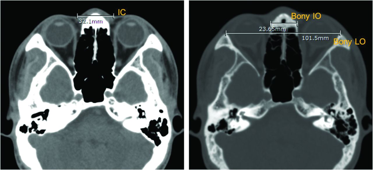
An axial CT of the brain. Left, soft-tissue intercanthal (IC); right, bony interorbital (IO) and bony lateral orbital (LO) distances, which measure 32.10, 23.65, and 101.50 mm, respectively.

RESULTS

A total of 983 scans from 932 individuals were reviewed, of whom 554 were males (59.4%) and 378 were females (40.6%). The mean ± SD age for males was 9.0 ± 6.5 years and for females was 7.8 ± 6.4 years. The mean measurements of soft tissue IC, bony IO, and bony LO distances for both sexes, categorized by age group, are shown in Tables 1⇓–3. As expected, there was a rapid increase in the IC distance in the first 2 years of life. The average ± SD IC distance was 22.1 ± 2.8 mm among neonates (0–3 months). At 22–24 months, the average ± SD IC distance was 27.3 ± 3.8 mm or approximately 80% of the mean IC distances among adults ages > 18 years (Table 1 and On-line Fig 1). The average IC distances increased with age. By the age of 8 years, the mean IC distance was 29.2 mm or approximately 90% of the mean IC distance among adults aged > 18 years. Small differences between the sexes, between 1 and 2 mm, were also detected among those >15 years of age. On average, after the age of 15 years, males tended to have larger IC distances than did the females. By age 15 years, among the males, the average IC distance was approximately 90% of the mean IC distances among adult men at >18 years. For females, by age 15 years, the average IC distance was already similar to the mean IC distances among adult women at age >18 years (Table 1).

View this table:
  • View inline
  • View popup
Table 1:

Values for soft-tissue intercanthal distances by age group and sex

View this table:
  • View inline
  • View popup
Table 2:

Values for bony interorbital distances by age group and sex

View this table:
  • View inline
  • View popup
Table 3:

Values for bony lateral orbital (LO) distances by age group and sex

This pattern of rapid growth in early life and difference between males and females aged >15 years was also observed from IO distances and LO distances. Among neonates (ages, 0–3 months), the average IO distance was 16.4 mm and was 18.6 mm by age 2 years. By the age of 8 years, the average IO distance as 22.5 mm or 90% of the mean IO distance in adulthood. Among those >15 years of age, the mean IO distance was 1–2 mm longer for men compared with women (Table 2 and On-line Fig 2). As demonstrated in Table 3, by 2 years of age, the average LO distance (77.3 mm) was also nearly 80% of the average LO distances among adults >18 years (90.0 mm). By the age of 8 years, the average LO distance (88.1 mm) was also almost 90% of the average adult LO distance at the age of 18 years. Similar to IC and IO distances, after the age of 15 years, the average LO size was 3–5 mm longer among men compared with women (On-line Fig 3). Although there is evidence for rapid growth and small differences between the sexes in IO and LO distances after the age of 15 years, the mean ratios between IO/LO were relatively stable, at 0.25, across age groups and between the sexes (Table 4). Ninety percent of all the scans had an IO:LO ratio between 0.22 and 0.28 (Fig 3).

View this table:
  • View inline
  • View popup
Table 4:

Values for bony interorbital (IO) to lateral orbital (LO) distances by age group and sex

FIG 3.
  • Download figure
  • Open in new tab
  • Download powerpoint
FIG 3.

The ratio of the interorbital (IO) to bony lateral orbital (LO) distances by age and sex.

DISCUSSION

This study aimed to document normative facial parameters among children, adolescents, and young adults in Thailand by using CT. Consistent patterns emerged that facial parameters were approximately 80% developed within the first 2 years of life and 90% developed by the age of 8 years. However, small differences between the sexes in distances after the age of 15 years also existed. Despite different growth rates across different ages and sexes, the ratios between IO:LO were relatively constant. Compared with a study conducted in the United States that examined orbital measurements of healthy infants and children,12 our average soft-tissue IC distances were slightly lower, whereas, the average bony LO distances were of similar values and our bony IO average distance was longer. The findings were not surprising because facial parameters differ among difference ethnicities.1,21,22 Our facial parameters were in the line with a recent report from Thailand.20

Similar to previous literature, there were no significant differences in our measured parameters between males and females from birth until the early teenage years.23,24 However, we noticed differences in our measured parameters between the sexes at ages ≥15 years. A previous study conducted in Thailand by using direct anthropometric measurements also found significant differences between the sexes in their binocular width, which was concordant with our bony LO distance in the adolescent age group.22 Another study, from Malaysia, that used 3D facial imaging and anthropometric techniques also found some differences in LO and IC distances between the sexes in late adolescence.25 This is not surprising because studies documented that, unlike males, facial growth is usually completed by the age of 14–15 years among females.26,27

Our study has some limitations with regard to using samples from a single referral center in northern Thailand and of using a hospital-based sample. However, our results were in line with the recent study conducted in Bangkok, which indicated that their sample was representative of the normal Thai population.20 There were limited longitudinal data because most of the scans used were of unique individuals rather than repeated follow-up. There is literature that indicates that the cross-sectional design is likely to underestimate growth rates but is useful for determining cutoff normative values because they are more likely to be representative of the healthy population.20,28 The strength of our study is the large number of scans, which allowed more-detailed age- and sex-specific normative values not previously published in an Asian population.

Based on the strengths of our study, our findings have several clinical implications. Establishing normative values is important because a diagnosis of craniofacial abnormality, for example, telecanthus, relies on age- and sex-specific means and SDs. To avoid or reduce the number of unnecessary postoperative CTs, the values of soft-tissue IC can be used for follow-up of patients after craniofacial surgery, such as frontoethmoidal encephalomeningocele correction, craniofacial cleft correction, orbital translocation, and facial bipartition. The understanding of normal growth development also helps identify the timing of surgery of craniofacial anomalies, such as frontonasal dysplasia, hypertelorism, and craniofacial cleft because correction of the orbital complex or glabella region should be performed when the bones are nearly fully grown. Results of our study suggest that the facial parameters are almost fully developed around the age of 8 years in this population. The finding that the IO:LO ratio is relatively constant, at 0.25, across ages and sexes helps inform surgeons on the aesthetic or cosmetic facial ratio for pre-, intra-, and postoperative evaluations.

CONCLUSIONS

This study provides detailed age-and sex-specific normative data of the craniofacial measurement in children and adolescents in Thailand based on CT imaging. These data can be used for evaluating individual patients with craniofacial abnormalities as well as determining the treatment in Thai and Asian populations where craniofacial abnormalities, for example, frontoethmoidal encephalomeningocele, are common.

Indicates open access to non-subscribers at www.ajnr.org

References

  1. 1.↵
    1. Farkas LG,
    2. Katic MJ,
    3. Forrest CR
    . Comparison of craniofacial measurements of young adult African-American and North American white males and females. Ann Plast Surg 2007;59:692–98 doi:10.1097/01.sap.0000258954.55068.b4 pmid:18046155
    CrossRefPubMed
  2. 2.↵
    1. Patil SB,
    2. Kale SM,
    3. Math M, et al
    . Anthropometry of the eyelid and palpebral fissure in an Indian population. Aesthet Surg J 2011;31:290–94 doi:10.1177/1090820X11398475 pmid:21385738
    CrossRefPubMed
  3. 3.↵
    1. Richards CG
    . Frontoethmoidal meningoencephalocele: a common and severe congenital abnormality in South East Asia. Arch Dis Child 1992;67:717–19 doi:10.1136/adc.67.6.717 pmid:1626991
    Abstract/FREE Full Text
  4. 4.↵
    1. Dumrongpisutikul N,
    2. Triampo A,
    3. Janthanimi P, et al
    . Incidence of associated brain and ophthalmic anomalies in frontoethmoidal encephalomeningocele evaluated by multidetector computed tomography facial bone imaging. J Craniofac Surg 2017;28:454–58 doi:10.1097/SCS.0000000000003357 pmid:28045833
    CrossRefPubMed
  5. 5.↵
    1. Meling TR,
    2. Due-Tønnessen BJ,
    3. Helseth E, et al
    . Frontoethmoidal meningoencephaloceles. Tidsskr Nor Laegeforen 2000;120:2250–52 pmid:10997082
    PubMed
  6. 6.↵
    1. Chowchuen B,
    2. Thanapaisal C,
    3. Chowchuen P, et al
    . Frontoethmoidal meningoencephalocele: challenges and the Tawanchai center’s long-term integrated management. J Med Assoc Thai 2011;94(Suppl):S129–40 pmid:22423428
    PubMed
  7. 7.↵
    1. Harris R,
    2. Nagarkar P,
    3. Amirlak B
    . Varied definitions of nasolabial angle: searching for consensus among rhinoplasty surgeons and an algorithm for selecting the ideal method. Plast Reconstr Surg Glob Open 2016;4:e752 doi:10.1097/GOX.0000000000000729 pmid:27482491
    CrossRefPubMed
  8. 8.↵
    1. Le TT,
    2. Farkas LG,
    3. Ngim RCK, et al
    . Proportionality in Asian and North American Caucasian faces using neoclassical facial canons as criteria. Aesthetic Plast Surg 2002;26:64–69 doi:10.1007/s00266-001-0033-7 pmid:11891603
    CrossRefPubMed
  9. 9.↵
    1. Farkas LG,
    2. Tompson BD,
    3. Katic MJ, et al
    . Differences between direct (anthropometric) and indirect (cephalometric) measurements of the skull. J Craniofac Surg 2002;13:105–10; discussion 109–10 doi:10.1097/00001665-200201000-00024 pmid:11887005
    CrossRefPubMed
  10. 10.↵
    1. Park DH,
    2. Choi WS,
    3. Yoon SH, et al
    . Anthropometry of Asian eyelids by age. Plast Reconstr Surg 2008;121:1405–13 doi:10.1097/01.prs.0000304608.33432.67 pmid:18349663
    CrossRefPubMed
  11. 11.↵
    1. Laestadius ND,
    2. Aase JM,
    3. Smith DW
    . Normal inner canthal and outer orbital dimensions. J Pediatr 1969;74:465–68 doi:10.1016/S0022-3476(69)80206-4 pmid:5764779
    CrossRefPubMed
  12. 12.↵
    1. Pool GM,
    2. Didier RA,
    3. Bardo D, et al
    . Computed tomography-generated anthropometric measurements of orbital relationships in normal infants and children. J Neurosurg Pediatr 2016;18:201–06 doi:10.3171/2016.2.PEDS15475 pmid:27129093
    CrossRefPubMed
  13. 13.↵
    1. Farkas LG,
    2. Hreczko TM,
    3. Katic MJ, et al
    . Proportion indices in the craniofacial regions of 284 healthy North American white children between 1 and 5 years of age. J Craniofac Surg 2003;14:13–28 doi:10.1097/00001665-200301000-00004 pmid:12544216
    CrossRefPubMed
  14. 14.↵
    1. Matteson SR,
    2. Bechtold W,
    3. Phillips C, et al
    . A method for three-dimensional image reformation for quantitative cephalometric analysis. J Oral Maxillofac Surg 1989;47:1053–61 doi:10.1016/0278-2391(89)90180-8 pmid:2795298
    CrossRefPubMed
  15. 15.↵
    1. Christiansen EL,
    2. Thompson JR,
    3. Kopp S
    . Intra- and inter-observer variability and accuracy in the determination of linear and angular measurements in computed tomography: an in vitro and in situ study of human mandibles. Acta Odontol Scand 1986;44:221–29 doi:10.3109/00016358608997724 pmid:3465194
    CrossRefPubMed
  16. 16.↵
    1. Waitzman AA,
    2. Posnick JC,
    3. Armstrong DC, et al
    . Craniofacial skeletal measurements based on computed tomography: part I. Accuracy and reproducibility. Cleft Palate Craniofac J 1992;29:112–17 doi:10.1597/1545-1569_1992_029_0112_csmboc_2.3.co_2 pmid:1571344
    CrossRefPubMed
  17. 17.↵
    1. Farkas LG
    . Accuracy of anthropometric measurements: past, present, and future. Cleft Palate Craniofac J 1996;33:10–18; discussion 19–22 doi:10.1597/1545-1569_1996_033_0010_aoampp_2.3.co_2 pmid:8849854
    CrossRefPubMed
  18. 18.↵
    1. Richtsmeier JT,
    2. Paik CH,
    3. Elfert PC, et al
    . Precision, repeatability, and validation of the localization of cranial landmarks using computed tomography scans. Cleft Palate Craniofac J 1995;32:217–27 doi:10.1597/1545-1569_1995_032_0217_pravot_2.3.co_2 pmid:7605789
    CrossRefPubMed
  19. 19.↵
    1. Gupta V,
    2. Prabhakar A,
    3. Yadav M, et al
    . Computed tomography imaging-based normative orbital measurement in Indian population. Indian J Ophthalmol 2019;67:659–63 doi:10.4103/ijo.IJO_1187_18 pmid:31007233
    CrossRefPubMed
  20. 20.↵
    1. Chatdokmaiprai C,
    2. Kiranantawat K,
    3. Lertsithichai P, et al
    . Normative data of the interorbital distance in Thai population. J Craniofac Surg 2018;29:1939–44 doi:10.1097/SCS.0000000000005050 pmid:30234716
    CrossRefPubMed
  21. 21.↵
    1. Erian A,
    2. Shiffman MA
    1. Prendergast PM
    . Facial proportions. In: Erian A, Shiffman MA, eds. Advanced Surgical Facial Rejuvenation. New York: Springer-Verlag Berlin Heidelberg; 2012:15–22
  22. 22.↵
    1. Farkas LG,
    2. Katic MJ,
    3. Forrest CR, et al
    . International anthropometric study of facial morphology in various ethnic groups/races. J Craniofac Surg 2005;16:615–46 doi:10.1097/01.scs.0000171847.58031.9e pmid:16077306
    CrossRefPubMed
  23. 23.↵
    1. Freihofer HP
    . Inner intercanthal and interorbital distances. J Maxillofac Surg 1980;8:324–26 doi:10.1016/S0301-0503(80)80121-4 pmid:6936515
    CrossRefPubMed
  24. 24.↵
    1. Méhes K,
    2. Kitzvéger E
    . Inner canthal and intermamillary indices in the newborn infant. J Pediatr 1974;85:90–92 doi:10.1016/S0022-3476(74)80295-7 pmid:4852493
    CrossRefPubMed
  25. 25.↵
    1. Othman SA,
    2. Majawit LP,
    3. Wan Hassan WN, et al
    . Anthropometric study of three-dimensional facial morphology in Malay adults. PLoS One 2016;11:e0164180 doi:10.1371/journal.pone.0164180 pmid:27706220
    CrossRefPubMed
  26. 26.↵
    1. Palmert MR,
    2. Dunkel L
    . Clinical practice. Delayed puberty. N Engl J Med 2012;366:443–53 doi:10.1056/NEJMcp1109290 pmid:22296078
    CrossRefPubMed
  27. 27.↵
    1. Ferrario VF,
    2. Sforza C,
    3. Poggio CE, et al
    . Craniofacial growth: a three-dimensional soft-tissue study from 6 years to adulthood. J Craniofac Genet Dev Biol 1998;18:138–49 pmid:9785218
    PubMed
  28. 28.↵
    1. Preece MA
    . Standardization of growth. Acta Paediatr Scand Suppl 1989;78:57–64; discussion 81–83 doi:10.1111/j.1651-2227.1989.tb17170.x pmid:2665420
    CrossRefPubMed
  • Received March 17, 2020.
  • Accepted after revision June 23, 2020.
  • © 2020 by American Journal of Neuroradiology
View Abstract
PreviousNext
Back to top
Advertisement
Print
Download PDF
Email Article

Thank you for your interest in spreading the word on American Journal of Neuroradiology.

NOTE: We only request your email address so that the person you are recommending the page to knows that you wanted them to see it, and that it is not junk mail. We do not capture any email address.

Enter multiple addresses on separate lines or separate them with commas.
CT-Based Measurements of Facial Parameters of Healthy Children and Adolescents in Thailand
(Your Name) has sent you a message from American Journal of Neuroradiology
(Your Name) thought you would like to see the American Journal of Neuroradiology web site.
CAPTCHA
This question is for testing whether or not you are a human visitor and to prevent automated spam submissions.
Cite this article
N. Jullabussapa, K. Khwanngern, C. Pateekhum, C. Angkurawaranon, S. Angkurawaranon
CT-Based Measurements of Facial Parameters of Healthy Children and Adolescents in Thailand
American Journal of Neuroradiology Aug 2020, DOI: 10.3174/ajnr.A6731

Citation Manager Formats

  • BibTeX
  • Bookends
  • EasyBib
  • EndNote (tagged)
  • EndNote 8 (xml)
  • Medlars
  • Mendeley
  • Papers
  • RefWorks Tagged
  • Ref Manager
  • RIS
  • Zotero
0 Responses
Respond to this article
Share
Bookmark this article
CT-Based Measurements of Facial Parameters of Healthy Children and Adolescents in Thailand
N. Jullabussapa, K. Khwanngern, C. Pateekhum, C. Angkurawaranon, S. Angkurawaranon
American Journal of Neuroradiology Aug 2020, DOI: 10.3174/ajnr.A6731
del.icio.us logo Twitter logo Facebook logo Mendeley logo
  • Tweet Widget
  • Facebook Like
  • Google Plus One
Purchase

Jump to section

  • Article
    • Abstract
    • ABBREVIATIONS:
    • MATERIALS AND METHODS
    • RESULTS
    • DISCUSSION
    • CONCLUSIONS
    • References
  • Figures & Data
  • Supplemental
  • Info & Metrics
  • Responses
  • References
  • PDF

Related Articles

  • PubMed
  • Google Scholar

Cited By...

  • Outcomes of enucleation and porous polyethylene orbital implant insertion in patients with paediatric retinoblastoma: a long-term follow-up study
  • Crossref (8)
  • Google Scholar

This article has been cited by the following articles in journals that are participating in Crossref Cited-by Linking.

  • An optimal deep learning framework for multi-type hemorrhagic lesions detection and quantification in head CT images for traumatic brain injury
    Aniwat Phaphuangwittayakul, Yi Guo, Fangli Ying, Ahmad Yahya Dawod, Salita Angkurawaranon, Chaisiri Angkurawaranon
    Applied Intelligence 2022 52 7
  • Craniofacial and Upper Airway Development in Patients With Treacher Collins Syndrome
    Yanxian Lin, Xiaoyang Ma, Yuanliang Huang, Lin Mu, Liya Yang, Minghao Zhao, Fang Xie, Chao Zhang, Jiajie Xu, Jianjian Lu, Li Teng
    Journal of Craniofacial Surgery 2021 32 7
  • Outcomes of enucleation and porous polyethylene orbital implant insertion in patients with paediatric retinoblastoma: a long-term follow-up study
    Min Kyu Yang, Min Joung Lee, Namju Kim, Hokyung Choung, Sang In Khwarg
    British Journal of Ophthalmology 2022 106 4
  • Anthropometric orbital measurements in a paediatric population
    Nivana Mohan, Brenda Zola De Gama, Lelika Lazarus
    Translational Research in Anatomy 2022 29
  • Age and sex estimation by cranial and paranasal sinus metric measurements of Turkish citizens using PLS methods
    Mirac Ozdemır, Nursen Turan Yurtsever, Esra Akdenız, Onur Bugdaycı
    Canadian Society of Forensic Science Journal 2025 58 1
  • Imaging of Pfeiffer Syndrome: A Case Report
    Deni Setiawan, Audy Sarah Putrini Adibrata, Puspita P. Sari, Atta Kuntara, Gery P. Yogaswara
    Open Access Macedonian Journal of Medical Sciences 2022 10 C
  • Large Isolated Nasoorbital Type of Frontoethmoidal Encephalomeningocele
    Chirakan Charoenvicha, Krit Khwanngern, Wimon Sirimaharaj
    Annals of Plastic Surgery 2022 89 1
  • Practical Considerations in Computerized Surgical Planning for Frontofacial Surgery
    Mario S. Blondin, Blake Dunson, Christopher M. Runyan
    Seminars in Plastic Surgery 2024 38 03

More in this TOC Section

  • FRACTURE MR in Congenital Vertebral Anomalies
  • Neuroimaging Delineation and Progression of SLSMD
  • fetal brain development of 10 weeks gestation
Show more PEDIATRIC NEUROIMAGING

Similar Articles

Advertisement

Indexed Content

  • Current Issue
  • Accepted Manuscripts
  • Article Preview
  • Past Issues
  • Editorials
  • Editor's Choice
  • Fellows' Journal Club
  • Letters to the Editor
  • Video Articles

Cases

  • Case Collection
  • Archive - Case of the Week
  • Archive - Case of the Month
  • Archive - Classic Case

More from AJNR

  • Trainee Corner
  • Imaging Protocols
  • MRI Safety Corner
  • Book Reviews

Multimedia

  • AJNR Podcasts
  • AJNR Scantastics

Resources

  • Turnaround Time
  • Submit a Manuscript
  • Submit a Video Article
  • Submit an eLetter to the Editor/Response
  • Manuscript Submission Guidelines
  • Statistical Tips
  • Fast Publishing of Accepted Manuscripts
  • Graphical Abstract Preparation
  • Imaging Protocol Submission
  • Evidence-Based Medicine Level Guide
  • Publishing Checklists
  • Author Policies
  • Become a Reviewer/Academy of Reviewers
  • News and Updates

About Us

  • About AJNR
  • Editorial Board
  • Editorial Board Alumni
  • Alerts
  • Permissions
  • Not an AJNR Subscriber? Join Now
  • Advertise with Us
  • Librarian Resources
  • Feedback
  • Terms and Conditions
  • AJNR Editorial Board Alumni

American Society of Neuroradiology

  • Not an ASNR Member? Join Now

© 2025 by the American Society of Neuroradiology All rights, including for text and data mining, AI training, and similar technologies, are reserved.
Print ISSN: 0195-6108 Online ISSN: 1936-959X

Powered by HighWire