Advertisement

Main menu

  • Home
  • Content
    • Current Issue
    • Accepted Manuscripts
    • Article Preview
    • Past Issue Archive
    • Video Articles
    • AJNR Case Collection
    • Case of the Week Archive
    • Case of the Month Archive
    • Classic Case Archive
  • Special Collections
    • AJNR Awards
    • Low-Field MRI
    • Alzheimer Disease
    • ASNR Foundation Special Collection
    • Photon-Counting CT
    • View All
  • Multimedia
    • AJNR Podcasts
    • AJNR SCANtastic
    • Trainee Corner
    • MRI Safety Corner
    • Imaging Protocols
  • For Authors
    • Submit a Manuscript
    • Submit a Video Article
    • Submit an eLetter to the Editor/Response
    • Manuscript Submission Guidelines
    • Statistical Tips
    • Fast Publishing of Accepted Manuscripts
    • Graphical Abstract Preparation
    • Imaging Protocol Submission
    • Author Policies
  • About Us
    • About AJNR
    • Editorial Board
    • Editorial Board Alumni
  • More
    • Become a Reviewer/Academy of Reviewers
    • Subscribers
    • Permissions
    • Alerts
    • Feedback
    • Advertisers
    • ASNR Home

User menu

  • Alerts
  • Log in

Search

  • Advanced search
American Journal of Neuroradiology

American Journal of Neuroradiology

ASHNR American Society of Functional Neuroradiology ASHNR American Society of Pediatric Neuroradiology ASSR
  • Alerts
  • Log in

Advanced Search

  • Home
  • Content
    • Current Issue
    • Accepted Manuscripts
    • Article Preview
    • Past Issue Archive
    • Video Articles
    • AJNR Case Collection
    • Case of the Week Archive
    • Case of the Month Archive
    • Classic Case Archive
  • Special Collections
    • AJNR Awards
    • Low-Field MRI
    • Alzheimer Disease
    • ASNR Foundation Special Collection
    • Photon-Counting CT
    • View All
  • Multimedia
    • AJNR Podcasts
    • AJNR SCANtastic
    • Trainee Corner
    • MRI Safety Corner
    • Imaging Protocols
  • For Authors
    • Submit a Manuscript
    • Submit a Video Article
    • Submit an eLetter to the Editor/Response
    • Manuscript Submission Guidelines
    • Statistical Tips
    • Fast Publishing of Accepted Manuscripts
    • Graphical Abstract Preparation
    • Imaging Protocol Submission
    • Author Policies
  • About Us
    • About AJNR
    • Editorial Board
    • Editorial Board Alumni
  • More
    • Become a Reviewer/Academy of Reviewers
    • Subscribers
    • Permissions
    • Alerts
    • Feedback
    • Advertisers
    • ASNR Home
  • Follow AJNR on Twitter
  • Visit AJNR on Facebook
  • Follow AJNR on Instagram
  • Join AJNR on LinkedIn
  • RSS Feeds

AJNR Awards, New Junior Editors, and more. Read the latest AJNR updates

LetterLetter

Susceptibility-Weighted Imaging in Cranial Dural Arteriovenous Fistulas

J. Saini, B. Thomas, N.K. Bodhey, A. Periakaruppan and J.M. Babulal
American Journal of Neuroradiology January 2009, 30 (1) e6; DOI: https://doi.org/10.3174/ajnr.A1265
J. Saini
  • Find this author on Google Scholar
  • Find this author on PubMed
  • Search for this author on this site
B. Thomas
  • Find this author on Google Scholar
  • Find this author on PubMed
  • Search for this author on this site
N.K. Bodhey
  • Find this author on Google Scholar
  • Find this author on PubMed
  • Search for this author on this site
A. Periakaruppan
  • Find this author on Google Scholar
  • Find this author on PubMed
  • Search for this author on this site
J.M. Babulal
  • Find this author on Google Scholar
  • Find this author on PubMed
  • Search for this author on this site
  • Article
  • Figures & Data
  • Info & Metrics
  • Responses
  • References
  • PDF
Loading

Susceptibility-weighted imaging (SWI) increases the conspicuity of venous structures within the brain.1,2 However, there are no reports of its usefulness in the detection of cranial dural arteriovenous fistulas (DAVF), which may be less conspicuous on conventional MR imaging sequences. We report the case of a patient with DAVF, which was imaged with SWI in addition to the conventional MR images.

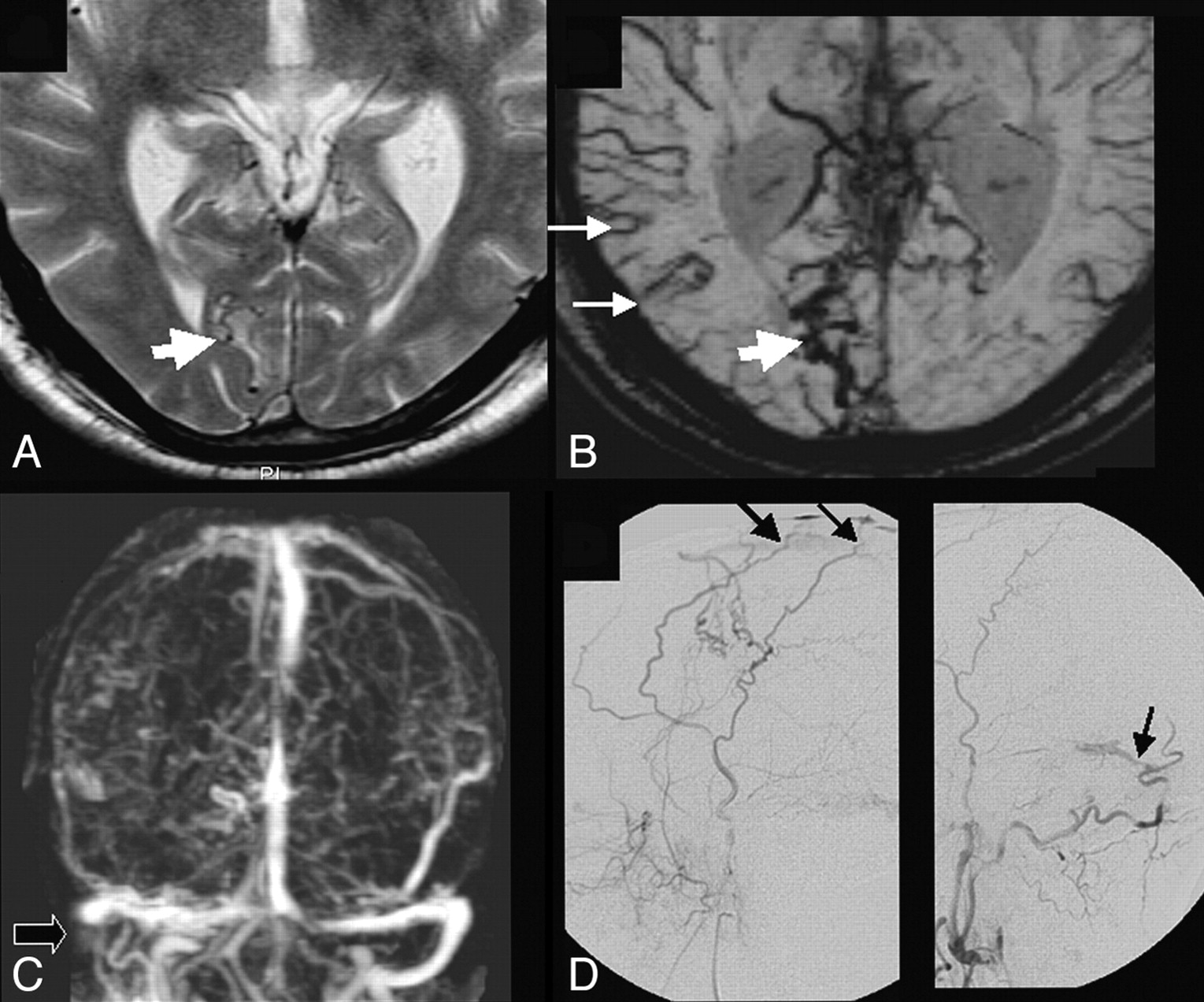
A 63-year-old man presented with gradual-onset, slowly progressive, painless bilateral visual loss of 4 months’ duration. There was no history of tinnitus. On examination, only perception of light was present bilaterally, and the fundus showed papilledema on either side. Ocular movements revealed weak abduction bilaterally. Pupils were equal and sluggishly reacting to light. No other localizing signs could be identified, and no cranial bruit was detected. Axial T2-weighted MR imaging was equivocal (Fig 1A), but SWI with minimal intensity projection showed very conspicuous venous vasculature of the entire brain, more on the right, with prominent collateral venous channels in the right occipital region (Fig 1B). Dynamic contrast-enhanced MR venographic study demonstrated occlusion of the right distal transverse and sigmoid sinuses (Fig 1C). A digital subtraction angiography (DSA) study revealed multiple DAVF in the superior sagittal sinus and the right transverse sinus, with feeders noted from right middle meningeal, superficial temporal, and occipital arteries with delayed cerebral venous drainage (Fig 1D).

SWI correlated very well with the DSA findings. Diffuse prominence of the venous vasculature noted on the SWI could be explained by the prolonged passage time of the blood in the intracranial circulation from the functional obstruction caused by DAVF at the level of the venous sinuses. Therefore, this prominence possibly allows for increased oxygen extraction, which results in more desaturation of blood in the veins, which makes them stand out on SWI. Venous engorgement, which occurs in cases of DAVF, may also be contributing to this imaging appearance. The possible site of the fistula may also be inferred from the presence of a cluster of vessels, which can be very well visualized on SWI. Moreover, in this case, we noticed, after particulate embolization of the external carotid feeders, a significant reduction in the prominence of the cortical veins with improved cerebral circulation time and better filling of the venous sinuses on follow-up imaging after 1 week.

The clinical features of DAVF range from vague symptoms such as headache, vertigo, or tinnitus to disabling neurologic deficits or a life-threatening intracranial hemorrhage.3 It may be difficult to diagnose DAVF at an early stage because of its nonspecific clinical and imaging findings. Therefore, the use of SWI may be helpful in the early diagnosis and prompt treatment in such cases. It may also help to differentiate between other causes of raised intracranial tension and may aid in assessing the posttherapeutic improvement in cerebral circulation.

  Fig 1.
  • Download figure
  • Open in new tab
  • Download powerpoint
Fig 1.

A, Axial T2-weighted image showing subtle tortuous flow voids in the right occipital region (thick white arrow). B, SWI minimal intensity projection image is showing abnormally conspicuous cortical veins more on the right side (thin white arrows) with a cluster of vessels in the right occipital region, thought to represent collateral venous flow (thick white arrow). C, Dynamic contrast-enhanced MR venographic study is showing the occluded right distal transverse and sigmoid venous sinuses (black arrow). D, DSA study revealed multiple dural arteriovenous fistulas (thin black arrows) in the region of the superior sagittal sinus and transverse sinuses.

References

  1. ↵
    Seghal V, Delproposto Z, Haacke EM, et al. Clinical applications of neuroimaging with susceptibility-weighted imaging. J Magn Reson Imaging 2005;22:439–50
    CrossRefPubMed
  2. ↵
    Thomas B, Somasundaram S, Thamburaj K, et al. Clinical applications of susceptibility weighted MR imaging of the brain: a pictorial review. Neuroradiology 2008;50:105–16
    CrossRefPubMed
  3. ↵
    Kwon BJ, Han MH, Kang HS, et al. MR imaging findings of intracranial dural arteriovenous fistulas: relations with venous drainage patterns. AJNR Am J Neuroradiol 2005;26:2500–07
    Abstract/FREE Full Text
  • Copyright © American Society of Neuroradiology
PreviousNext
Back to top

In this issue

American Journal of Neuroradiology: 30 (1)
American Journal of Neuroradiology
Vol. 30, Issue 1
January 2009
  • Table of Contents
  • Index by author
Advertisement
Print
Download PDF
Email Article

Thank you for your interest in spreading the word on American Journal of Neuroradiology.

NOTE: We only request your email address so that the person you are recommending the page to knows that you wanted them to see it, and that it is not junk mail. We do not capture any email address.

Enter multiple addresses on separate lines or separate them with commas.
Susceptibility-Weighted Imaging in Cranial Dural Arteriovenous Fistulas
(Your Name) has sent you a message from American Journal of Neuroradiology
(Your Name) thought you would like to see the American Journal of Neuroradiology web site.
CAPTCHA
This question is for testing whether or not you are a human visitor and to prevent automated spam submissions.
Cite this article
J. Saini, B. Thomas, N.K. Bodhey, A. Periakaruppan, J.M. Babulal
Susceptibility-Weighted Imaging in Cranial Dural Arteriovenous Fistulas
American Journal of Neuroradiology Jan 2009, 30 (1) e6; DOI: 10.3174/ajnr.A1265

Citation Manager Formats

  • BibTeX
  • Bookends
  • EasyBib
  • EndNote (tagged)
  • EndNote 8 (xml)
  • Medlars
  • Mendeley
  • Papers
  • RefWorks Tagged
  • Ref Manager
  • RIS
  • Zotero
0 Responses
Respond to this article
Share
Bookmark this article
Susceptibility-Weighted Imaging in Cranial Dural Arteriovenous Fistulas
J. Saini, B. Thomas, N.K. Bodhey, A. Periakaruppan, J.M. Babulal
American Journal of Neuroradiology Jan 2009, 30 (1) e6; DOI: 10.3174/ajnr.A1265
del.icio.us logo Twitter logo Facebook logo Mendeley logo
  • Tweet Widget
  • Facebook Like
  • Google Plus One
Purchase

Jump to section

  • Article
    • References
  • Figures & Data
  • Info & Metrics
  • Responses
  • References
  • PDF

Related Articles

  • No related articles found.
  • PubMed
  • Google Scholar

Cited By...

  • No citing articles found.
  • Crossref (33)
  • Google Scholar

This article has been cited by the following articles in journals that are participating in Crossref Cited-by Linking.

  • Susceptibility-weighted imaging for differential diagnosis of cerebral vascular pathology: A pictorial review
    Yu-Kun Tsui, Fong Y. Tsai, Anton N. Hasso, Fred Greensite, Binh V. Nguyen
    Journal of the Neurological Sciences 2009 287 1-2
  • New MR sequences in daily practice: susceptibility weighted imaging. A pictorial essay
    Roberto Gasparotti, Lorenzo Pinelli, Roberto Liserre
    Insights into Imaging 2011 2 3
  • Accuracy of Susceptibility-Weighted Imaging for the Detection of Arteriovenous Shunting in Vascular Malformations of the Brain
    Bharathi D. Jagadeesan, Josser E. Delgado Almandoz, Christopher J. Moran, Tammie L.S. Benzinger
    Stroke 2011 42 1
  • Imaging of Cerebral Arteriovenous Malformations and Dural Arteriovenous Fistulas
    Mahmud Mossa-Basha, James Chen, Dheeraj Gandhi
    Neurosurgery Clinics of North America 2012 23 1
  • Susceptibility-weighted angiography (SWAN) of cerebral veins and arteries compared to TOF-MRA
    T. Boeckh-Behrens, J. Lutz, N. Lummel, M. Burke, T. Wesemann, V. Schöpf, H. Brückmann, J. Linn
    European Journal of Radiology 2012 81 6
  • Simultaneous Arteriovenous Shunting and Venous Congestion Identification in Dural Arteriovenous Fistulas Using Susceptibility-Weighted Imaging: Initial Experience
    L. Letourneau-Guillon, T. Krings
    American Journal of Neuroradiology 2012 33 2
  • Susceptibility-Weighted Imaging in Migraine with Aura
    E. Karaarslan, S. Ulus, M. Kürtüncü
    American Journal of Neuroradiology 2011 32 1
  • Intracranial Dural Arteriovenous Fistula with Retrograde Cortical Venous Drainage: Use of Susceptibility-Weighted Imaging in Combination with Dynamic Susceptibility Contrast Imaging
    K. Noguchi, N. Kuwayama, M. Kubo, Y. Kamisaki, K. Kameda, G. Tomizawa, H. Kawabe, H. Seto
    American Journal of Neuroradiology 2010 31 10
  • The Use of Susceptibility-Weighted Imaging as an Indicator of Retrograde Leptomeningeal Venous Drainage and Venous Congestion With Dural Arteriovenous Fistula
    Ichiro Nakagawa, Toshiaki Taoka, Takeshi Wada, Hiroyuki Nakagawa, Masahiko Sakamoto, Kimihiko Kichikawa, Yasuo Hironaka, Yasushi Motoyama, Young-Su Park, Hiroyuki Nakase
    Neurosurgery 2013 72 1
  • Endovascular treatment of 170 consecutive cranial dural arteriovenous fistulae: results and complications
    Gerasimos Baltsavias, Anton Valavanis
    Neurosurgical Review 2014 37 1

More in this TOC Section

  • Letter to the Editor regarding “Automated Volumetric Software in Dementia: Help or Hindrance to the Neuroradiologist?”
  • Reply:
  • Brain AVM’s Nidus: What if We Hadn’t Understood Anything?
Show more LETTERS

Similar Articles

Advertisement

Indexed Content

  • Current Issue
  • Accepted Manuscripts
  • Article Preview
  • Past Issues
  • Editorials
  • Editor's Choice
  • Fellows' Journal Club
  • Letters to the Editor
  • Video Articles

Cases

  • Case Collection
  • Archive - Case of the Week
  • Archive - Case of the Month
  • Archive - Classic Case

More from AJNR

  • Trainee Corner
  • Imaging Protocols
  • MRI Safety Corner

Multimedia

  • AJNR Podcasts
  • AJNR Scantastics

Resources

  • Turnaround Time
  • Submit a Manuscript
  • Submit a Video Article
  • Submit an eLetter to the Editor/Response
  • Manuscript Submission Guidelines
  • Statistical Tips
  • Fast Publishing of Accepted Manuscripts
  • Graphical Abstract Preparation
  • Imaging Protocol Submission
  • Evidence-Based Medicine Level Guide
  • Publishing Checklists
  • Author Policies
  • Become a Reviewer/Academy of Reviewers
  • News and Updates

About Us

  • About AJNR
  • Editorial Board
  • Editorial Board Alumni
  • Alerts
  • Permissions
  • Not an AJNR Subscriber? Join Now
  • Advertise with Us
  • Librarian Resources
  • Feedback
  • Terms and Conditions
  • AJNR Editorial Board Alumni

American Society of Neuroradiology

  • Not an ASNR Member? Join Now

© 2025 by the American Society of Neuroradiology All rights, including for text and data mining, AI training, and similar technologies, are reserved.
Print ISSN: 0195-6108 Online ISSN: 1936-959X

Powered by HighWire