Advertisement

Main menu

  • Home
  • Content
    • Current Issue
    • Accepted Manuscripts
    • Article Preview
    • Past Issue Archive
    • Video Articles
    • AJNR Case Collection
    • Case of the Week Archive
    • Case of the Month Archive
    • Classic Case Archive
  • Special Collections
    • AJNR Awards
    • Low-Field MRI
    • Alzheimer Disease
    • ASNR Foundation Special Collection
    • Photon-Counting CT
    • View All
  • Multimedia
    • AJNR Podcasts
    • AJNR SCANtastic
    • Trainee Corner
    • MRI Safety Corner
    • Imaging Protocols
  • For Authors
    • Submit a Manuscript
    • Submit a Video Article
    • Submit an eLetter to the Editor/Response
    • Manuscript Submission Guidelines
    • Statistical Tips
    • Fast Publishing of Accepted Manuscripts
    • Graphical Abstract Preparation
    • Imaging Protocol Submission
    • Author Policies
  • About Us
    • About AJNR
    • Editorial Board
    • Editorial Board Alumni
  • More
    • Become a Reviewer/Academy of Reviewers
    • Subscribers
    • Permissions
    • Alerts
    • Feedback
    • Advertisers
    • ASNR Home

User menu

  • Alerts
  • Log in

Search

  • Advanced search
American Journal of Neuroradiology

American Journal of Neuroradiology

ASHNR American Society of Functional Neuroradiology ASHNR American Society of Pediatric Neuroradiology ASSR
  • Alerts
  • Log in

Advanced Search

  • Home
  • Content
    • Current Issue
    • Accepted Manuscripts
    • Article Preview
    • Past Issue Archive
    • Video Articles
    • AJNR Case Collection
    • Case of the Week Archive
    • Case of the Month Archive
    • Classic Case Archive
  • Special Collections
    • AJNR Awards
    • Low-Field MRI
    • Alzheimer Disease
    • ASNR Foundation Special Collection
    • Photon-Counting CT
    • View All
  • Multimedia
    • AJNR Podcasts
    • AJNR SCANtastic
    • Trainee Corner
    • MRI Safety Corner
    • Imaging Protocols
  • For Authors
    • Submit a Manuscript
    • Submit a Video Article
    • Submit an eLetter to the Editor/Response
    • Manuscript Submission Guidelines
    • Statistical Tips
    • Fast Publishing of Accepted Manuscripts
    • Graphical Abstract Preparation
    • Imaging Protocol Submission
    • Author Policies
  • About Us
    • About AJNR
    • Editorial Board
    • Editorial Board Alumni
  • More
    • Become a Reviewer/Academy of Reviewers
    • Subscribers
    • Permissions
    • Alerts
    • Feedback
    • Advertisers
    • ASNR Home
  • Follow AJNR on Twitter
  • Visit AJNR on Facebook
  • Follow AJNR on Instagram
  • Join AJNR on LinkedIn
  • RSS Feeds

AJNR Awards, New Junior Editors, and more. Read the latest AJNR updates

OtherHead and Neck Imaging

Expansile Organized Maxillary Sinus Hematoma: MR and CT Findings and Review of Literature

T. Nishiguchi, A. Nakamura, K. Mochizuki, Y. Tokuhara, H. Yamane and Y. Inoue
American Journal of Neuroradiology August 2007, 28 (7) 1375-1377; DOI: https://doi.org/10.3174/ajnr.A0629
T. Nishiguchi
  • Find this author on Google Scholar
  • Find this author on PubMed
  • Search for this author on this site
A. Nakamura
  • Find this author on Google Scholar
  • Find this author on PubMed
  • Search for this author on this site
K. Mochizuki
  • Find this author on Google Scholar
  • Find this author on PubMed
  • Search for this author on this site
Y. Tokuhara
  • Find this author on Google Scholar
  • Find this author on PubMed
  • Search for this author on this site
H. Yamane
  • Find this author on Google Scholar
  • Find this author on PubMed
  • Search for this author on this site
Y. Inoue
  • Find this author on Google Scholar
  • Find this author on PubMed
  • Search for this author on this site
  • Article
  • Figures & Data
  • Info & Metrics
  • Responses
  • References
  • PDF
Loading

Abstract

SUMMARY: An organized hematoma is a chronic state of fibrotic tissue surrounding a hemorrhage. A mass lesion resulting from hematoma in the maxillary sinus was first reported in 1917, and the term, “blood boil,” was clinically coined from such features as encapsulated blood and locally aggressive behavior. Subsequently, others have reported lesions with a similar appearance and clinical course, and now, in Japan, blood boil is used as a clinical term for such lesions. Factors that may predispose a patient to hematoma formation vary, and the pathogenesis of the mass is still uncertain. The lesions are mainly composed of an organized hematoma, regardless of their origin. We present 2 cases of organized maxillary sinus hematomas that have unusual radiologic findings and correlate these findings with the histopathologic findings.

Case Reports

Case 1

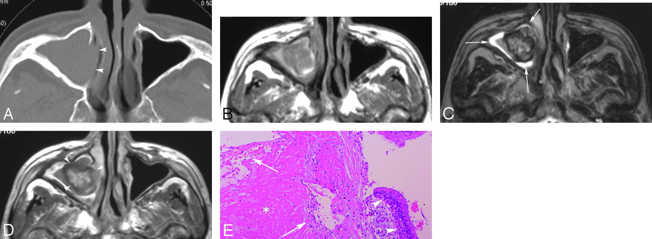
A 22-year-old man was referred to the otolaryngology department with a 6-month history of nasal congestion and swelling in his left cheek. When he was examined in our hospital, no epistaxis or neurologic deficit was noticed. There was no history of hematopoietic disorder. Endonasal examination revealed a pinkish mass in the left nasal cavity. The nasal septum was deviated to the right. A CT scan showed an 8-cm mass in the left maxillary sinus, which was primarily expansile with focal areas of bone loss (Fig 1A). On MR imaging, the mass had a slightly high T1-weighted signal intensity, heterogeneous low-to-high T2-weighted signal intensity, and scattered areas of well-demarcated enhancement on contrast-enhanced T1-weighted images (Fig 1B–D). A high signal intensity zone surrounded the mass on T2-weighted images (Fig 1C). A malignant tumor was suspected because of the size of the mass and the bony changes. Caldwell-Luc approach allowed en bloc resection of the mass. Pathology showed a fibrous encapsulated organized hematoma with surrounding inflamed mucosa without a neoplastic tissue (Fig 1E).

Fig 1.
  • Download figure
  • Open in new tab
  • Download powerpoint
Fig 1.

CT, MR imaging, and histopathologic findings in case 1.

A, The axial image of CT (bone algorithm) shows expanding mass in the left maxillary sinus. Expansile bone destruction (arrowheads) of the medial wall and bone defects (asterisks) of the anterior and posterior wall are observed.

B, T1-weighted spin-echo (TR/TE, 620 ms/15 ms) axial image depicts slightly high intensity areas on the mass. The left pterygopalatine fossa fat and the left retroantral fat are replaced by the mass (arrowheads).

C, T2-weighted fast spin-echo (TR/TE, 5000 ms/120 ms) axial image depicts curvilinear hyperintensity areas around the mass (arrows).

D, Contrast-enhanced T1-weighted spin-echo (TR/TE, 620 ms/15 ms) axial image clearly shows the antral and nasal parts of the mass (arrowheads). Markedly enhanced portions, which are clearly demarcated from nonenhanced portions, are observed in the mass.

E, Hematoxylin-eosin stains (low-power field, original magnification ×4) reveal the hematoma with organization in case 1. There is a reactive hyperplasia of endothelial cells in the hematoma (asterisks). Scattered fibroblasts are observed in the fibrous tissue margin (arrows). In the margin of the specimen, an inflamed columnar epithelium is confirmed (arrowheads). No tumoral cells are found.

Case 2

A 76-year-old man presented with a 5-month history of repeated nasal bleeding. He has been followed for cirrhosis for several years; however, no coagulopathy was present. Endonasal examination demonstrated a pinkish mass in the right nasal cavity, with medial displacement of the lateral nasal wall. On CT, a 2- × 3-cm mass displaced and thinned the medial antral wall (Fig 2A). On MR imaging, the mass was primarily isointense to muscle on T1-weighted images but contained areas of high signal intensity (Fig 2B). On T2-weighted images, there was a central region of high signal intensity surrounded by a low signal intensity zone, and a thinner rim of high signal intensity surrounded the entire mass (Fig 2C). The mass was markedly enhanced in a multinodular manner on contrast-enhanced T1-weighted images (Fig 2D). A malignant tumor was initially suspected because of a history of repeated epistaxis and the age of the patient. During surgery, the mass was totally resected by a transnasal approach. Histolopathology showed only a fibrous encapsulated organized hematoma (Fig 2E).

Fig 2.
  • Download figure
  • Open in new tab
  • Download powerpoint
Fig 2.

CT, MR imaging, and histopathologic findings in case 2.

A, The axial image of CT (bone algorithm) shows the mass in the right maxillary sinus. The pressure on the medial maxillary sinus wall has caused medial bowing of the wall (arrowheads).

B, T1-weighted spin-echo (TR/TE, 620 ms/15 ms) axial image depicts punctuate high signals on the mass.

C, T2-weighted fast spin-echo (TR/TE, 5000 ms/120 ms) axial image depicts a curvilinear hyperintensity area around the mass (arrows).

D, Contrast enhanced T1-weighted spin-echo (TR/TE, 620 ms/15 ms) axial image shows the mass (arrowheads). A central enhanced area corresponding with the area of central heterogeneous hyperintensity on T2-weighted image is observed in the mass.

E, Hematoxylin-eosin stains (low-power field, original magnification × 40) reveal the hematoma with organization in case 2. An admixture of fibrin network and hemorrhage (asterisk) is observed. There is a reactive hyperplasia of endothelial cells (arrows). An inflamed columnar epithelium is confirmed (arrowheads). No tumoral cells are found.

Discussion

An organized hematoma in the maxillary sinus is a rare lesion. Fewer than 100 cases1–8 have been reported to our knowledge. The mass usually originates in a single maxillary sinus and is generally located in the medial antral wall near the sinus ostium. Patients range in age between 20 and 76 years; no sex predominance is observed. It occurs in patients with and without a bleeding diathesis. The patient often presents with various symptoms that are caused by the mass, depending on the size and location. The most frequent symptom is nasal congestion and rhinorrhea because of the obstruction and secondary inflammatory process by the mass. Repeated epistaxis is reported in 70% of patients.2 Pain or facial swelling occurs when the mass compresses the adjacent structures. The mass gradually enlarges and causes pressure remodeling of the antral walls with some focal areas of apparent bone destruction. CT scans show an expansile maxillary sinus mass with some areas of bone resorption (Fig 1). On MR imaging, the mass usually has a variable signal intensity on T1- and T2-weighted images, ranging from low to high. After contrast administration, discrete areas of enhancement are present within the mass. The surrounding inflamed sinus mucosa, despite the bony changes, suggest a benign process on imaging.

The pathogenesis of organized hematomas has not been elucidated. However, Lee et al3 suggest a possible mechanism for the formation of an organized hematoma as follows: 1) repeated hemorrhage in the semiclosed lumen (maxillary sinus) forms a hematoma encapsulated by fibrosis; 2) the encapsulation prevents the absorption of the hematoma and induces vascularization, which causes rebleeding and increasing pressure within the hematoma; and 3) the progressive expansion of a hematoma causes the demineralization of adjacent structures. A variety of benign lesions or clinical states have been described in association with the development of an organized hematoma and they display similar conditions clinically and radiologically. Ozaki et al4 suggest that a hematoma originates from a hemangioma or an inflammatory vascular injury, and they define an organized hematoma without an underlying hemangioma as an inflammatory pseudotumor. A bleeding diathesis, an aggressive fungal infection, radiation therapy, a postoperative complication, or trauma are also potential causes of a similar condition.5–9 The photomicrograph of the histopathologic specimen in our subjects shows excessive endothelial proliferation with adjacent subacute and chronic hematoma and surrounding fibrous tissue margin. Histopathologic findings and clinical course in our subjects resemble those from other reports of hematomas of the maxillary sinus.10 We speculate that the same pathophysiologic mechanism may be responsible for the hematomas seen in conjunction with the entities described above. In our histopathologic specimens, there is no cholesterol cleft with reactive foreign-body giant cells seen in cholesterol granuloma. No mucous retention or infiltration of foamy histiocytes seen in the mucocele is observed. A hematoma is subepithelially located; thus, a hemorrhagic cyst is ruled out. We suggest that an organized maxillary sinus hematoma is different from these entities.

Conclusion

Benign hematoma of the sinus is a rare lesion of uncertain pathophysiology that may mimic a neoplasm. It is important to include this in the differential diagnosis of an expansile maxillary sinus mass containing a hemorrhagic product so that inappropriate surgery can be avoided.

Acknowledgments

We thank Dr Kenichi Wakasa for helpful comments on our article.

References

  1. ↵
    Tadokoro K. Jyougakudou Ketsuryu ni tsuite. Dainichi Jibi 1917;23:359–60
  2. ↵
    Fujii M. The diagnosis and therapy of paranasal blood boil. JOHNS 1995;11:705–08
  3. ↵
    Lee PK, Wu JK, Ludemann JP. Hemorrhagic pseudotumor of the maxillary sinus: J Otolaryngol 2004;33:206–08
    PubMed
  4. ↵
    Ozaki M, Sakai T, Ikeda K. Hemangioma of the nasal cavity and sinuses. Jikou 1977;49:53–58
  5. ↵
    Gupta S, Mohapatra BB, Ghai S, et al. Haemophilic pseudotumor of the paranasal sinuses. Haemophilia 2001;7:595–99
    CrossRefPubMed
  6. Unlu HH, Mutlu C, Ayhan S, et al. Organized hematoma of the maxillary sinus mimicking tumor. Auris Nasus Larynx 2001;28:253–55
    CrossRefPubMed
  7. Tabaee A, Kacker A. Hematoma of the maxillary sinus presenting as a mass. Int J Pediatr Otorhinolaryngol 2002;65:153–57
    CrossRefPubMed
  8. ↵
    Segev Y, Landsberg R, Fliss DM. MR imaging appearance of frontal sinus barotrauma. AJNR Am J Neuroradiol 2003;24:346–47
    Abstract/FREE Full Text
  9. ↵
    Bong-JL, Hyo-Jin P, Seong-Cheol H. Organized hematoma of the maxillary sinus. Acta Otolaryngol 2003;123:869–72
    CrossRefPubMed
  10. ↵
    Yagisawa M, Ishitoya J, Tsukuda M. Hematoma-like mass of the maxillary sinus. Acta Otolaryngol 2006;126:277–81
    PubMed
  • Received July 30, 2006.
  • Accepted after revision November 22, 2006.
  • Copyright © American Society of Neuroradiology
View Abstract
PreviousNext
Back to top

In this issue

American Journal of Neuroradiology: 28 (7)
American Journal of Neuroradiology
Vol. 28, Issue 7
August 2007
  • Table of Contents
  • Index by author
Advertisement
Print
Download PDF
Email Article

Thank you for your interest in spreading the word on American Journal of Neuroradiology.

NOTE: We only request your email address so that the person you are recommending the page to knows that you wanted them to see it, and that it is not junk mail. We do not capture any email address.

Enter multiple addresses on separate lines or separate them with commas.
Expansile Organized Maxillary Sinus Hematoma: MR and CT Findings and Review of Literature
(Your Name) has sent you a message from American Journal of Neuroradiology
(Your Name) thought you would like to see the American Journal of Neuroradiology web site.
CAPTCHA
This question is for testing whether or not you are a human visitor and to prevent automated spam submissions.
Cite this article
T. Nishiguchi, A. Nakamura, K. Mochizuki, Y. Tokuhara, H. Yamane, Y. Inoue
Expansile Organized Maxillary Sinus Hematoma: MR and CT Findings and Review of Literature
American Journal of Neuroradiology Aug 2007, 28 (7) 1375-1377; DOI: 10.3174/ajnr.A0629

Citation Manager Formats

  • BibTeX
  • Bookends
  • EasyBib
  • EndNote (tagged)
  • EndNote 8 (xml)
  • Medlars
  • Mendeley
  • Papers
  • RefWorks Tagged
  • Ref Manager
  • RIS
  • Zotero
0 Responses
Respond to this article
Share
Bookmark this article
Expansile Organized Maxillary Sinus Hematoma: MR and CT Findings and Review of Literature
T. Nishiguchi, A. Nakamura, K. Mochizuki, Y. Tokuhara, H. Yamane, Y. Inoue
American Journal of Neuroradiology Aug 2007, 28 (7) 1375-1377; DOI: 10.3174/ajnr.A0629
del.icio.us logo Twitter logo Facebook logo Mendeley logo
  • Tweet Widget
  • Facebook Like
  • Google Plus One
Purchase

Jump to section

  • Article
    • Abstract
    • Case Reports
    • Discussion
    • Conclusion
    • Acknowledgments
    • References
  • Figures & Data
  • Info & Metrics
  • Responses
  • References
  • PDF

Related Articles

  • No related articles found.
  • PubMed
  • Google Scholar

Cited By...

  • No citing articles found.
  • Crossref (30)
  • Google Scholar

This article has been cited by the following articles in journals that are participating in Crossref Cited-by Linking.

  • Acute invasive fungal rhinosinusitis: MR imaging features and their impact on prognosis
    Ye Ra Choi, Ji-hoon Kim, Hye Sook Min, Jae-Kyung Won, Hyun Jik Kim, Roh-Eul Yoo, Koung Mi Kang, Sun-Won Park, Tae Jin Yun, Seung Hong Choi, Chul-Ho Sohn, Jung Hyo Rhim, Soo Chin Kim, Jae Wook Ryu, Dong Gyu Na
    Neuroradiology 2018 60 7
  • Imaging features of benign mass lesions in the nasal cavity and paranasal sinuses according to the 2017 WHO classification
    Hiroyuki Tatekawa, Taro Shimono, Masahiko Ohsawa, Satoshi Doishita, Shinichi Sakamoto, Yukio Miki
    Japanese Journal of Radiology 2018 36 6
  • Sinonasal organized Hematoma: Clinical features of seventeen cases and a systematic review
    Soo Jung Choi, Sung Tae Seo, Ki‐Sang Rha, Yong Min Kim
    The Laryngoscope 2015 125 9
  • Diagnostic characteristics of sinonasal organizing hematomas: avoiding misdiagnosis
    Arthur W. Wu, Jonathan Y. Ting, Roderick C. Borgie, Nicolas Y. Busaba, Peter M. Sadow, Amy F. Juliano, Stacey T. Gray, Eric H. Holbrook
    International Forum of Allergy & Rhinology 2013 3 7
  • Organized Hematoma
    Wenhui Pang, Li Hu, Huan Wang, Yan Sha, Na Ma, Shuyi Wang, Quan Liu, Xicai Sun, Dehui Wang
    Otolaryngology–Head and Neck Surgery 2016 154 4
  • Endoscopic Sinus Surgery for the Treatment of Organized Hematoma of the Maxillary Sinus
    HIDEAKI SUZUKI, TSUYOSHI INABA, NOBUAKI HIRAKI, KOICHI HASHIDA, TETSURO WAKASUGI, YOHEI KADOKAWA, TSUYOSHI UDAKA
    The Kurume Medical Journal 2008 55 3/4
  • CT and MR imaging findings of sinonasal angiomatous polyps
    Jing Zou, Fengyuan Man, Kai Deng, Yuanyuan Zheng, Dapeng Hao, Wenjian Xu
    European Journal of Radiology 2014 83 3
  • Imaging characteristics of sinonasal organized hematoma
    Joonho Hur, Jae Kyun Kim, Jun Soo Byun, Woong Jae Lee
    Acta Radiologica 2015 56 8
  • Organised haematoma of the sphenoid sinus mimicking a pituitary tumour
    T Nakagawa, Y Kawai, T Sakamoto, J Ito
    The Journal of Laryngology & Otology 2010 124 1
  • Evaluation of multiparametric MRI differentiating sinonasal angiomatous polyp from malignant tumors
    Xinyan Wang, Ying Liu, Qinghua Chen, Junfang Xian
    Neuroradiology 2019 61 8

More in this TOC Section

  • Contrast Enhanced Pituitary CISS/FIESTA
  • Temporal Evolution of Vestibular schwannoma
  • FDG Uptake and Thyroid Cancer Post-BRAF Therapy
Show more Head and Neck Imaging

Similar Articles

Advertisement

Indexed Content

  • Current Issue
  • Accepted Manuscripts
  • Article Preview
  • Past Issues
  • Editorials
  • Editor's Choice
  • Fellows' Journal Club
  • Letters to the Editor
  • Video Articles

Cases

  • Case Collection
  • Archive - Case of the Week
  • Archive - Case of the Month
  • Archive - Classic Case

More from AJNR

  • Trainee Corner
  • Imaging Protocols
  • MRI Safety Corner
  • Book Reviews

Multimedia

  • AJNR Podcasts
  • AJNR Scantastics

Resources

  • Turnaround Time
  • Submit a Manuscript
  • Submit a Video Article
  • Submit an eLetter to the Editor/Response
  • Manuscript Submission Guidelines
  • Statistical Tips
  • Fast Publishing of Accepted Manuscripts
  • Graphical Abstract Preparation
  • Imaging Protocol Submission
  • Evidence-Based Medicine Level Guide
  • Publishing Checklists
  • Author Policies
  • Become a Reviewer/Academy of Reviewers
  • News and Updates

About Us

  • About AJNR
  • Editorial Board
  • Editorial Board Alumni
  • Alerts
  • Permissions
  • Not an AJNR Subscriber? Join Now
  • Advertise with Us
  • Librarian Resources
  • Feedback
  • Terms and Conditions
  • AJNR Editorial Board Alumni

American Society of Neuroradiology

  • Not an ASNR Member? Join Now

© 2025 by the American Society of Neuroradiology All rights, including for text and data mining, AI training, and similar technologies, are reserved.
Print ISSN: 0195-6108 Online ISSN: 1936-959X

Powered by HighWire