Advertisement

Main menu

  • Home
  • Content
    • Current Issue
    • Accepted Manuscripts
    • Article Preview
    • Past Issue Archive
    • Video Articles
    • AJNR Case Collection
    • Case of the Week Archive
    • Case of the Month Archive
    • Classic Case Archive
  • Special Collections
    • AJNR Awards
    • Low-Field MRI
    • Alzheimer Disease
    • ASNR Foundation Special Collection
    • Photon-Counting CT
    • View All
  • Multimedia
    • AJNR Podcasts
    • AJNR SCANtastic
    • Trainee Corner
    • MRI Safety Corner
    • Imaging Protocols
  • For Authors
    • Submit a Manuscript
    • Submit a Video Article
    • Submit an eLetter to the Editor/Response
    • Manuscript Submission Guidelines
    • Statistical Tips
    • Fast Publishing of Accepted Manuscripts
    • Graphical Abstract Preparation
    • Imaging Protocol Submission
    • Author Policies
  • About Us
    • About AJNR
    • Editorial Board
    • Editorial Board Alumni
  • More
    • Become a Reviewer/Academy of Reviewers
    • Subscribers
    • Permissions
    • Alerts
    • Feedback
    • Advertisers
    • ASNR Home

User menu

  • Alerts
  • Log in

Search

  • Advanced search
American Journal of Neuroradiology

American Journal of Neuroradiology

ASHNR American Society of Functional Neuroradiology ASHNR American Society of Pediatric Neuroradiology ASSR
  • Alerts
  • Log in

Advanced Search

  • Home
  • Content
    • Current Issue
    • Accepted Manuscripts
    • Article Preview
    • Past Issue Archive
    • Video Articles
    • AJNR Case Collection
    • Case of the Week Archive
    • Case of the Month Archive
    • Classic Case Archive
  • Special Collections
    • AJNR Awards
    • Low-Field MRI
    • Alzheimer Disease
    • ASNR Foundation Special Collection
    • Photon-Counting CT
    • View All
  • Multimedia
    • AJNR Podcasts
    • AJNR SCANtastic
    • Trainee Corner
    • MRI Safety Corner
    • Imaging Protocols
  • For Authors
    • Submit a Manuscript
    • Submit a Video Article
    • Submit an eLetter to the Editor/Response
    • Manuscript Submission Guidelines
    • Statistical Tips
    • Fast Publishing of Accepted Manuscripts
    • Graphical Abstract Preparation
    • Imaging Protocol Submission
    • Author Policies
  • About Us
    • About AJNR
    • Editorial Board
    • Editorial Board Alumni
  • More
    • Become a Reviewer/Academy of Reviewers
    • Subscribers
    • Permissions
    • Alerts
    • Feedback
    • Advertisers
    • ASNR Home
  • Follow AJNR on Twitter
  • Visit AJNR on Facebook
  • Follow AJNR on Instagram
  • Join AJNR on LinkedIn
  • RSS Feeds

AJNR Awards, New Junior Editors, and more. Read the latest AJNR updates

OtherHead and Neck Imaging

Bisphosphonate-Associated Mandibular Osteonecrosis

E. Carneiro, P. Vibhute, A. Montazem and P.M. Som
American Journal of Neuroradiology May 2006, 27 (5) 1096-1097;
E. Carneiro
  • Find this author on Google Scholar
  • Find this author on PubMed
  • Search for this author on this site
P. Vibhute
  • Find this author on Google Scholar
  • Find this author on PubMed
  • Search for this author on this site
A. Montazem
  • Find this author on Google Scholar
  • Find this author on PubMed
  • Search for this author on this site
P.M. Som
  • Find this author on Google Scholar
  • Find this author on PubMed
  • Search for this author on this site
  • Article
  • Figures & Data
  • Info & Metrics
  • Responses
  • References
  • PDF
Loading

Abstract

SUMMARY: A 66-year-old man with multiple myeloma treated with zoledronic acid, melphalan, and prednisone after a tooth extraction developed severe osteonecrosis of the mandible that was nonresponsive to antibiotic therapy. A CT scan showed innumerable fragmented sequestra in the mandible and some adjacent soft tissue fullness around the mandible. The association between the use of bisphosphonates and osteonecrosis of the jaw has been described only recently.

Multiple myeloma (MM) is a disseminated plasma cell neoplasm characterized clinically by osteolytic bone lesions, monoclonal immunoglobulin detectable in the serum and/or urine (in more than 90% of patients), renal insufficiency, and amyloidosis (in 10% of patients). MM is the third-most-common hematologic malignancy, accounting for nearly 10% of all such neoplasia.1,2 It is often inaccurately viewed as a rare disease because of its short survival (median survival, 2.5–3 years) and its uniformly fatal outcome. Bone involvement is the hallmark of MM, and these findings are manifest in most patients at the time of diagnosis as lytic lesions, osteoporosis, and/or fractures.3

Improved understanding of myeloma-associated bone disease has led to the use of prophylactic bisphosphonate therapy. Apart from delaying the onset of skeletal events, the new generation of bisphosphonates—in particular pamidronate and zoledronic acid—exerts antimyeloma effects indirectly by inducing osteoclast apoptosis or directly by inducing myeloma cell apoptosis.4 Recent reports have shown an increasing association between the use of bisphosphonates and osteonecrosis of the jaw (ONJ).5–8 We report the case of a patient with MM who received chronic bisphosphonate therapy and developed osteomyelitis of the mandible a few days after a tooth extraction. The mandibular disease is documented with CT for the first time in the radiologic literature. The clinical literature contains 2 previous CT images, both cases being far less extensively involved than our case.7,8

Case Report

A 66-year-old man was diagnosed with MM in 2002. Shortly after his diagnosis, treatment with zoledronic acid, melphalan, and prednisone was initiated. After approximately 2–3 months, he developed a loose mandibular molar, which was removed. The extraction socket never healed, and the patient underwent biopsy of the exposed bone, which was consistent with osteomyelitis. He was placed on multiple courses of antibiotics without resolution of the exposed bone. During that interval, the teeth adjacent to the nonhealing socket also became loose and were subsequently removed sequentially.

In October 2003, the patient complained of jaw pain and numbness of his lower lip and chin. On physical examination, the patient had large areas of exposed and seemingly nonvital bone in the mandible bilaterally. This was especially prominent along the mylohyoid ridge areas. There was purulent exudate within the open wounds and a fetid odor. There were multiple mobile teeth within the mandible. The patient had anesthesia along the dermatome distribution of the right inferior alveolar nerve. There were no lesions present within the maxilla, and all of the maxillary teeth were stable. Panoramic radiographs showed areas of patchy radiolucency within the mandible, widened periodontal ligament spaces around the mandibular dentition, and periosteal bone deposition along the inferior border of the mandible. Aggressive local wound care was initiated, and the patient had a satisfactory response with near resolution of the infection and decrease in pain. Reparative granulation tissue in intervening sites along the open wounds was noted during a 2-month period.

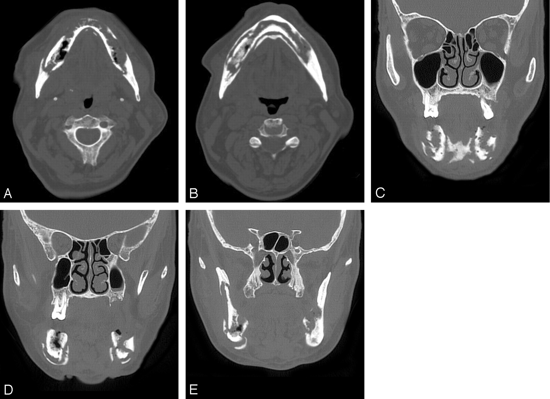
The patient was then lost to follow-up until February 2005, when he returned to the office complaining of severe pain. He had stopped treatment. On intraoral examination, the patient had copious purulence from large areas of exposed, necrotic bone along most of the mandibular alveolar ridge. Thin intervening bands of normal-appearing mucosa were present within the anterior mandible. He was noted to have 2 draining fistulas in the submental area with active purulent exudate. Palpation of the exposed bone in the right posterior mandible resulted in mobility of the exposed bone in the anterior and left mandible. A panoramic radiograph showed large areas of bone destruction throughout the mandible with proliferative bone deposition along the inferior border. A CT scan showed (Fig. 1) innumerable fragmented sequestra in the mandible, starting at the level of the coronoid process on the right and the midramus on the left, extending through the angle and body bilaterally and including the symphysis. There was some soft tissue fullness adjacent to the posterior body of the mandible on the right side and some cellulitic/edematous type changes in the soft tissues around the mandible.

  Fig 1.
  • Download figure
  • Open in new tab
  • Download powerpoint
Fig 1.

Serial axial (A and B) and coronal (C–E) CT scans of the mandible show extensive fragmentation and sequestration of this edendulous mandible. There was mild adjacent soft tissue fullness. This degree of osteonecrosis would be excessive for the more common radiation-related disease.

Discussion

ONJ has multiple etiologies and is most commonly seen following radiation therapy for the head and neck malignancies.6 Rare cases have been described in association with herpes zoster and in a patient with osteopetrosis.9 The jaw findings in MM are present in 5%–39% of cases and are the first manifestation of the disease in 17% of patients. Patients typically present with painless mandibular swelling. The radiologic features are variable, but most commonly noted are solitary or multiple punched-out lytic lesions, occasionally with septation.10

Bisphosphonates are nonmetabolizated analogues of pyrophosphate that play an important role in bone reabsorption. They inhibit osteoclastic action, have an antiangiogenesis effect, and are helpful in the maintenance of the calcium and phosphate equilibrium. Although the exact mechanism of this bisphosphonate-mediated osteoclast inhibition has not been completely elucidated, their role in decreasing osteoclast-mediated lysis of bone has been well established in clinical trials in patients with MM, breast cancer, Paget disease of bone, and postmenopausal osteoporosis.8 These drugs are effective in reducing bone pain, hypercalcemia, and skeletal-related events in patients with osteolytic metastasis. In fact, according to the American Society of Clinical Oncology Practice Guidelines, the use of intravenous pamidronate and zoledronic acid is recommended for patients with pain due to osteolytic bone disease and as adjunctive treatment for patients receiving radiation therapy, analgesics, or surgical intervention to stabilize fractures or impending fractures.11

ONJ associated with bisphosphonates most commonly occurs in the mandible but can also occur in the maxilla. Most patients present with jaw pain and exposed necrotic bone, which usually starts at the site of a recently extracted tooth (70%–77%), similar to our patient.6,8 The more or less unique involvement of the jaw bones by osteonecrosis has been attributed to their direct exposure to the external environment via the teeth, which in turn are frequently associated with periodontal disease and trauma. The clinical presentation may simulate a dental abscess and osteomyelitis and radiographs usually show regions of mottled bone, consistent with sequestrum formation.6

The differential diagnosis of ONJ should include neuralgia-inducing cavitatory osteonecrosis (NICO). Unlike patients with ONJ associated with bisphosphonate use, patients with NICO present with atypical facial pain or trigeminal neuralgia, which often has been present for years. Tooth loosening and tooth loss because of dissolution of the alveolar medullary bone are also common findings in NICO. The radiographic changes associated with NICO are variable but primarily show a poorly demarcated, nonexpansile radiolucency of the affected alveolus, often with irregular vertical remnants of lamina dura associated with old dental extraction sites.12

Bisphosphonate-associated ONJ has only recently been described in the clinical literature. To the best of our knowledge, imaging features were mentioned in only 2 of these reports, and they were in patients with far more subtle disease than seen in our patient.7,8 Although the exact mechanism of bisphosphonate-associated ONJ is not fully understood, there many patients with MM, metastatic cancers, and osteoporosis who are receiving chronic bisphosphonate therapy. Because imaging studies will be conducted for many of these patients, the radiologist should be aware of this unusual entity.

References

  1. ↵
    American Cancer Society. Cancer: facts & figures 2002. Atlanta: American Cancer Society;2002 :4
  2. ↵
    Kinney MKC, Swerdlow SH. Hematopoietic and lymphoid disorders. In: Barnes L, ed. Surgical pathology of the head and neck. New York: Marcel Dekker;2001 :1326–29
  3. ↵
    Kyle RA, Rajkumar SV. Multiple myeloma. N Eng J Med 2004;351:1860–73
    CrossRefPubMed
  4. ↵
    Berensen JR, Rosen LS, Howell A, et al. Zoledronic acid reduces skeletal-related events in patients with osteolitic metastases. Cancer 2001;91:1191–200
    CrossRefPubMed
  5. ↵
    Magliorati CA. Bisphosphonates and oral cavity avascular bone necrosis. J Clin Oncol 2003;21:4253–54
    FREE Full Text
  6. ↵
    Marx RE. Pamidronate (Aredia) and zoledronic acid (zometa) induced avascular necrosis of the jaws: a growing epidemic. J Oral Maxillofac Surg 2003;61:1115–18
    CrossRefPubMed
  7. ↵
    Wang J, Godger NM, Pogrel MA. Osteonecrosis of the jaws associated with cancer chemotherapy. J Oral Maxillofac Surg 2003;61:1104–107
    CrossRefPubMed
  8. ↵
    Ruggiero SL, Mehotra MBBS, Rosenberg TJ, et al. Osteonecrosis of the jaws associated with the use of bisphosphonates: a review of 63 cases. J Oral Maxillofac Surg 2004;62:527–34
    CrossRefPubMed
  9. ↵
    Mostofi R, Marchmont-Robinson H, Freije S. Spontaneous tooth exfoliation and osteonecrosis following a herpes zooster infection of the fifth cranial nerve. J Oral Maxillofac Surg 1987;45:264
    CrossRefPubMed
  10. ↵
    Witt C, Borges AC, Klein K, et al. Radiographic manifestations of multiple myeloma in the mandible: a retrospective study of 77 patients. J Oral Maxillofac Surg 1997;55:450–53
    CrossRefPubMed
  11. ↵
    Berenson JR, Hillner E, Kyle RA, et al. American Society of Clinical Oncology Practice Guidelines: the role of bisphosphonates in multiple myeloma. J Clin Oncol 2002;20:3719–36
    Abstract/FREE Full Text
  12. ↵
    Bouquot JE, Roberts AM, Person P, et al. Neuralgia-inducing cavitational osteonecrosis (NICO): osteomyelitis in 224 jawbone samples from patients with facial neuralgia. Oral Surg Oral Med Oral Pathol 1992;73:307–09
    PubMed
  • Copyright © American Society of Neuroradiology
View Abstract
PreviousNext
Back to top

In this issue

American Journal of Neuroradiology: 27 (5)
American Journal of Neuroradiology
Vol. 27, Issue 5
May 2006
  • Table of Contents
  • Index by author
Advertisement
Print
Download PDF
Email Article

Thank you for your interest in spreading the word on American Journal of Neuroradiology.

NOTE: We only request your email address so that the person you are recommending the page to knows that you wanted them to see it, and that it is not junk mail. We do not capture any email address.

Enter multiple addresses on separate lines or separate them with commas.
Bisphosphonate-Associated Mandibular Osteonecrosis
(Your Name) has sent you a message from American Journal of Neuroradiology
(Your Name) thought you would like to see the American Journal of Neuroradiology web site.
CAPTCHA
This question is for testing whether or not you are a human visitor and to prevent automated spam submissions.
Cite this article
E. Carneiro, P. Vibhute, A. Montazem, P.M. Som
Bisphosphonate-Associated Mandibular Osteonecrosis
American Journal of Neuroradiology May 2006, 27 (5) 1096-1097;

Citation Manager Formats

  • BibTeX
  • Bookends
  • EasyBib
  • EndNote (tagged)
  • EndNote 8 (xml)
  • Medlars
  • Mendeley
  • Papers
  • RefWorks Tagged
  • Ref Manager
  • RIS
  • Zotero
0 Responses
Respond to this article
Share
Bookmark this article
Bisphosphonate-Associated Mandibular Osteonecrosis
E. Carneiro, P. Vibhute, A. Montazem, P.M. Som
American Journal of Neuroradiology May 2006, 27 (5) 1096-1097;
del.icio.us logo Twitter logo Facebook logo Mendeley logo
  • Tweet Widget
  • Facebook Like
  • Google Plus One

Jump to section

  • Article
    • Abstract
    • Case Report
    • Discussion
    • References
  • Figures & Data
  • Info & Metrics
  • Responses
  • References
  • PDF

Related Articles

  • No related articles found.
  • PubMed
  • Google Scholar

Cited By...

  • Onco-Nephrology: The Pathophysiology and Treatment of Malignancy-Associated Hypercalcemia
  • Crossref
  • Google Scholar

This article has not yet been cited by articles in journals that are participating in Crossref Cited-by Linking.

More in this TOC Section

  • ASL Sensitivity for Head and Neck Paraganglioma
  • Post SRS Peritumoral Hyperintense Signal of VSs
  • Contrast Enhanced Pituitary CISS/FIESTA
Show more Head and Neck Imaging

Similar Articles

Advertisement

Indexed Content

  • Current Issue
  • Accepted Manuscripts
  • Article Preview
  • Past Issues
  • Editorials
  • Editor's Choice
  • Fellows' Journal Club
  • Letters to the Editor
  • Video Articles

Cases

  • Case Collection
  • Archive - Case of the Week
  • Archive - Case of the Month
  • Archive - Classic Case

More from AJNR

  • Trainee Corner
  • Imaging Protocols
  • MRI Safety Corner
  • Book Reviews

Multimedia

  • AJNR Podcasts
  • AJNR Scantastics

Resources

  • Turnaround Time
  • Submit a Manuscript
  • Submit a Video Article
  • Submit an eLetter to the Editor/Response
  • Manuscript Submission Guidelines
  • Statistical Tips
  • Fast Publishing of Accepted Manuscripts
  • Graphical Abstract Preparation
  • Imaging Protocol Submission
  • Evidence-Based Medicine Level Guide
  • Publishing Checklists
  • Author Policies
  • Become a Reviewer/Academy of Reviewers
  • News and Updates

About Us

  • About AJNR
  • Editorial Board
  • Editorial Board Alumni
  • Alerts
  • Permissions
  • Not an AJNR Subscriber? Join Now
  • Advertise with Us
  • Librarian Resources
  • Feedback
  • Terms and Conditions
  • AJNR Editorial Board Alumni

American Society of Neuroradiology

  • Not an ASNR Member? Join Now

© 2025 by the American Society of Neuroradiology All rights, including for text and data mining, AI training, and similar technologies, are reserved.
Print ISSN: 0195-6108 Online ISSN: 1936-959X

Powered by HighWire