Advertisement

Main menu

  • Home
  • Content
    • Current Issue
    • Accepted Manuscripts
    • Article Preview
    • Past Issue Archive
    • Video Articles
    • AJNR Case Collection
    • Case of the Week Archive
    • Case of the Month Archive
    • Classic Case Archive
  • Special Collections
    • AJNR Awards
    • Low-Field MRI
    • Alzheimer Disease
    • ASNR Foundation Special Collection
    • Photon-Counting CT
    • View All
  • Multimedia
    • AJNR Podcasts
    • AJNR SCANtastic
    • Trainee Corner
    • MRI Safety Corner
    • Imaging Protocols
  • For Authors
    • Submit a Manuscript
    • Submit a Video Article
    • Submit an eLetter to the Editor/Response
    • Manuscript Submission Guidelines
    • Statistical Tips
    • Fast Publishing of Accepted Manuscripts
    • Graphical Abstract Preparation
    • Imaging Protocol Submission
    • Author Policies
  • About Us
    • About AJNR
    • Editorial Board
    • Editorial Board Alumni
  • More
    • Become a Reviewer/Academy of Reviewers
    • Subscribers
    • Permissions
    • Alerts
    • Feedback
    • Advertisers
    • ASNR Home

User menu

  • Alerts
  • Log in

Search

  • Advanced search
American Journal of Neuroradiology

American Journal of Neuroradiology

ASHNR American Society of Functional Neuroradiology ASHNR American Society of Pediatric Neuroradiology ASSR
  • Alerts
  • Log in

Advanced Search

  • Home
  • Content
    • Current Issue
    • Accepted Manuscripts
    • Article Preview
    • Past Issue Archive
    • Video Articles
    • AJNR Case Collection
    • Case of the Week Archive
    • Case of the Month Archive
    • Classic Case Archive
  • Special Collections
    • AJNR Awards
    • Low-Field MRI
    • Alzheimer Disease
    • ASNR Foundation Special Collection
    • Photon-Counting CT
    • View All
  • Multimedia
    • AJNR Podcasts
    • AJNR SCANtastic
    • Trainee Corner
    • MRI Safety Corner
    • Imaging Protocols
  • For Authors
    • Submit a Manuscript
    • Submit a Video Article
    • Submit an eLetter to the Editor/Response
    • Manuscript Submission Guidelines
    • Statistical Tips
    • Fast Publishing of Accepted Manuscripts
    • Graphical Abstract Preparation
    • Imaging Protocol Submission
    • Author Policies
  • About Us
    • About AJNR
    • Editorial Board
    • Editorial Board Alumni
  • More
    • Become a Reviewer/Academy of Reviewers
    • Subscribers
    • Permissions
    • Alerts
    • Feedback
    • Advertisers
    • ASNR Home
  • Follow AJNR on Twitter
  • Visit AJNR on Facebook
  • Follow AJNR on Instagram
  • Join AJNR on LinkedIn
  • RSS Feeds

AJNR Awards, New Junior Editors, and more. Read the latest AJNR updates

Letter

Caution; Confusion Ahead…

R. Capocci, E. Shotar, N.-A. Sourour, I. Haffaf, B. Bartolini and F. Clarençon
American Journal of Neuroradiology June 2017, 38 (6) E40-E43; DOI: https://doi.org/10.3174/ajnr.A5179
R. Capocci
aDepartment of Interventional Neuroradiology Pitié-Salpêtrière Hospital, APHP Paris, France
  • Find this author on Google Scholar
  • Find this author on PubMed
  • Search for this author on this site
  • ORCID record for R. Capocci
E. Shotar
bDepartment of Interventional Neuroradiology Pitié-Salpêtrière Hospital, APHP Paris, France
cParis VI University Pierre et Marie Curie Paris, France
  • Find this author on Google Scholar
  • Find this author on PubMed
  • Search for this author on this site
  • ORCID record for E. Shotar
N.-A. Sourour
dDepartment of Interventional Neuroradiology Pitié-Salpêtrière Hospital, APHP Paris, France
  • Find this author on Google Scholar
  • Find this author on PubMed
  • Search for this author on this site
  • ORCID record for N.-A. Sourour
I. Haffaf
eDepartment of Interventional Neuroradiology Pitié-Salpêtrière Hospital, APHP Paris, France
  • Find this author on Google Scholar
  • Find this author on PubMed
  • Search for this author on this site
  • ORCID record for I. Haffaf
B. Bartolini
fDepartment of Interventional Neuroradiology Pitié-Salpêtrière Hospital, APHP Paris, France
  • Find this author on Google Scholar
  • Find this author on PubMed
  • Search for this author on this site
  • ORCID record for B. Bartolini
F. Clarençon
gDepartment of Interventional Neuroradiology Pitié-Salpêtrière Hospital, APHP Paris, France
hParis VI University Pierre et Marie Curie Paris, France
  • Find this author on Google Scholar
  • Find this author on PubMed
  • Search for this author on this site
  • ORCID record for F. Clarençon
  • Article
  • Figures & Data
  • Info & Metrics
  • Responses
  • References
  • PDF
Loading

We read with great interest the article entitled “Endovascular Therapy of M2 Occlusion in IMS III: Role of M2 Segment Definition and Location on Clinical and Revascularization Outcomes” by Tomsick et al.1 This study is a post hoc subgroup analysis of the patients randomized in the endovascular arm of the Interventional Management of Stroke (IMS) III study who underwent a mechanical thrombectomy (MT) for an MCA M2 segment occlusion. This article provides interesting data on distal (ie, M2) occlusions treated by endovascular means. Indeed, the recent randomized controlled trials (RCTs)2⇓⇓⇓⇓–7 that showed the effectiveness of MT in acute ischemic stroke with large vessel occlusion included very few cases of M2 occlusion (Table). Consequently, the American Heart Association (AHA) guidelines,8 according to the results of RCTs, suggest that only M1 and more proximal arterial occlusions should be safely treated by MT. Scant data (only nonrandomized, retrospective, monocenter series) on the safety and effectiveness of MT in M2 occlusions are available in the literature.9⇓⇓⇓⇓⇓–15 Despite the potential interest of this paper, we would like to raise some comments on its methods.

View this table:
  • View inline
  • View popup

Number of patients with M2 occlusion in the recent randomized controlled trials on MT

First, we would like to underline the fact that a subgroup analysis, as mentioned in many papers and letters,16⇓–18 is prone to bias in the statistical analysis. Consequently, the results of such post hoc analyses on small volume subgroups should be interpreted with caution.

Second, we found it very questionable to perform a post hoc analysis of a study19 that showed such a low recanalization rate, due to the use of obsolete devices like “sonography-assisted thrombolysis” (EKOS system; EKOS, Bothell, Washington) and the “Merci retriever” (Concentric Medical, Mountain View, California), in the era of stent retrievers and aspiration devices. Indeed, the overall recanalization rate of M2 occlusion in this series was only 40%. Recent monocenter retrospective studies using more recent devices showed a recanalization rate over 75%.9,13,15 Our center's experience with distal artery occlusions treated by endovascular means shows a recanalization rate of 76%.20

Third, we would like to report our disagreement with the MCA segmentation used in this paper. Indeed, the MCA segmentation commonly used is the one described in 1938 by Fischer21 (Fig 1) and further used in anatomic22 and angiographic23 descriptive studies. In Fischer's21 paper (written in the German language), the MCA segments are clearly defined:

Fig 1.
  • Download figure
  • Open in new tab
  • Download powerpoint
Fig 1.

Original drawings of the intracranial arteries by Fischer.21 A, Lateral view; B, frontal view. Reprinted with permission from Fischer E. Die Lageabweichungen der vorderen Hirnarterie im Gefässbild. Zentralbl Neurochir 1938;3:300–13.

Der Verlauf der A. cerebri media zerfällt in folgende Unterabschnitte:

  1. Den horizontalen Anfangsteil (M1), von der Teilungsstelle der Carotis int. Bis zu dem etwa rechtwinkligen Knie der A. cerebri media reichend,

  2. Den nach hinten zu ansteigenden Inselabschnitt (M2), welcher mit 2–3 Hauptästen dem Inselgebiet dicht aufliegt, im Seitenbild in der arteriellen Gefäßachse (Moniz) des Gehirns verläuft und im Vorderbild nahezu vertikal ansteigt,

  3. Gefäßverzweigungen (M3) der vorgenannten Hauptäste der Fossa Sylvii mit dem Kandelaber (Foix) und charakteristischen Schleifenbildungen der Aa. Frontales asc. Im Seitenbild. Auf der Vorderaufnahme bilden diese zusammen mit der folgenden Gruppe ein charakteristisches, nach oben zu scharf begrenztes Fächerbild (M3–4), das bei Tumoren der Zentral- oder Parietalregion eine typische Kompression nach unten erfährt,

  4. Gefäßverzweigungen (M4) im hintersten Teil der Fissura Sylvii (Gyrus angularis-Gebiet), die im Seitenbild deutlich hervortreten, dagegen auf der Vorderaufnahme mit dem Fächer (M3–4), zusammenfallen,

  5. Endausbreitungen (M5) der mittleren hirnarterie, Sie sind zum Teil auf der Vorderaufnahme als feinere und mehr lockere Gefäßmaschen unmittelbar über dem dichteren und etwas gröber gezeichneten Fächerbild sichtbar, besonders klar jedoch im Seitenbild als divergiende Endäste (M5) zu erkennen (Aa. Parietalis post., angularis und temporalis post.) Bei Tumoren der Hinterhauptlappens können diese Äste von unten her eine Zusammendrängung und Parallelverlagerung nach oben oder aber, bei Entwicklung des Tumors mehr von dorsal her, eine stärkere Auseinanderdrängung in rechtwinkliger bis gerader Form erfahren.

Our translation of this article reports that:

“The course of the middle cerebral artery is decomposed in the following subsections:

  1. The horizontal initial part (M1), from the internal carotid bifurcation to the distal genu of the middle cerebral artery.

  2. On the lateral view, the insular section progresses along the axis of the brain arteries toward the rear and upwardly (M2) and gives birth to 2–3 main branches lying on the insula, and, on the front view, it is ascending almost vertically.

  3. The junction of the above-mentioned main branches of the Sylvian fissure (M3) with the candelabra (Foix) shows the typical loop aspect of the ascending frontal artery on the lateral view. On the frontal view, these branches form and limit sharply with the following group a typical image of a fan turned upward (M3–4), translated downward in case of a central or parietal lobe tumor.

  4. Vessel intersection (M4) at the rear part of the Sylvian fissure (gyrus angularis), which clearly stands out on the lateral view, whereas they coincide with the fan on the frontal view.

  5. At the terminal section (M5) of the middle cerebral artery, there are, on the frontal view, fine and looser vascular stitches immediately above the attenuated and more visibly marked fan; however, on the lateral view, these appear particularly clear as the segments are divergent (M5) (posterior parietal, angular, and posterior temporal arteries). With occipital lobe tumors, these branches can be pushed together from downward and be translated upwardly, but with the development of more dorsal tumors, a stronger compression can shift these structures frontally.”

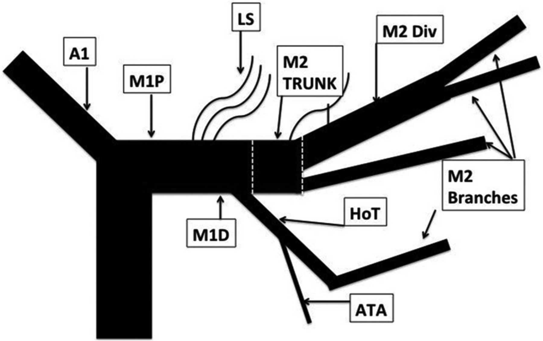
We think that using a classification without respecting criteria and landmarks that define these different segments is very confusing. Indeed, in their paper, the authors artificially created what they called an “M2 trunk” (Fig 2)1 that definitively belongs to the M1 segment according to Fischer's classification (horizontal segment, before the MCA genu). This imprecise interpretation of a segmentation commonly used worldwide may lead to substantial misunderstandings and may render the results published in this series noncomparable with other studies dealing with M2 occlusions. Recently, Goyal et al24 made an effort to clarify what should be considered as the M1 segment and detailed the M1 and M2 segment anatomic variations. In particular, they proposed to assimilate large anterior temporal artery (ATA) variation (ie, ATA supplying more than the anterior aspect of the temporal lobe) to an M2 segment.

Fig 2.
  • Download figure
  • Open in new tab
  • Download powerpoint
Fig 2.

Drawing summarizing the MCA segmentation used in Tomsick et al's1 article.

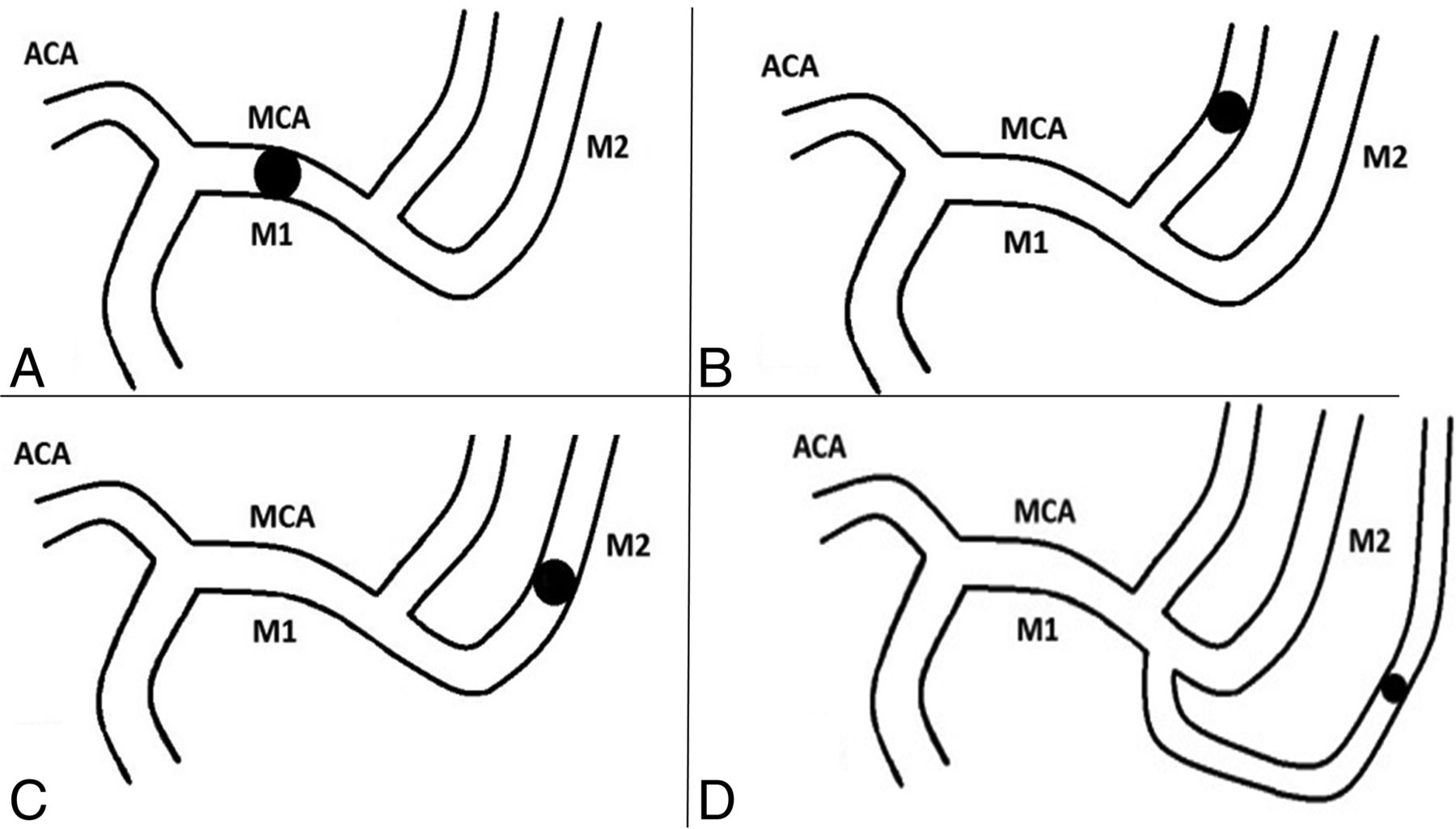
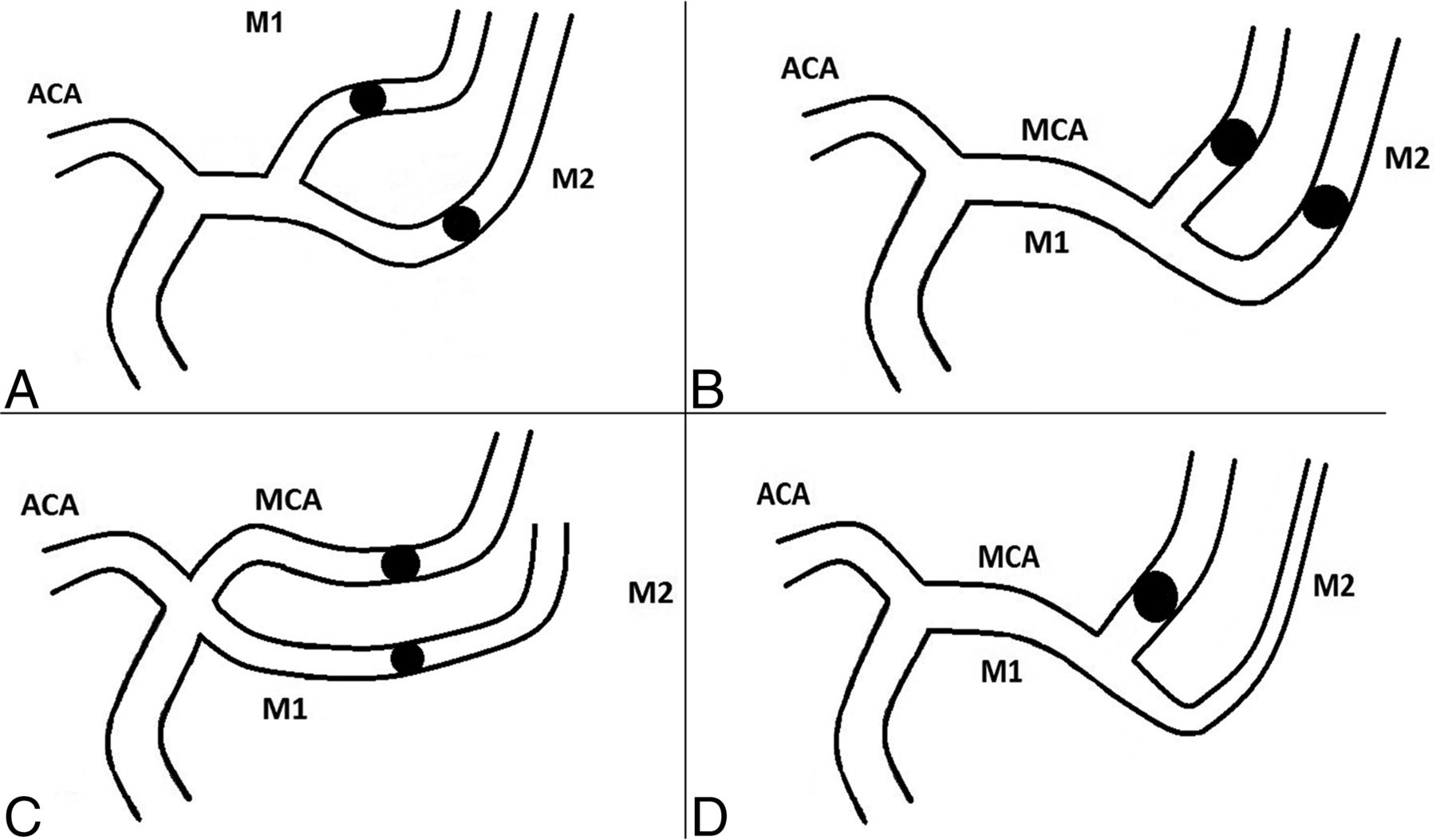
To definitively clarify what is an M1 or M2 occlusion, we suggest using a classification such as the one presented below. In this classification, in addition to true M1 or M2 occlusions (Fig 3), we describe “M1-like” (Fig 4) occlusions that comprise:

  • Occlusion of both branches after MCA division, proximal (short M1 segment) (Fig 4A) or distal to the MCA's genu (Fig 4B);

  • Occlusion of both branches of a duplicated or accessory MCA (Fig 4C); and,

  • Occlusion of either the superior or inferior division of the MCA, if it is a dominant branch (ie, division branch feeding ≥75% of the MCA's cortical territory) (Fig 4D).

Fig 3.
  • Download figure
  • Open in new tab
  • Download powerpoint
Fig 3.

Drawings summarizing true M1 and M2 occlusions. A, M1 occlusion; B, superior M2 occlusion; C, inferior M2 occlusion; D, M2 trifurcation occlusion. ACA indicates anterior cerebral artery.

Fig 4.
  • Download figure
  • Open in new tab
  • Download powerpoint
Fig 4.

Drawings summarizing “M1-like” occlusions. A, Occlusion of both branches after MCA division, short M1 segment; B, occlusion of both branches after MCA division, distal to the MCA's genu; C, occlusion of both branches of a duplicated or accessory MCA; D, occlusion of either the superior or inferior division of the MCA if it is a dominant branch (ie, division branch feeding ≥75% of the MCA's cortical territory). ACA indicates anterior cerebral artery.

We also describe “M2-like” (Fig 5) occlusions that comprise:

  • Occlusion of 1 branch after MCA division, proximal (short M1 segment) or distal to the MCA's genu (Fig 5A);

  • Occlusion of 1 branch of a duplicated or accessory MCA (Fig 5B); and,

  • Occlusion of the ATA if its trunk is large (ie, as big as M2) (Fig 5C).

Fig 5.
  • Download figure
  • Open in new tab
  • Download powerpoint
Fig 5.

Drawings summarizing “M2-like” occlusions. A, Occlusion of 1 branch after MCA division, proximal (short M1 segment) or distal to the MCA's genu; B, occlusion of 1 branch of a duplicated or accessory MCA; C, occlusion of the anterior temporal artery if its trunk is large (ie, as big as M2). ACA indicates anterior cerebral artery.

To conclude, we think that speaking the same language, by using the classifications in a common way, is the only manner to provide comparable results.

Footnotes

  • Disclosures: Nader Sourour—UNRELATED: Consultancy: Medtronic; Payment for Development of Educational Presentations: Medtronic, Comments: workshop; Stock/Stock Options: Medina, Comments: former investor. Bruno Bartolini—UNRELATED: Consultancy: Stryker. Frédéric Clarençon—UNRELATED: Consultancy: Codman, Medtronic.

References

  1. 1.↵
    1. Tomsick TA,
    2. Carrozzella J,
    3. Foster L, et al
    . Endovascular therapy of M2 occlusion in IMS III: role of M2 segment definition and location on clinical and revascularization outcomes. AJNR Am J Neuroradiol 2017;38:84–89 doi:10.3174/ajnr.A4979 pmid:27765740
    Abstract/FREE Full Text
  2. 2.↵
    1. Berkhemer OA,
    2. Fransen PS,
    3. Beumer D, et al
    . A randomized trial of intraarterial treatment for acute ischemic stroke. N Engl J Med 2015;372:11–20 doi:10.1056/NEJMoa1411587 pmid:25517348
    CrossRefPubMed
  3. 3.↵
    1. Saver JL,
    2. Goyal M,
    3. Bonafe A, et al
    . Stent-retriever thrombectomy after intravenous t-PA vs. t-PA alone in stroke. N Engl J Med 2015;372:2285–95 doi:10.1056/NEJMoa1415061 pmid:25882376
    CrossRefPubMed
  4. 4.↵
    1. Jovin TG,
    2. Chamorro A,
    3. Cobo E, et al
    . Thrombectomy within 8 hours after symptom onset in ischemic stroke. N Engl J Med 2015;372:2296–306 doi:10.1056/NEJMoa1503780 pmid:25882510
    CrossRefPubMed
  5. 5.↵
    1. Campbell BC,
    2. Mitchell PJ,
    3. Kleinig TJ, et al
    . Endovascular therapy for ischemic stroke with perfusion-imaging selection. N Engl J Med 2015;372:1009–18 doi:10.1056/NEJMoa1414792 pmid:25671797
    CrossRefPubMed
  6. 6.↵
    1. Goyal M,
    2. Demchuk AM,
    3. Menon BK, et al
    . Randomized assessment of rapid endovascular treatment of ischemic stroke. N Engl J Med 2015;372:1019–30 doi:10.1056/NEJMoa1414905 pmid:25671798
    CrossRefPubMed
  7. 7.↵
    1. Bracard S,
    2. Ducrocq X,
    3. Mas JL, et al
    . Mechanical thrombectomy after intravenous alteplase versus alteplase alone after stroke (THRACE): a randomised controlled trial. Lancet Neurol 2016;15:1138–47 doi:10.1016/S1474-4422(16)30177-6 pmid:27567239
    CrossRefPubMed
  8. 8.↵
    1. Powers WJ,
    2. Derdeyn CP,
    3. Biller J, et al
    . 2015 AHA/ASA focused update of the 2013 guidelines for the early management of patients with acute ischemic stroke regarding endovascular treatment: a guideline for healthcare professionals from the American Heart Association/American Stroke Association. Stroke 2015;46:3020–35 doi:10.1161/STR.0000000000000074 pmid:26123479
    Abstract/FREE Full Text
  9. 9.↵
    1. Park JS,
    2. Kwak HS
    . Manual aspiration thrombectomy using penumbra catheter in patients with acute M2 occlusion: a single-center analysis. J Korean Neurosurg Soc 2016;59:352–56 doi:10.3340/jkns.2016.59.4.352 pmid:27446515
    CrossRefPubMed
  10. 10.↵
    1. Kim YW,
    2. Son S,
    3. Kang DH, et al
    . Endovascular thrombectomy for M2 occlusions: comparison between forced arterial suction thrombectomy and stent retriever thrombectomy. J Neurointerv Surg 2016 Jul 5. [Epub ahead of print] doi:10.1136/neurintsurg-2016-012466 pmid:27382124
    CrossRefPubMed
  11. 11.↵
    1. Wong JH,
    2. Do HM,
    3. Telischak NA, et al
    . Initial experience with SOFIA as an intermediate catheter in mechanical thrombectomy for acute ischemic stroke. J Neurointerv Surg 2016 Oct 27. [Epub ahead of print] doi:10.1136/neurintsurg-2016-012750 pmid:27789787
    CrossRefPubMed
  12. 12.↵
    1. Coutinho JM,
    2. Liebeskind DS,
    3. Slater LA, et al
    . Mechanical thrombectomy for isolated M2 occlusions: a post hoc analysis of the STAR, SWIFT, and SWIFT PRIME studies. AJNR Am J Neuroradiol 2016;37:667–72 doi:10.3174/ajnr.A4591 pmid:26564442
    Abstract/FREE Full Text
  13. 13.↵
    1. Dorn F,
    2. Lockau H,
    3. Stetefeld H, et al
    . Mechanical thrombectomy of M2-occlusion. J Stroke Cerebrovasc Dis 2015;24:1465–70 doi:10.1016/j.jstrokecerebrovasdis.2015.04.013 pmid:259952748
    CrossRefPubMed
  14. 14.↵
    1. Sung SM,
    2. Lee TH,
    3. Lee SW, et al
    . Emergent intracranial stenting for acute M2 occlusion of middle cerebral artery. Clin Neurol Neurosurg 2014;119:110–15 doi:10.1016/j.clineuro.2014.01.027 pmid:24635938
    CrossRefPubMed
  15. 15.↵
    1. Flores A,
    2. Tomasello A,
    3. Cardona P, et al
    . Endovascular treatment for M2 occlusions in the era of stentrievers: a descriptive multicenter experience. J Neurointerventional Surg 2015;7:234–37 doi:10.1136/neurintsurg-2014-011100 pmid:24578483
    Abstract/FREE Full Text
  16. 16.↵
    1. Ewald B
    . Post hoc choice of cut points introduced bias to diagnostic research. J Clin Epidemiol 2006;59:798–801 doi:10.1016/j.jclinepi.2005.11.025 pmid:16828672
    CrossRefPubMed
  17. 17.↵
    1. Kivimäki M,
    2. Singh-Manoux A,
    3. Ferrie JE, et al
    . Post hoc decision-making in observational epidemiology–is there need for better research standards? Int J Epidemiol 2013;42:367–70 doi:10.1093/ije/dyt036 pmid:23569177
    FREE Full Text
  18. 18.↵
    Post-hoc analysis in clinical trials. Research Supervisor Connect, University of Sydney, Australia. http://sydney.edu.au/research/opportunities/opportunities/2133. Accessed February 2, 2017.
  19. 19.↵
    1. Broderick JP,
    2. Palesch YY,
    3. Demchuk AM, et al
    . Endovascular therapy after intravenous t-PA versus t-PA alone for stroke. N Engl J Med 2013;368:893–903 doi:10.1056/NEJMoa1214300 pmid:23390923
    CrossRefPubMed
  20. 20.↵
    1. Premat K,
    2. Bartolini B,
    3. Di Maria F, et al
    . Single-center experience using the 3MAX reperfusion catheter for the treatment of acute ischemic stroke with distal arterial occlusion. In: Proceedings of the 29th European Congress of Radiology, Vienna, Austria. March 1–5, 2017
  21. 21.↵
    1. Fischer E
    . Die Lageabweichungen der vorderen Hirnarterie im Gefäßbild. Zentralbl Neurochir 1938;3:300–13
  22. 22.↵
    1. Gibo H,
    2. Carver CC,
    3. Rhoton AL Jr., et al
    . Microsurgical anatomy of the middle cerebral artery. J Neurosurg 1981;54:151–69 doi:10.3171/jns.1981.54.2.0151 pmid:7452329
    CrossRefPubMed
  23. 23.↵
    1. Krayenbuhl HA
    . Cerebral Angiography (2nd revised edition). London: Butterworth & Co; 1968
  24. 24.↵
    1. Goyal M,
    2. Menon BK,
    3. Krings T, et al
    . What constitutes the M1 segment of the middle cerebral artery? J Neurointerv Surg 2016 Jan 11. [Epub ahead of print] doi:10.1136/neurintsurg-2015-012191 pmid:26863104
    Abstract/FREE Full Text
  • © 2017 by American Journal of Neuroradiology
PreviousNext
Back to top

In this issue

American Journal of Neuroradiology: 38 (6)
American Journal of Neuroradiology
Vol. 38, Issue 6
1 Jun 2017
  • Table of Contents
  • Index by author
  • Complete Issue (PDF)
Advertisement
Print
Download PDF
Email Article

Thank you for your interest in spreading the word on American Journal of Neuroradiology.

NOTE: We only request your email address so that the person you are recommending the page to knows that you wanted them to see it, and that it is not junk mail. We do not capture any email address.

Enter multiple addresses on separate lines or separate them with commas.
Caution; Confusion Ahead…
(Your Name) has sent you a message from American Journal of Neuroradiology
(Your Name) thought you would like to see the American Journal of Neuroradiology web site.
CAPTCHA
This question is for testing whether or not you are a human visitor and to prevent automated spam submissions.
Cite this article
R. Capocci, E. Shotar, N.-A. Sourour, I. Haffaf, B. Bartolini, F. Clarençon
Caution; Confusion Ahead…
American Journal of Neuroradiology Jun 2017, 38 (6) E40-E43; DOI: 10.3174/ajnr.A5179

Citation Manager Formats

  • BibTeX
  • Bookends
  • EasyBib
  • EndNote (tagged)
  • EndNote 8 (xml)
  • Medlars
  • Mendeley
  • Papers
  • RefWorks Tagged
  • Ref Manager
  • RIS
  • Zotero
0 Responses
Respond to this article
Share
Bookmark this article
Caution; Confusion Ahead…
R. Capocci, E. Shotar, N.-A. Sourour, I. Haffaf, B. Bartolini, F. Clarençon
American Journal of Neuroradiology Jun 2017, 38 (6) E40-E43; DOI: 10.3174/ajnr.A5179
del.icio.us logo Twitter logo Facebook logo Mendeley logo
  • Tweet Widget
  • Facebook Like
  • Google Plus One
Purchase

Jump to section

  • Article
    • Footnotes
    • References
  • Figures & Data
  • Info & Metrics
  • Responses
  • References
  • PDF

Related Articles

  • Reply:
  • PubMed
  • Google Scholar

Cited By...

  • Association of Carotid Artery Disease with Collateralization and Infarct Growth in Patients with Acute Middle Cerebral Artery Occlusion
  • Early neurological deterioration in patients with minor stroke due to isolated M2 occlusion undergoing medical management: a retrospective multicenter study
  • Mechanical thrombectomy in minor stroke due to isolated M2 occlusion: a multicenter retrospective matched analysis
  • Mechanical thrombectomy for acute ischemic stroke with occlusion of the M2 segment of the middle cerebral artery: a meta-analysis
  • Crossref
  • Google Scholar

This article has not yet been cited by articles in journals that are participating in Crossref Cited-by Linking.

Similar Articles

Advertisement

Indexed Content

  • Current Issue
  • Accepted Manuscripts
  • Article Preview
  • Past Issues
  • Editorials
  • Editor's Choice
  • Fellows' Journal Club
  • Letters to the Editor
  • Video Articles

Cases

  • Case Collection
  • Archive - Case of the Week
  • Archive - Case of the Month
  • Archive - Classic Case

More from AJNR

  • Trainee Corner
  • Imaging Protocols
  • MRI Safety Corner
  • Book Reviews

Multimedia

  • AJNR Podcasts
  • AJNR Scantastics

Resources

  • Turnaround Time
  • Submit a Manuscript
  • Submit a Video Article
  • Submit an eLetter to the Editor/Response
  • Manuscript Submission Guidelines
  • Statistical Tips
  • Fast Publishing of Accepted Manuscripts
  • Graphical Abstract Preparation
  • Imaging Protocol Submission
  • Evidence-Based Medicine Level Guide
  • Publishing Checklists
  • Author Policies
  • Become a Reviewer/Academy of Reviewers
  • News and Updates

About Us

  • About AJNR
  • Editorial Board
  • Editorial Board Alumni
  • Alerts
  • Permissions
  • Not an AJNR Subscriber? Join Now
  • Advertise with Us
  • Librarian Resources
  • Feedback
  • Terms and Conditions
  • AJNR Editorial Board Alumni

American Society of Neuroradiology

  • Not an ASNR Member? Join Now

© 2025 by the American Society of Neuroradiology All rights, including for text and data mining, AI training, and similar technologies, are reserved.
Print ISSN: 0195-6108 Online ISSN: 1936-959X

Powered by HighWire