Advertisement

Main menu

  • Home
  • Content
    • Current Issue
    • Accepted Manuscripts
    • Article Preview
    • Past Issue Archive
    • Video Articles
    • AJNR Case Collection
    • Case of the Week Archive
    • Case of the Month Archive
    • Classic Case Archive
  • Special Collections
    • Advancing NeuroMRI with High-Relaxivity Contrast Agents
    • AJNR Awards
    • Low-Field MRI
    • Alzheimer Disease
    • ASNR Foundation Special Collection
    • Photon-Counting CT
    • View All
  • Multimedia
    • AJNR Podcasts
    • AJNR SCANtastic
    • Trainee Corner
    • MRI Safety Corner
    • Imaging Protocols
  • For Authors
    • Submit a Manuscript
    • Submit a Video Article
    • Submit an eLetter to the Editor/Response
    • Manuscript Submission Guidelines
    • Statistical Tips
    • Fast Publishing of Accepted Manuscripts
    • Graphical Abstract Preparation
    • Imaging Protocol Submission
    • Author Policies
  • About Us
    • About AJNR
    • Editorial Board
    • Editorial Board Alumni
  • More
    • Become a Reviewer/Academy of Reviewers
    • Subscribers
    • Permissions
    • Alerts
    • Feedback
    • Advertisers
    • ASNR Home

User menu

  • Alerts
  • Log in
  • Log out

Search

  • Advanced search
American Journal of Neuroradiology

American Journal of Neuroradiology

ASHNR American Society of Functional Neuroradiology ASHNR American Society of Pediatric Neuroradiology ASSR
  • Alerts
  • Log in
  • Log out

Advanced Search

  • Home
  • Content
    • Current Issue
    • Accepted Manuscripts
    • Article Preview
    • Past Issue Archive
    • Video Articles
    • AJNR Case Collection
    • Case of the Week Archive
    • Case of the Month Archive
    • Classic Case Archive
  • Special Collections
    • Advancing NeuroMRI with High-Relaxivity Contrast Agents
    • AJNR Awards
    • Low-Field MRI
    • Alzheimer Disease
    • ASNR Foundation Special Collection
    • Photon-Counting CT
    • View All
  • Multimedia
    • AJNR Podcasts
    • AJNR SCANtastic
    • Trainee Corner
    • MRI Safety Corner
    • Imaging Protocols
  • For Authors
    • Submit a Manuscript
    • Submit a Video Article
    • Submit an eLetter to the Editor/Response
    • Manuscript Submission Guidelines
    • Statistical Tips
    • Fast Publishing of Accepted Manuscripts
    • Graphical Abstract Preparation
    • Imaging Protocol Submission
    • Author Policies
  • About Us
    • About AJNR
    • Editorial Board
    • Editorial Board Alumni
  • More
    • Become a Reviewer/Academy of Reviewers
    • Subscribers
    • Permissions
    • Alerts
    • Feedback
    • Advertisers
    • ASNR Home
  • Follow AJNR on Twitter
  • Visit AJNR on Facebook
  • Follow AJNR on Instagram
  • Join AJNR on LinkedIn
  • RSS Feeds

AJNR Awards, New Junior Editors, and more. Read the latest AJNR updates


Improved Turnaround Times | Median time to first decision: 12 days

Letter
Open Access

Recent Advances in Understanding Gadolinium Retention in the Brain

T. Kanda, H. Oba, K. Toyoda and S. Furui
American Journal of Neuroradiology January 2016, 37 (1) E1-E2; DOI: https://doi.org/10.3174/ajnr.A4586
T. Kanda
aDepartment of Radiology Teikyo University School of Medicine Itabashi-ku, Japan
  • Find this author on Google Scholar
  • Find this author on PubMed
  • Search for this author on this site
  • ORCID record for T. Kanda
H. Oba
aDepartment of Radiology Teikyo University School of Medicine Itabashi-ku, Japan
  • Find this author on Google Scholar
  • Find this author on PubMed
  • Search for this author on this site
  • ORCID record for H. Oba
K. Toyoda
aDepartment of Radiology Teikyo University School of Medicine Itabashi-ku, Japan
  • Find this author on Google Scholar
  • Find this author on PubMed
  • Search for this author on this site
  • ORCID record for K. Toyoda
S. Furui
aDepartment of Radiology Teikyo University School of Medicine Itabashi-ku, Japan
  • Find this author on Google Scholar
  • Find this author on PubMed
  • Search for this author on this site
  • ORCID record for S. Furui
  • Article
  • Figures & Data
  • Info & Metrics
  • Responses
  • References
  • PDF
Loading

We wish to comment on the August 2015 article of Adin et al1 in the American Journal of Neuroradiology (AJNR) entitled “Hyperintense Dentate Nuclei on T1-Weighted MRI: Relation to Repeat Gadolinium Administration.” The authors reported the relationship between the hyperintense dentate nucleus on unenhanced T1WI and past gadolinium based–contrast agent (GBCA) administration. This relationship was first reported by our group on December 7, 2013.2 Since then, several important reports have been published, and knowledge regarding gadolinium deposition has increased remarkably.

Our group3 and Radbruch et al4 evaluated the difference in the signal change between patients repeatedly administered linear GBCA and macrocyclic GBCA. A change in the signal intensity of the dentate nucleus was observed in the former, but not in the latter. McDonald et al5 and our group6 evaluated the brain tissue from postmortem specimens, and gadolinium deposition was verified from the brain tissue.7 Robert et al8 injected GBCA 20 times into rats and evaluated the signal-intensity change of the dentate nucleus on T1WI and gadolinium concentration in the brain. A hyperintense dentate nucleus was observed in rats with repeat linear GBCA administration, but not with repeat macrocyclic GBCA administration. The gadolinium concentration of the brain with repeat linear GBCA administration was 14 times greater than that with repeat macrocyclic GBCA administration.8 The work of Adin et al1 was confirmed in our first study. It was accepted by AJNR on February 19, 2015, and was published on-line on August 20, 2015. In this short period, studies on gadolinium deposition advance so rapidly, a more prompt publication schedule from AJNR would be desirable.

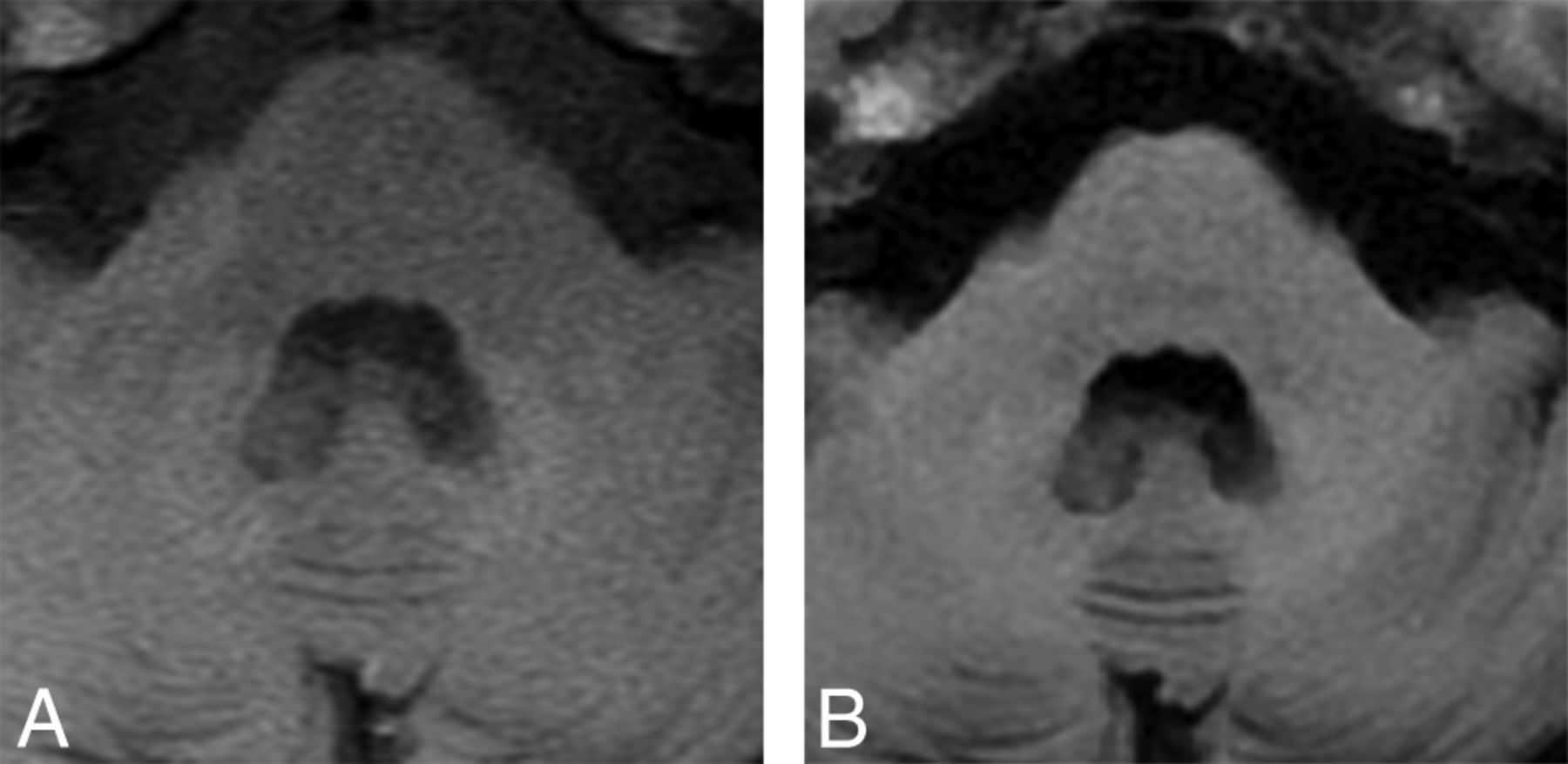
In previous studies (our study), a hyperintense dentate nucleus on T1WI was detected in subjects with >5 previous administrations of gadodiamide or gadopentetate dimeglumine. In contrast, in this study, some of the subjects with >12 previous administrations of GBCA did not show hyperintensity in the dentate nucleus. One reason may be the use of macrocyclic GBCAs in these subjects. In addition, the detectability of the hyperintense dentate nucleus on various sequences of T1WI may have influenced their results. According to our experience with several cases, the detectability of high signal intensity in the dentate nucleus differs between spin-echo T1WI and T1 FLAIR (Fig 1). Adin et al1 evaluated the hyperintense dentate nucleus with various T1WI sequences, such as MPRAGE, spin-echo, and T1 FLAIR. The different detectabilities of hyperintense dentate nuclei on these sequences may have influenced their results.

Fig 1.
  • Download figure
  • Open in new tab
  • Download powerpoint
Fig 1.

Images in a 41-year-old woman with a history of malignant lymphoma and 7 administrations of gadopentetate dimeglumine. The dentate nucleus is hyperintense on spin-echo T1WI (A), but not on T1 FLAIR (B).

Indicates open access to non-subscribers at www.ajnr.org

References

  1. 1.↵
    1. Adin ME,
    2. Kleinberg L,
    3. Vaidya D, et al
    . Hyperintense dentate nuclei on T1-weighted MRI: relation to repeat gadolinium administration. AJNR Am J Neuroradiol 2015 Aug 20. [Epub ahead of print] doi:10.3174/ajnr.A4378 pmid:26294649
    Abstract/FREE Full Text
  2. 2.↵
    1. Kanda T,
    2. Ishii K,
    3. Kawaguchi H, et al
    . High signal intensity in the dentate nucleus and globus pallidus on unenhanced T1-weighted MR images: relationship with increasing cumulative dose of a gadolinium-based contrast material. Radiology 2014;270:834–41 doi:10.1148/radiol.13131669 pmid:24475844
    CrossRefPubMed
  3. 3.↵
    1. Kanda T,
    2. Osawa M,
    3. Oba H, et al
    . High signal intensity in dentate nucleus on unenhanced T1-weighted MR images: association with linear versus macrocyclic gadolinium chelate administration. Radiology 2015;275:803–09 doi:10.1148/radiol.14140364 pmid:25633504
    CrossRefPubMed
  4. 4.↵
    1. Radbruch A,
    2. Weberling LD,
    3. Kieslich PJ, et al
    . Gadolinium retention in the dentate nucleus and globus pallidus is dependent on the class of contrast agent. Radiology 2015;275:783–91 doi:10.1148/radiol.2015150337 pmid:25848905
    CrossRefPubMed
  5. 5.↵
    1. McDonald RJ,
    2. McDonald JS,
    3. Kallmes DF, et al
    . Intracranial gadolinium deposition after contrast-enhanced MR imaging. Radiology 2015;275:772–82 doi:10.1148/radiol.15150025 pmid:25742194
    CrossRefPubMed
  6. 6.↵
    1. Kanda T,
    2. Fukusato T,
    3. Matsuda M, et al
    . Gadolinium-based contrast agent accumulates in the brain even in subjects without severe renal dysfunction: evaluation of autopsy brain specimens with inductively coupled plasma mass spectroscopy. Radiology 2015;276:228–32 doi:10.1148/radiol.2015142690 pmid:25942417
    CrossRefPubMed
  7. 7.↵
    1. Kanal E,
    2. Tweedle MF
    . Residual or retained gadolinium: practical implications for radiologists and our patients. Radiology 2015;275:630–34 doi:10.1148/radiol.2015150805 pmid:25942418
    CrossRefPubMed
  8. 8.↵
    1. Robert P,
    2. Lehericy S,
    3. Grand S, et al
    . T1-weighted hypersignal in the deep cerebellar nuclei after repeated administrations of gadolinium-based contrast agents in healthy rats: difference between linear and macrocyclic agents. Invest Radiol 2015;50:473–80 doi:10.1097/RLI.0000000000000181 pmid:26107651
    CrossRefPubMed
  • © 2016 by American Journal of Neuroradiology
PreviousNext
Back to top

In this issue

American Journal of Neuroradiology: 37 (1)
American Journal of Neuroradiology
Vol. 37, Issue 1
1 Jan 2016
  • Table of Contents
  • Index by author
  • Complete Issue (PDF)
Advertisement
Print
Download PDF
Email Article

Thank you for your interest in spreading the word on American Journal of Neuroradiology.

NOTE: We only request your email address so that the person you are recommending the page to knows that you wanted them to see it, and that it is not junk mail. We do not capture any email address.

Enter multiple addresses on separate lines or separate them with commas.
Recent Advances in Understanding Gadolinium Retention in the Brain
(Your Name) has sent you a message from American Journal of Neuroradiology
(Your Name) thought you would like to see the American Journal of Neuroradiology web site.
CAPTCHA
This question is for testing whether or not you are a human visitor and to prevent automated spam submissions.
Cite this article
T. Kanda, H. Oba, K. Toyoda, S. Furui
Recent Advances in Understanding Gadolinium Retention in the Brain
American Journal of Neuroradiology Jan 2016, 37 (1) E1-E2; DOI: 10.3174/ajnr.A4586

Citation Manager Formats

  • BibTeX
  • Bookends
  • EasyBib
  • EndNote (tagged)
  • EndNote 8 (xml)
  • Medlars
  • Mendeley
  • Papers
  • RefWorks Tagged
  • Ref Manager
  • RIS
  • Zotero
0 Responses
Respond to this article
Share
Bookmark this article
Recent Advances in Understanding Gadolinium Retention in the Brain
T. Kanda, H. Oba, K. Toyoda, S. Furui
American Journal of Neuroradiology Jan 2016, 37 (1) E1-E2; DOI: 10.3174/ajnr.A4586
del.icio.us logo Twitter logo Facebook logo Mendeley logo
  • Tweet Widget
  • Facebook Like
  • Google Plus One
Purchase

Jump to section

  • Article
    • References
  • Figures & Data
  • Info & Metrics
  • Responses
  • References
  • PDF

Related Articles

  • Reply:
  • PubMed
  • Google Scholar

Cited By...

  • Arterial Wall Imaging in Pediatric Stroke
  • Deep Brain Nuclei T1 Shortening after Gadobenate Dimeglumine in Children: Influence of Radiation and Chemotherapy
  • T1 Signal-Intensity Increase in the Dentate Nucleus after Multiple Exposures to Gadodiamide: Intraindividual Comparison between 2 Commonly Used Sequences
  • Crossref
  • Google Scholar

This article has not yet been cited by articles in journals that are participating in Crossref Cited-by Linking.

Similar Articles

Advertisement

Indexed Content

  • Current Issue
  • Accepted Manuscripts
  • Article Preview
  • Past Issues
  • Editorials
  • Editor's Choice
  • Fellows' Journal Club
  • Letters to the Editor
  • Video Articles

Cases

  • Case Collection
  • Archive - Case of the Week
  • Archive - Case of the Month
  • Archive - Classic Case

More from AJNR

  • Trainee Corner
  • Imaging Protocols
  • MRI Safety Corner
  • Book Reviews

Multimedia

  • AJNR Podcasts
  • AJNR Scantastics

Resources

  • Turnaround Time
  • Submit a Manuscript
  • Submit a Video Article
  • Submit an eLetter to the Editor/Response
  • Manuscript Submission Guidelines
  • Statistical Tips
  • Fast Publishing of Accepted Manuscripts
  • Graphical Abstract Preparation
  • Imaging Protocol Submission
  • Evidence-Based Medicine Level Guide
  • Publishing Checklists
  • Author Policies
  • Become a Reviewer/Academy of Reviewers
  • News and Updates

About Us

  • About AJNR
  • Editorial Board
  • Editorial Board Alumni
  • Alerts
  • Permissions
  • Not an AJNR Subscriber? Join Now
  • Advertise with Us
  • Librarian Resources
  • Feedback
  • Terms and Conditions
  • AJNR Editorial Board Alumni

American Society of Neuroradiology

  • Not an ASNR Member? Join Now

© 2025 by the American Society of Neuroradiology All rights, including for text and data mining, AI training, and similar technologies, are reserved.
Print ISSN: 0195-6108 Online ISSN: 1936-959X

Powered by HighWire