Advertisement

Main menu

  • Home
  • Content
    • Current Issue
    • Accepted Manuscripts
    • Article Preview
    • Past Issue Archive
    • Video Articles
    • AJNR Case Collection
    • Case of the Week Archive
    • Case of the Month Archive
    • Classic Case Archive
  • Special Collections
    • AJNR Awards
    • Low-Field MRI
    • Alzheimer Disease
    • ASNR Foundation Special Collection
    • Photon-Counting CT
    • View All
  • Multimedia
    • AJNR Podcasts
    • AJNR SCANtastic
    • Trainee Corner
    • MRI Safety Corner
    • Imaging Protocols
  • For Authors
    • Submit a Manuscript
    • Submit a Video Article
    • Submit an eLetter to the Editor/Response
    • Manuscript Submission Guidelines
    • Statistical Tips
    • Fast Publishing of Accepted Manuscripts
    • Graphical Abstract Preparation
    • Imaging Protocol Submission
    • Author Policies
  • About Us
    • About AJNR
    • Editorial Board
    • Editorial Board Alumni
  • More
    • Become a Reviewer/Academy of Reviewers
    • Subscribers
    • Permissions
    • Alerts
    • Feedback
    • Advertisers
    • ASNR Home

User menu

  • Alerts
  • Log in

Search

  • Advanced search
American Journal of Neuroradiology

American Journal of Neuroradiology

ASHNR American Society of Functional Neuroradiology ASHNR American Society of Pediatric Neuroradiology ASSR
  • Alerts
  • Log in

Advanced Search

  • Home
  • Content
    • Current Issue
    • Accepted Manuscripts
    • Article Preview
    • Past Issue Archive
    • Video Articles
    • AJNR Case Collection
    • Case of the Week Archive
    • Case of the Month Archive
    • Classic Case Archive
  • Special Collections
    • AJNR Awards
    • Low-Field MRI
    • Alzheimer Disease
    • ASNR Foundation Special Collection
    • Photon-Counting CT
    • View All
  • Multimedia
    • AJNR Podcasts
    • AJNR SCANtastic
    • Trainee Corner
    • MRI Safety Corner
    • Imaging Protocols
  • For Authors
    • Submit a Manuscript
    • Submit a Video Article
    • Submit an eLetter to the Editor/Response
    • Manuscript Submission Guidelines
    • Statistical Tips
    • Fast Publishing of Accepted Manuscripts
    • Graphical Abstract Preparation
    • Imaging Protocol Submission
    • Author Policies
  • About Us
    • About AJNR
    • Editorial Board
    • Editorial Board Alumni
  • More
    • Become a Reviewer/Academy of Reviewers
    • Subscribers
    • Permissions
    • Alerts
    • Feedback
    • Advertisers
    • ASNR Home
  • Follow AJNR on Twitter
  • Visit AJNR on Facebook
  • Follow AJNR on Instagram
  • Join AJNR on LinkedIn
  • RSS Feeds

AJNR Awards, New Junior Editors, and more. Read the latest AJNR updates

LetterLetter

Expounding on the Distinction between Lateral Dural Tears and Leaking Meningeal Diverticula in Spontaneous Intracranial Hypotension

Ajay A. Madhavan, Richard I. Farb, Waleed Brinjikji, Jeremy K. Cutsforth-Gregory and Wouter I. Schievink
American Journal of Neuroradiology June 2024, DOI: https://doi.org/10.3174/ajnr.A8308
Ajay A. Madhavan
aDivision of NeuroradiologyDepartment of RadiologyMayo ClinicRochester, Minnesota
  • Find this author on Google Scholar
  • Find this author on PubMed
  • Search for this author on this site
  • ORCID record for Ajay A. Madhavan
Richard I. Farb
bDivision of NeuroradiologyDepartment of RadiologyUniversity of TorontoToronto, Ontario, Canada
  • Find this author on Google Scholar
  • Find this author on PubMed
  • Search for this author on this site
  • ORCID record for Richard I. Farb
Waleed Brinjikji
cDivision of NeuroradiologyDepartment of RadiologyMayo ClinicRochester, Minnesota
  • Find this author on Google Scholar
  • Find this author on PubMed
  • Search for this author on this site
  • ORCID record for Waleed Brinjikji
Jeremy K. Cutsforth-Gregory
dDepartment of NeurologyMayo ClinicRochester, Minnesota
  • Find this author on Google Scholar
  • Find this author on PubMed
  • Search for this author on this site
  • ORCID record for Jeremy K. Cutsforth-Gregory
Wouter I. Schievink
eDepartment of NeurosurgeryCedars-Sinai Medical CenterLos Angeles, California
  • Find this author on Google Scholar
  • Find this author on PubMed
  • Search for this author on this site
  • ORCID record for Wouter I. Schievink
  • Article
  • Figures & Data
  • Info & Metrics
  • Responses
  • References
  • PDF
Loading

Spontaneous intracranial hypotension is caused by a spinal CSF leak. Currently, various classification schemes for CSF leaks exist, with 1 system dividing leaks into ventral dural tears (type 1a), lateral/posterior dural tears (type 1b), leaks from meningeal diverticula (type 2), and CSF-venous fistulas (type 3).1 Farb et al2 subsequently classified these leaks in a similar fashion. Ventral and lateral dural tears essentially always result in extradural CSF collections that can be detected on spine MR imaging or CT myelography.3 These are usually intraspinal epidural collections, though CSF can extend through the foramina in some instances. Leaks from meningeal diverticula, in contrast, usually result in only paraspinal CSF accumulation (Fig 1A), though intraspinal extradural CSF can rarely be seen. CSF-venous fistulas essentially never have associated extradural CSF.

FIG 1.
  • Download figure
  • Open in new tab
  • Download powerpoint
FIG 1.

Lateral CSF leaks in 3 patients (A, Leaking meningeal diverticulum. B and C, Lateral dural tears with herniated arachnoid diverticula). In the first patient, 30-minute delayed left lateral decubitus CT myelography (CTM) shows paraspinal contrast accumulation at L2 (A, solid arrow) adjacent to a small meningeal diverticulum (A, dashed arrow), compatible with a leaking diverticulum. In the second patient, a coronal image from decubitus photon-counting CTM (B) shows a left T12 lateral dural tear with an arachnoid diverticulum herniating through the dural defect (B, solid arrow) and separate extradural contrast accumulation (B, dashed arrows). In the third patient, axial image from a decubitus photon-counting CTM (C) shows a right T11 lateral dural tear, again with an arachnoid diverticulum herniating through the dural tear (C, solid arrow) and separately leaking extradural contrast (C, dashed arrow). In this case, the precise site of the dural defect was clearly seen (C, arrowhead).

Although these different types of CSF leaks are generally well-understood, lateral dural tears can be difficult to discriminate from leaking meningeal diverticula. Lateral dural tears most commonly occur along the axilla of nerve root sleeves, but they also less frequently occur at the nerve root sleeve shoulders or at the level of the pedicle.3 They are commonly, though not always, associated with an arachnoid diverticulum that herniates or billows through the dural defect (Fig 1B, -C). This herniated diverticulum involves only the arachnoid and therefore differs from the typical perineural meningeal diverticula, which are lined by the dura and arachnoid. The cause of arachnoid herniation from lateral dural tears is unclear, but it is possible that the more capacious lateral epidural space (compared with the ventral epidural space) and lack of penetrating osseous spicules causing lateral leaks play a role. Most important, the arachnoid diverticulum is a secondary manifestation of the lateral dural tear and not the source of CSF leak. Rather, CSF usually leaks from the margins of the lateral dural tear.3 On decubitus DSA or CT myelography, contrast opacification of this diverticulum is usually seen concurrently with epidural contrast leakage.4 The dural defect itself is usually not seen. Thus, lateral dural tears may falsely appear to represent a “ruptured” or “leaking” meningeal diverticulum on myelography. Many lateral dural tears are likely inadvertently diagnosed as leaks from meningeal diverticula, because both result in a lateral leak and harbor a diverticulum at the site of the leak.

High-resolution modalities such photon-counting CT may sometimes reveal the precise site of lateral dural tears, clarifying the diagnosis (Fig 1C). Often, surgical exploration is the only way to visualize the actual dural tear (Fig 2). Further study with surgical correlation will be necessary to determine how frequently lateral leaks diagnosed on myelography represent lateral dural tears with secondarily herniated arachnoid diverticula versus primarily leaking meningeal diverticula. Although 1 initial study on this topic suggested that leaks from meningeal diverticula were common (up to 42% of CSF leaks), this was before the routine use of decubitus myelography to diagnose CSF-venous fistulas and lateral dural tears.1 We suspect that lateral dural tears and CSF-venous fistulas are far more common than initially believed.

FIG 2.
  • Download figure
  • Open in new tab
  • Download powerpoint
FIG 2.

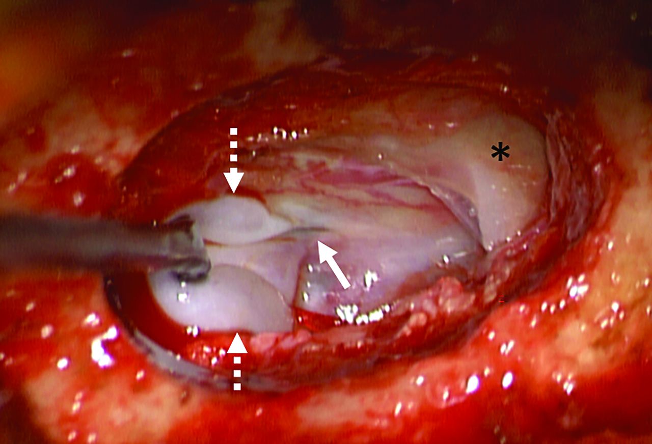
Intraoperative photograph demonstrating the typical appearance of a lateral dural tear inferior to the nerve root sleeve (asterisk). A dural tear (solid arrow) is seen with a thin arachnoid diverticulum (dashed arrows) herniating or “billowing” out from the defect. The actual site of CSF leak was from the edges of the dural tear itself (solid arrow) rather than the diverticulum.

The distinction between lateral dural tears and leaks from meningeal diverticula is important for 2 reasons: First, accurate epidemiologic data about these leaks are needed to understand their pathophysiology; second, it is important to determine how these different leak types respond to treatment, such as blood patching or surgery. To conclude, we encourage close attention to the distinction between lateral dural tears associated with herniated arachnoid diverticula versus leaking meningeal diverticula. While these 2 leak types share some similarities on myelography, lateral dural tears are generally more common and essentially always associated with intraspinal extradural CSF collections. Leaking diverticula, by contrast, are relatively uncommon and usually cause paraspinal CSF accumulation rather than intraspinal extradural collections. Further study with radiographic and surgical correlation will be helpful to corroborate these suppositions.

Footnotes

  • Disclosure forms provided by the authors are available with the full text and PDF of this article at www.ajnr.org.

References

  1. 1.↵
    1. Schievink WI,
    2. Maya MM,
    3. Jean-Pierre S, et al
    . A classification system of spontaneous spinal CSF leaks. Neurology 2016;87:673–79 doi:10.1212/WNL.0000000000002986 pmid:27440149
    CrossRefPubMed
  2. 2.↵
    1. Farb RI,
    2. Nicholsobn PJ,
    3. Massicotte EM, et al
    . Spontaneous intracranial hypotension: a systematic imaging approach for CSF leak localization and management based on MRI and digital subtraction myelography. AJNR Am J Neuroradiol 2019;40:745–53 doi:10.3174/ajnr.A6016 pmid:3092308
    Abstract/FREE Full Text
  3. 3.↵
    1. Schievink WI,
    2. Maya MM,
    3. Tay AS-MS, et al
    . Lateral spinal CSF leaks in patients with spontaneous intracranial hypotension: radiologic-anatomic study of different variants. AJNR Am J Neuroradiol 2024 June 13. [Epub ahead of print] doi:10.3174/ajnr.A8261
    CrossRef
  4. 4.↵
    1. Madhavan AA,
    2. Verdoorn JT,
    3. Shlapak DP, et al
    . Lateral decubitus dynamic CT myelography for fast cerebrospinal fluid leak localization. Neuroradiology 2022;64:1897–903 doi:10.1007/s00234-022-02985-y pmid:35614322
    CrossRefPubMed
  • © 2024 by American Journal of Neuroradiology
PreviousNext
Back to top
Advertisement
Print
Download PDF
Email Article

Thank you for your interest in spreading the word on American Journal of Neuroradiology.

NOTE: We only request your email address so that the person you are recommending the page to knows that you wanted them to see it, and that it is not junk mail. We do not capture any email address.

Enter multiple addresses on separate lines or separate them with commas.
Expounding on the Distinction between Lateral Dural Tears and Leaking Meningeal Diverticula in Spontaneous Intracranial Hypotension
(Your Name) has sent you a message from American Journal of Neuroradiology
(Your Name) thought you would like to see the American Journal of Neuroradiology web site.
CAPTCHA
This question is for testing whether or not you are a human visitor and to prevent automated spam submissions.
Cite this article
Ajay A. Madhavan, Richard I. Farb, Waleed Brinjikji, Jeremy K. Cutsforth-Gregory, Wouter I. Schievink
Expounding on the Distinction between Lateral Dural Tears and Leaking Meningeal Diverticula in Spontaneous Intracranial Hypotension
American Journal of Neuroradiology Jun 2024, DOI: 10.3174/ajnr.A8308

Citation Manager Formats

  • BibTeX
  • Bookends
  • EasyBib
  • EndNote (tagged)
  • EndNote 8 (xml)
  • Medlars
  • Mendeley
  • Papers
  • RefWorks Tagged
  • Ref Manager
  • RIS
  • Zotero
0 Responses
Respond to this article
Share
Bookmark this article
Expounding on the Distinction between Lateral Dural Tears and Leaking Meningeal Diverticula in Spontaneous Intracranial Hypotension
Ajay A. Madhavan, Richard I. Farb, Waleed Brinjikji, Jeremy K. Cutsforth-Gregory, Wouter I. Schievink
American Journal of Neuroradiology Jun 2024, DOI: 10.3174/ajnr.A8308
del.icio.us logo Twitter logo Facebook logo Mendeley logo
  • Tweet Widget
  • Facebook Like
  • Google Plus One
Purchase

Jump to section

  • Article
    • Footnotes
    • References
  • Figures & Data
  • Info & Metrics
  • Responses
  • References
  • PDF

Related Articles

  • No related articles found.
  • PubMed
  • Google Scholar

Cited By...

  • Advanced Imaging of Type 2 Spinal CSF Leaks with Ultra-High-Resolution Conebeam CT Myelography
  • Additional Diagnostic Value of Conebeam CT Myelography Performed after Digital Subtraction Myelography for Detecting CSF-Venous Fistulas
  • Spontaneous Intracranial Hypotension in Children: A Multi-Institutional Review of Spinal CSF Leaks Localized on Advanced Myelography
  • Spinal CSF Leaks: The Neuroradiologist Transforming Care
  • Crossref
  • Google Scholar

This article has not yet been cited by articles in journals that are participating in Crossref Cited-by Linking.

More in this TOC Section

  • Reply:
  • Brain AVM’s Nidus: What if We Hadn’t Understood Anything?
  • Letter to the Editor regarding “Automated Volumetric Software in Dementia: Help or Hindrance to the Neuroradiologist?”
Show more LETTERS

Similar Articles

Advertisement

Indexed Content

  • Current Issue
  • Accepted Manuscripts
  • Article Preview
  • Past Issues
  • Editorials
  • Editor's Choice
  • Fellows' Journal Club
  • Letters to the Editor
  • Video Articles

Cases

  • Case Collection
  • Archive - Case of the Week
  • Archive - Case of the Month
  • Archive - Classic Case

More from AJNR

  • Trainee Corner
  • Imaging Protocols
  • MRI Safety Corner
  • Book Reviews

Multimedia

  • AJNR Podcasts
  • AJNR Scantastics

Resources

  • Turnaround Time
  • Submit a Manuscript
  • Submit a Video Article
  • Submit an eLetter to the Editor/Response
  • Manuscript Submission Guidelines
  • Statistical Tips
  • Fast Publishing of Accepted Manuscripts
  • Graphical Abstract Preparation
  • Imaging Protocol Submission
  • Evidence-Based Medicine Level Guide
  • Publishing Checklists
  • Author Policies
  • Become a Reviewer/Academy of Reviewers
  • News and Updates

About Us

  • About AJNR
  • Editorial Board
  • Editorial Board Alumni
  • Alerts
  • Permissions
  • Not an AJNR Subscriber? Join Now
  • Advertise with Us
  • Librarian Resources
  • Feedback
  • Terms and Conditions
  • AJNR Editorial Board Alumni

American Society of Neuroradiology

  • Not an ASNR Member? Join Now

© 2025 by the American Society of Neuroradiology All rights, including for text and data mining, AI training, and similar technologies, are reserved.
Print ISSN: 0195-6108 Online ISSN: 1936-959X

Powered by HighWire