Advertisement

Main menu

  • Home
  • Content
    • Current Issue
    • Accepted Manuscripts
    • Article Preview
    • Past Issue Archive
    • Video Articles
    • AJNR Case Collection
    • Case of the Week Archive
    • Case of the Month Archive
    • Classic Case Archive
  • Special Collections
    • AJNR Awards
    • Low-Field MRI
    • Alzheimer Disease
    • ASNR Foundation Special Collection
    • Photon-Counting CT
    • View All
  • Multimedia
    • AJNR Podcasts
    • AJNR SCANtastic
    • Trainee Corner
    • MRI Safety Corner
    • Imaging Protocols
  • For Authors
    • Submit a Manuscript
    • Submit a Video Article
    • Submit an eLetter to the Editor/Response
    • Manuscript Submission Guidelines
    • Statistical Tips
    • Fast Publishing of Accepted Manuscripts
    • Graphical Abstract Preparation
    • Imaging Protocol Submission
    • Author Policies
  • About Us
    • About AJNR
    • Editorial Board
    • Editorial Board Alumni
  • More
    • Become a Reviewer/Academy of Reviewers
    • Subscribers
    • Permissions
    • Alerts
    • Feedback
    • Advertisers
    • ASNR Home

User menu

  • Alerts
  • Log in

Search

  • Advanced search
American Journal of Neuroradiology

American Journal of Neuroradiology

ASHNR American Society of Functional Neuroradiology ASHNR American Society of Pediatric Neuroradiology ASSR
  • Alerts
  • Log in

Advanced Search

  • Home
  • Content
    • Current Issue
    • Accepted Manuscripts
    • Article Preview
    • Past Issue Archive
    • Video Articles
    • AJNR Case Collection
    • Case of the Week Archive
    • Case of the Month Archive
    • Classic Case Archive
  • Special Collections
    • AJNR Awards
    • Low-Field MRI
    • Alzheimer Disease
    • ASNR Foundation Special Collection
    • Photon-Counting CT
    • View All
  • Multimedia
    • AJNR Podcasts
    • AJNR SCANtastic
    • Trainee Corner
    • MRI Safety Corner
    • Imaging Protocols
  • For Authors
    • Submit a Manuscript
    • Submit a Video Article
    • Submit an eLetter to the Editor/Response
    • Manuscript Submission Guidelines
    • Statistical Tips
    • Fast Publishing of Accepted Manuscripts
    • Graphical Abstract Preparation
    • Imaging Protocol Submission
    • Author Policies
  • About Us
    • About AJNR
    • Editorial Board
    • Editorial Board Alumni
  • More
    • Become a Reviewer/Academy of Reviewers
    • Subscribers
    • Permissions
    • Alerts
    • Feedback
    • Advertisers
    • ASNR Home
  • Follow AJNR on Twitter
  • Visit AJNR on Facebook
  • Follow AJNR on Instagram
  • Join AJNR on LinkedIn
  • RSS Feeds

AJNR Awards, New Junior Editors, and more. Read the latest AJNR updates

LetterLetter

Dilator-Dotter Technique for Acute Ischemic Stroke: Further Applications in the Vertebral Arteries

M. Cox, K. Layton and J. Hise
American Journal of Neuroradiology March 2022, 43 (3) E7-E8; DOI: https://doi.org/10.3174/ajnr.A7438
M. Cox
aSection of Neurointerventional RadiologyBaylor University Medical CenterDallas, Texas
  • Find this author on Google Scholar
  • Find this author on PubMed
  • Search for this author on this site
  • ORCID record for M. Cox
K. Layton
aSection of Neurointerventional RadiologyBaylor University Medical CenterDallas, Texas
  • Find this author on Google Scholar
  • Find this author on PubMed
  • Search for this author on this site
  • ORCID record for K. Layton
J. Hise
aSection of Neurointerventional RadiologyBaylor University Medical CenterDallas, Texas
  • Find this author on Google Scholar
  • Find this author on PubMed
  • Search for this author on this site
  • ORCID record for J. Hise

We read with great interest and appreciation the article titled, “The Dilator-Dotter Technique: A Modified Method of Rapid Internal Carotid Artery Revascularization in Acute Ischemic Stroke”1 published in an earlier issue of this journal. In this article, the authors detailed their experience with catheter-based angioplasty for tandem carotid and intracranial occlusions, with impressive results. They were able to achieve recanalization of the carotid artery in all 32 of their patients within a rapid timeframe (average case completion time, 25 minutes). The authors are to be commended for further refining and applying the Dotter technique to the carotid arteries for acute ischemic stroke and sharing their experience with the wider neuroradiology/neurointerventional community. We have also used the Dotter technique for acute ischemic stroke in our own practice, with good results. Recently, we expanded the dilator-Dotter technique to include patients with cervical vertebral artery occlusion and tandem basilar artery occlusion, with equally good results.

A 54-year-old woman with hypertension was evaluated at an outside hospital for a 1-day history of nausea, dizziness, and vomiting. A CT of the head was performed for new disconjugate gaze and progressive somnolence and showed an acute infarct in the left superior cerebellar hemisphere, but no hemorrhage. A CTA of the head and neck was also performed and showed occlusion of the dominant left vertebral artery in the neck and occlusive thrombus in the basilar tip extending into the left superior cerebellar artery. The right vertebral artery was small, with stenosis at the origin, and seemed to terminate as a posterior inferior cerebellar artery.

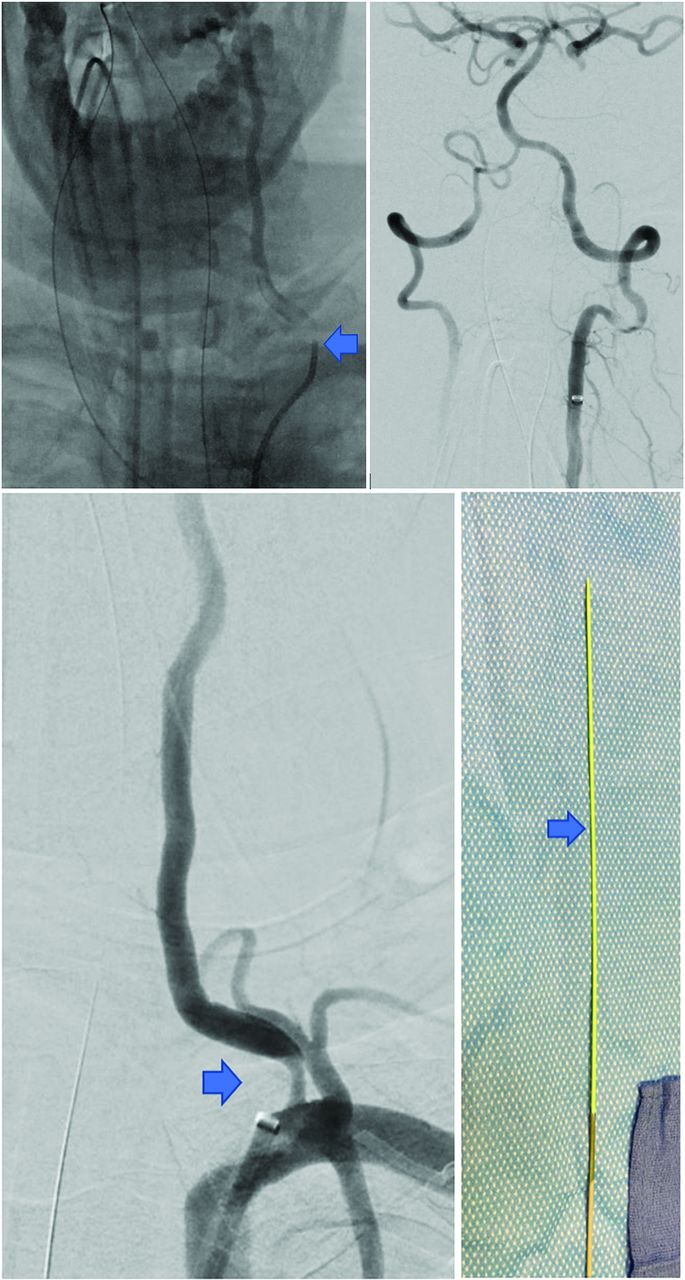
She was transferred for emergent mechanical thrombectomy. Angiography confirmed occlusion of the dominant left vertebral artery at its origin with intraluminal thrombus (Figure). An eptifibatide bolus was infused into the artery via the guide catheter. The area of occlusion was then traversed with an exchange-length 0.035-inch Glidewire (Terumo), and an 80-cm low-profile 6F Ballast sheath with a long, tapered dilator (Balt) was advanced smoothly through the stenosis for catheter-based angioplasty. The inner dilator and Glidewire were removed, resulting in thrombus extrusion from the Tuohy Borst valve.

FIGURE.
  • Download figure
  • Open in new tab
  • Download powerpoint
FIGURE.

The first image is a frontal projection during the diagnostic angiogram showing critical stenosis/occlusion of the dominant left vertebral artery, with intraluminal thrombus (arrow). The second image is a frontal projection immediately after Dotter angioplasty, with recanalization of the basilar tip thrombus seen on a prior CTA. Improvement of proximal flow may sometimes result in spontaneous resolution of the intracranial thrombus, with or without concomitant antithrombotics. The third image is a frontal-projection DSA showing moderate residual stenosis of the proximal left vertebral artery following Dotter angioplasty. The fourth image is a picture of the low-profile Ballast sheath, showing the long, tapered dilator (arrow), which allows a smooth transition of the sheath through a stenosis.

A run was performed and showed interval resolution of the thrombus at the basilar tip. The Ballast sheath was withdrawn into the left subclavian artery, and another run was performed, confirming interval improvement of proximal vertebral artery stenosis following Dotter angioplasty, now moderate. The entire case was performed in under 25 minutes. The patient’s mental status returned to her baseline during admission, and she was discharged to a rehabilitation facility with mild dizziness and ataxia.

In the properly selected patient, dilator-Dotter angioplasty is a powerful tool for achieving rapid recanalization in patients with acute ischemic stroke from tandem occlusions. When successful, this technique avoids the need for immediate antiplatelet therapy in patients who may need cranial decompressive surgery or CSF diversion during their hospitalization. Increasing recognition of this technique among neurointerventionalists has the potential to improve recanalization rates in the community, with wider benefits to patients at large.

Footnotes

  • Disclosure forms provided by the authors are available with the full text and PDF of this article at www.ajnr.org.

Reference

  1. 1.
    1. Amuluru K,
    2. Sahlein D,
    3. Al-Mufti F, et al
    . The dilator-Dotter technique: a modified method of rapid internal carotid artery revascularization in acute ischemic stroke. AJNR Am J Neuroradiol 2020;41:1863–68 doi:10.3174/ajnr.A6733 pmid:32883663
  • © 2022 by American Journal of Neuroradiology
Advertisement

Indexed Content

  • Current Issue
  • Accepted Manuscripts
  • Article Preview
  • Past Issues
  • Editorials
  • Editor's Choice
  • Fellows' Journal Club
  • Letters to the Editor
  • Video Articles

Cases

  • Case Collection
  • Archive - Case of the Week
  • Archive - Case of the Month
  • Archive - Classic Case

More from AJNR

  • Trainee Corner
  • Imaging Protocols
  • MRI Safety Corner
  • Book Reviews

Multimedia

  • AJNR Podcasts
  • AJNR Scantastics

Resources

  • Turnaround Time
  • Submit a Manuscript
  • Submit a Video Article
  • Submit an eLetter to the Editor/Response
  • Manuscript Submission Guidelines
  • Statistical Tips
  • Fast Publishing of Accepted Manuscripts
  • Graphical Abstract Preparation
  • Imaging Protocol Submission
  • Evidence-Based Medicine Level Guide
  • Publishing Checklists
  • Author Policies
  • Become a Reviewer/Academy of Reviewers
  • News and Updates

About Us

  • About AJNR
  • Editorial Board
  • Editorial Board Alumni
  • Alerts
  • Permissions
  • Not an AJNR Subscriber? Join Now
  • Advertise with Us
  • Librarian Resources
  • Feedback
  • Terms and Conditions
  • AJNR Editorial Board Alumni

American Society of Neuroradiology

  • Not an ASNR Member? Join Now

© 2025 by the American Society of Neuroradiology All rights, including for text and data mining, AI training, and similar technologies, are reserved.
Print ISSN: 0195-6108 Online ISSN: 1936-959X

Powered by HighWire