Advertisement

Main menu

  • Home
  • Content
    • Current Issue
    • Accepted Manuscripts
    • Article Preview
    • Past Issue Archive
    • Video Articles
    • AJNR Case Collection
    • Case of the Week Archive
    • Case of the Month Archive
    • Classic Case Archive
  • Special Collections
    • AJNR Awards
    • Low-Field MRI
    • Alzheimer Disease
    • ASNR Foundation Special Collection
    • Photon-Counting CT
    • View All
  • Multimedia
    • AJNR Podcasts
    • AJNR SCANtastic
    • Trainee Corner
    • MRI Safety Corner
    • Imaging Protocols
  • For Authors
    • Submit a Manuscript
    • Submit a Video Article
    • Submit an eLetter to the Editor/Response
    • Manuscript Submission Guidelines
    • Statistical Tips
    • Fast Publishing of Accepted Manuscripts
    • Graphical Abstract Preparation
    • Imaging Protocol Submission
    • Author Policies
  • About Us
    • About AJNR
    • Editorial Board
    • Editorial Board Alumni
  • More
    • Become a Reviewer/Academy of Reviewers
    • Subscribers
    • Permissions
    • Alerts
    • Feedback
    • Advertisers
    • ASNR Home

User menu

  • Alerts
  • Log in

Search

  • Advanced search
American Journal of Neuroradiology

American Journal of Neuroradiology

ASHNR American Society of Functional Neuroradiology ASHNR American Society of Pediatric Neuroradiology ASSR
  • Alerts
  • Log in

Advanced Search

  • Home
  • Content
    • Current Issue
    • Accepted Manuscripts
    • Article Preview
    • Past Issue Archive
    • Video Articles
    • AJNR Case Collection
    • Case of the Week Archive
    • Case of the Month Archive
    • Classic Case Archive
  • Special Collections
    • AJNR Awards
    • Low-Field MRI
    • Alzheimer Disease
    • ASNR Foundation Special Collection
    • Photon-Counting CT
    • View All
  • Multimedia
    • AJNR Podcasts
    • AJNR SCANtastic
    • Trainee Corner
    • MRI Safety Corner
    • Imaging Protocols
  • For Authors
    • Submit a Manuscript
    • Submit a Video Article
    • Submit an eLetter to the Editor/Response
    • Manuscript Submission Guidelines
    • Statistical Tips
    • Fast Publishing of Accepted Manuscripts
    • Graphical Abstract Preparation
    • Imaging Protocol Submission
    • Author Policies
  • About Us
    • About AJNR
    • Editorial Board
    • Editorial Board Alumni
  • More
    • Become a Reviewer/Academy of Reviewers
    • Subscribers
    • Permissions
    • Alerts
    • Feedback
    • Advertisers
    • ASNR Home
  • Follow AJNR on Twitter
  • Visit AJNR on Facebook
  • Follow AJNR on Instagram
  • Join AJNR on LinkedIn
  • RSS Feeds

AJNR Awards, New Junior Editors, and more. Read the latest AJNR updates

LetterLetter

Copper Deficiency Myelopathy Can Also Involve the Brain Stem

G. Kumar, M.K. Goyal, S. Lucchese and U. Dhand
American Journal of Neuroradiology January 2011, 32 (1) E14-E15; DOI: https://doi.org/10.3174/ajnr.A2261
G. Kumar
  • Find this author on Google Scholar
  • Find this author on PubMed
  • Search for this author on this site
M.K. Goyal
  • Find this author on Google Scholar
  • Find this author on PubMed
  • Search for this author on this site
S. Lucchese
  • Find this author on Google Scholar
  • Find this author on PubMed
  • Search for this author on this site
U. Dhand
  • Find this author on Google Scholar
  • Find this author on PubMed
  • Search for this author on this site

Copper deficiency is rare but can result from gastrointestinal surgery, excess zinc in the diet, parenteral nutrition, and malabsorption syndromes.1 Copper deficiency gives rise to a sensory ataxic myelopathy, symmetrically involving the pyramidal tracts and dorsal columns in the spinal cord,2 producing a clinical picture indistinguishable from subacute combined degeneration (SCD) associated with vitamin B12 deficiency.1 Brain stem involvement has not been previously reported with copper deficiency. Here we report the first patient with brain stem involvement in copper deficiency myelopathy, demonstrating that it can also be a rhombencephalopathy.

A 47-year-old man with a history of denture cream use insidiously developed bilateral painless vision loss, paresthesias, and quadriparesis over 4 weeks. Neurologic examination revealed left afferent pupillary defect and the presence of optic neuritis. Left upper motor neuron−type facial paralysis, quadriparesis, hyperreflexia, brisk jaw jerk, and a drop sensory level at T6 with graded loss of lower extremity sensation to all modalities were noted. Mild normocytic anemia was present; vitamin B12 and serum zinc were within normal levels. Serum copper (45 mcg/dL; reference range, 70–140 mcg/dL) and ceruloplasmin levels (9 mg/dL; reference range, 17–54 mg/dL) were low. MR imaging of the cervical spinal cord (Fig 1) and brain (Fig 2) is shown. At 6 months, the patient had not responded to copper supplementation and was wheelchair-bound; bilateral optic atrophy had also set in.

Fig 1.
  • Download figure
  • Open in new tab
  • Download powerpoint
Fig 1.

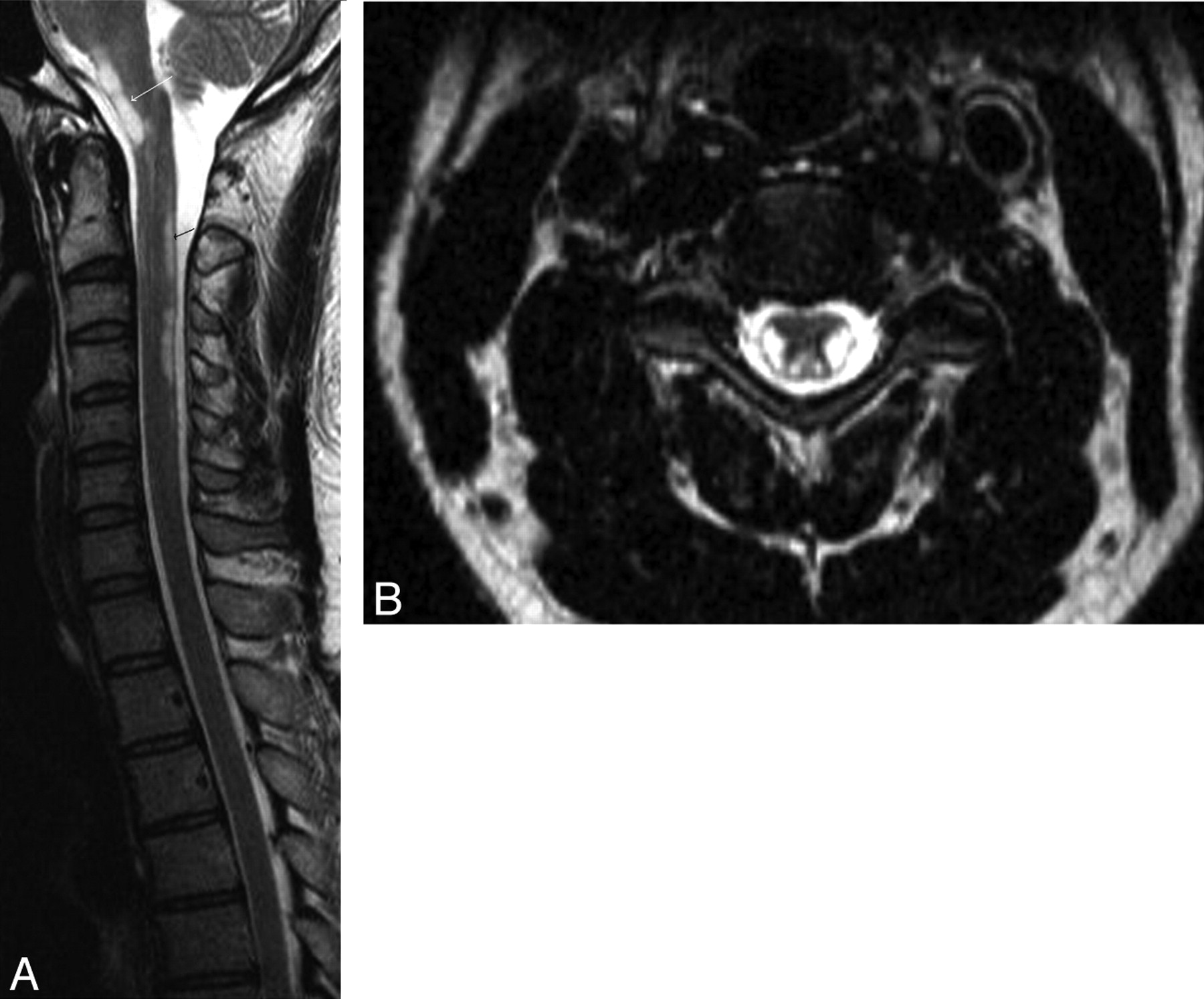
A, Sagittal view through the brain stem and cervical spinal cord shows the extent of T2 hyperintensities involving the pyramidal tract and posterior columns (black arrow), extending along the brain stem (white arrow). B, Axial section through the cervical spinal cord shows the strikingly symmetric T2 hyperintense lateral corticospinal tracts and posterior columns. Thoracic spine MR imaging findings were unremarkable (not shown).

Fig 2.
  • Download figure
  • Open in new tab
  • Download powerpoint
Fig 2.

MR images of the brain (axial sections, fluid-attenuated reversion recovery sequences) show the symmetric hyperintensities (arrows) involving the pyramidal tract in the (A) medulla, (B) pons, and (C) midbrain.

Copper, an essential trace element required by all life forms, is a component of key metalloenzymes (cytochrome c oxidase, superoxide dismutase, dopamine β hydroxylase), which have a critical role in the structure and function of the nervous system. Given its ubiquitous distribution, dietary copper deficiency is rare but results from causes such as gastrointestinal surgery, zinc excess, malabsorption syndromes, and parenteral nutrition.1 It is associated with symmetric involvement of the pyramidal tract and posterior columns, resulting in a clinical and radiologic picture indistinguishable from SCD2; rarely peripheral neuropathy and optic neuritis have been described.3 Clinical response to treatment is variable.1 To our knowledge, this is the first report of a case that shows striking involvement of the pyramidal tracts in the brain stem, in addition to illustrating typical MR imaging changes of copper deficiency myelopathy. It shows that copper deficiency can be a rhombencephalopathy.

References

  1. 1.
    1. Kumar N,
    2. Gross JB Jr.,
    3. Ahlskog JE
    . Copper deficiency myelopathy produces a clinical picture like subacute combined degeneration. Neurology 2004;63:33–39
  2. 2.
    1. Kumar N
    . Copper deficiency myelopathy (human swayback). Mayo Clin Proc 2006;81:1371–84
  3. 3.
    1. Gregg XT,
    2. Reddy V,
    3. Prchal JT
    . Copper deficiency masquerading as myelodysplastic syndrome. Blood 2002;100:1493–95
  • Copyright © American Society of Neuroradiology
Advertisement

Indexed Content

  • Current Issue
  • Accepted Manuscripts
  • Article Preview
  • Past Issues
  • Editorials
  • Editor's Choice
  • Fellows' Journal Club
  • Letters to the Editor
  • Video Articles

Cases

  • Case Collection
  • Archive - Case of the Week
  • Archive - Case of the Month
  • Archive - Classic Case

More from AJNR

  • Trainee Corner
  • Imaging Protocols
  • MRI Safety Corner
  • Book Reviews

Multimedia

  • AJNR Podcasts
  • AJNR Scantastics

Resources

  • Turnaround Time
  • Submit a Manuscript
  • Submit a Video Article
  • Submit an eLetter to the Editor/Response
  • Manuscript Submission Guidelines
  • Statistical Tips
  • Fast Publishing of Accepted Manuscripts
  • Graphical Abstract Preparation
  • Imaging Protocol Submission
  • Evidence-Based Medicine Level Guide
  • Publishing Checklists
  • Author Policies
  • Become a Reviewer/Academy of Reviewers
  • News and Updates

About Us

  • About AJNR
  • Editorial Board
  • Editorial Board Alumni
  • Alerts
  • Permissions
  • Not an AJNR Subscriber? Join Now
  • Advertise with Us
  • Librarian Resources
  • Feedback
  • Terms and Conditions
  • AJNR Editorial Board Alumni

American Society of Neuroradiology

  • Not an ASNR Member? Join Now

© 2025 by the American Society of Neuroradiology All rights, including for text and data mining, AI training, and similar technologies, are reserved.
Print ISSN: 0195-6108 Online ISSN: 1936-959X

Powered by HighWire