Advertisement

Main menu

  • Home
  • Content
    • Current Issue
    • Accepted Manuscripts
    • Article Preview
    • Past Issue Archive
    • Video Articles
    • AJNR Case Collection
    • Case of the Week Archive
    • Case of the Month Archive
    • Classic Case Archive
  • Special Collections
    • Advancing NeuroMRI with High-Relaxivity Contrast Agents
    • AJNR Awards
    • Low-Field MRI
    • Alzheimer Disease
    • ASNR Foundation Special Collection
    • Photon-Counting CT
    • View All
  • Multimedia
    • AJNR Podcasts
    • AJNR SCANtastic
    • Trainee Corner
    • MRI Safety Corner
    • Imaging Protocols
  • For Authors
    • Submit a Manuscript
    • Submit a Video Article
    • Submit an eLetter to the Editor/Response
    • Manuscript Submission Guidelines
    • Statistical Tips
    • Fast Publishing of Accepted Manuscripts
    • Graphical Abstract Preparation
    • Imaging Protocol Submission
    • Author Policies
  • About Us
    • About AJNR
    • Editorial Board
    • Editorial Board Alumni
  • More
    • Become a Reviewer/Academy of Reviewers
    • Subscribers
    • Permissions
    • Alerts
    • Feedback
    • Advertisers
    • ASNR Home

User menu

  • Alerts
  • Log in

Search

  • Advanced search
American Journal of Neuroradiology

American Journal of Neuroradiology

ASHNR American Society of Functional Neuroradiology ASHNR American Society of Pediatric Neuroradiology ASSR
  • Alerts
  • Log in

Advanced Search

  • Home
  • Content
    • Current Issue
    • Accepted Manuscripts
    • Article Preview
    • Past Issue Archive
    • Video Articles
    • AJNR Case Collection
    • Case of the Week Archive
    • Case of the Month Archive
    • Classic Case Archive
  • Special Collections
    • Advancing NeuroMRI with High-Relaxivity Contrast Agents
    • AJNR Awards
    • Low-Field MRI
    • Alzheimer Disease
    • ASNR Foundation Special Collection
    • Photon-Counting CT
    • View All
  • Multimedia
    • AJNR Podcasts
    • AJNR SCANtastic
    • Trainee Corner
    • MRI Safety Corner
    • Imaging Protocols
  • For Authors
    • Submit a Manuscript
    • Submit a Video Article
    • Submit an eLetter to the Editor/Response
    • Manuscript Submission Guidelines
    • Statistical Tips
    • Fast Publishing of Accepted Manuscripts
    • Graphical Abstract Preparation
    • Imaging Protocol Submission
    • Author Policies
  • About Us
    • About AJNR
    • Editorial Board
    • Editorial Board Alumni
  • More
    • Become a Reviewer/Academy of Reviewers
    • Subscribers
    • Permissions
    • Alerts
    • Feedback
    • Advertisers
    • ASNR Home
  • Follow AJNR on Twitter
  • Visit AJNR on Facebook
  • Follow AJNR on Instagram
  • Join AJNR on LinkedIn
  • RSS Feeds

AJNR Awards, New Junior Editors, and more. Read the latest AJNR updates


Improved Turnaround Times | Median time to first decision: 12 days

LetterLetter

Proton MR Spectroscopy of Progressive Multifocal Leukoencephalopathy−Immune Reconstitution Inflammatory Syndrome

V. Cuvinciuc, G. Martin-Blondel, B. Marchou and F. Bonneville
American Journal of Neuroradiology September 2010, 31 (8) E69-E70; DOI: https://doi.org/10.3174/ajnr.A2160
V. Cuvinciuc
  • Find this author on Google Scholar
  • Find this author on PubMed
  • Search for this author on this site
G. Martin-Blondel
  • Find this author on Google Scholar
  • Find this author on PubMed
  • Search for this author on this site
B. Marchou
  • Find this author on Google Scholar
  • Find this author on PubMed
  • Search for this author on this site
F. Bonneville
  • Find this author on Google Scholar
  • Find this author on PubMed
  • Search for this author on this site

We read with great interest the article by Bag et al1 entitled “JC Virus Infection of the Brain.” In this work, the authors dedicated a significant section to the inflammatory forms of progressive multifocal leukoencephalopathy (PML), referred to as PML-immune reconstitution inflammatory syndrome (IRIS). The authors underlined how this diagnosis may be challenging because there is no available biomarker of PML-IRIS and also because the suggestive MR imaging findings of PML-IRIS, namely the onset of contrast enhancement and/or mass effect of white matter T2 hyperintensities, are inconsistently present.1,2 Thus, proton MR (1H-MR) spectroscopy, a technique that studies the metabolism of the brain and lesions in vivo, could be of interest in demonstrating specific modifications in cases of PML-IRIS. However, as Bag et al1 noticed, while 1H-MR spectroscopy has already been reported in PML, the literature lacks spectroscopic descriptions of PML-IRIS. Herein, we wanted to briefly report our experience with serial 1H-MR spectroscopy in a patient with PML-IRIS.

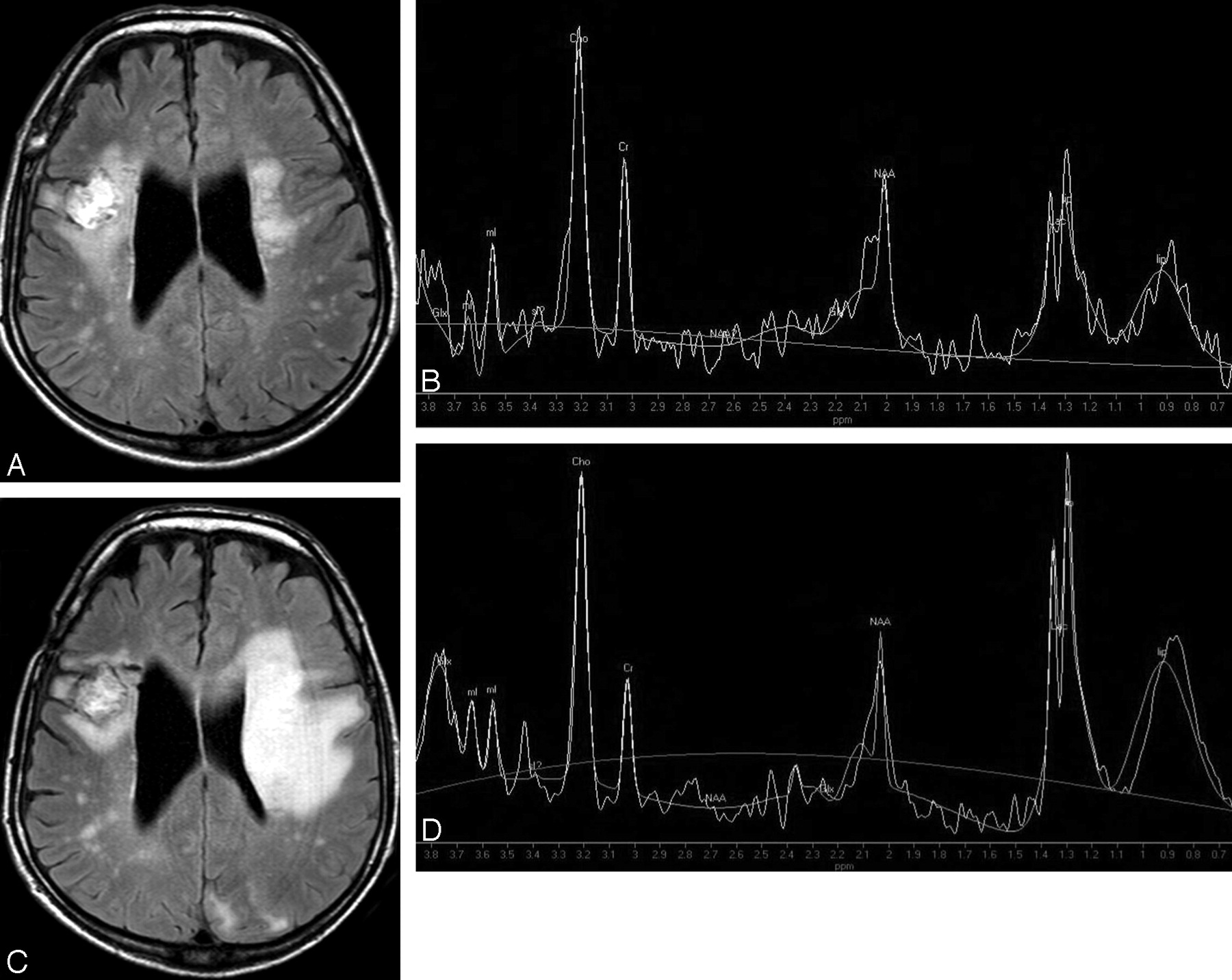
A 48-year-old man was admitted with a right frontal cerebral toxoplasmic abscess revealing human immunodeficiency virus-1 (HIV-1) infection. After the abscess was evacuated emergently, MR imaging was performed and showed confluent T2 hyperintensity in the left corona radiata, with no gadolinium uptake, compatible with PML (Fig 1A). Short TE 1H-MR spectroscopy of the left corona radiata lesion showed a mild elevation of choline (Cho), a diminution of N-acetylaspartate (NAA), the presence of lactate (Lac)/lipids at 1.3 ppm, and the presence of myo-inositol (mIns) (Fig 1B). Combined antiretroviral treatment was started. Two months later, the patient developed subacute right hemiplegia, while the HIV load had decreased by >2 log10 copies/mL. MR imaging showed a marked increase of the left white matter T2 hyperintensity, with mass effect (Fig 1C) and contrast enhancement. Subsequent short and long TE proton spectra showed an additional increase of Cho and Lac/lipid peaks (Fig 1D), compared with the spectrum of PML acquired before combined antiretroviral treatment initiation and immune restoration. Stereotactic brain biopsy further confirmed PML-IRIS.

Fig 1.
  • Download figure
  • Open in new tab
  • Download powerpoint
Fig 1.

A, Fluid-attenuated inversion recovery (FLAIR) axial image shows high signal intensity in the left corona radiata, compatible with PML. Note the right frontal postsurgical appearance after evacuation of a cerebral abscess. B, Short TE spectroscopy shows increased Cho, free lipids (and/or Lac), and diminished NAA. C, After 2 months of treatment, with adapted biologic response but paradoxic clinical worsening, axial FLAIR image reveals an enlargement of the left T2 hyperintensity with obvious mass effect on the ipsilateral ventricle. D, Short TE spectroscopy shows further increase in Cho, Lac, and free lipid peaks (long TE not shown). Glx indicates glutamine/glutamate; lip, lipid; mI, myo-inositol.

In conclusion and responding to Bag et al,1 we report a case of paradoxic PML-IRIS with serial MR spectroscopy features consistent with an increased inflammatory response. While the elevation of the mIns/creatine ratio has been shown to be associated with better survival rates in patients with PML,3 we propose that serial spectroscopy could be a diagnostic marker of PML-IRIS, deserving further study.

References

  1. 1.
    1. Bag AK,
    2. Cure JK,
    3. Chapman PR,
    4. et al
    . JC virus infection of the brain. AJNR Am J Neuroradiol 2010 Mar 18. [Epub ahead of print]
  2. 2.
    1. Tan K,
    2. Roda R,
    3. Ostrow L,
    4. et al
    . PML-IRIS in patients with HIV infection: clinical manifestations and treatment with steroids. Neurology 2009;72:1458–64
  3. 3.
    1. Katz-Brull R,
    2. Lenkinski RE,
    3. Du Pasquier RA,
    4. et al
    . Elevation of myoinositol is associated with disease containment in progressive multifocal leukoencephalopathy. Neurology 2004;63:897–900
  • Copyright © American Society of Neuroradiology
Advertisement

Indexed Content

  • Current Issue
  • Accepted Manuscripts
  • Article Preview
  • Past Issues
  • Editorials
  • Editor's Choice
  • Fellows' Journal Club
  • Letters to the Editor
  • Video Articles

Cases

  • Case Collection
  • Archive - Case of the Week
  • Archive - Case of the Month
  • Archive - Classic Case

More from AJNR

  • Trainee Corner
  • Imaging Protocols
  • MRI Safety Corner
  • Book Reviews

Multimedia

  • AJNR Podcasts
  • AJNR Scantastics

Resources

  • Turnaround Time
  • Submit a Manuscript
  • Submit a Video Article
  • Submit an eLetter to the Editor/Response
  • Manuscript Submission Guidelines
  • Statistical Tips
  • Fast Publishing of Accepted Manuscripts
  • Graphical Abstract Preparation
  • Imaging Protocol Submission
  • Evidence-Based Medicine Level Guide
  • Publishing Checklists
  • Author Policies
  • Become a Reviewer/Academy of Reviewers
  • News and Updates

About Us

  • About AJNR
  • Editorial Board
  • Editorial Board Alumni
  • Alerts
  • Permissions
  • Not an AJNR Subscriber? Join Now
  • Advertise with Us
  • Librarian Resources
  • Feedback
  • Terms and Conditions
  • AJNR Editorial Board Alumni

American Society of Neuroradiology

  • Not an ASNR Member? Join Now

© 2025 by the American Society of Neuroradiology All rights, including for text and data mining, AI training, and similar technologies, are reserved.
Print ISSN: 0195-6108 Online ISSN: 1936-959X

Powered by HighWire