Advertisement

Main menu

  • Home
  • Content
    • Current Issue
    • Accepted Manuscripts
    • Article Preview
    • Past Issue Archive
    • Video Articles
    • AJNR Case Collection
    • Case of the Week Archive
    • Case of the Month Archive
    • Classic Case Archive
  • Special Collections
    • AJNR Awards
    • Low-Field MRI
    • Alzheimer Disease
    • ASNR Foundation Special Collection
    • Photon-Counting CT
    • View All
  • Multimedia
    • AJNR Podcasts
    • AJNR SCANtastic
    • Trainee Corner
    • MRI Safety Corner
    • Imaging Protocols
  • For Authors
    • Submit a Manuscript
    • Submit a Video Article
    • Submit an eLetter to the Editor/Response
    • Manuscript Submission Guidelines
    • Statistical Tips
    • Fast Publishing of Accepted Manuscripts
    • Graphical Abstract Preparation
    • Imaging Protocol Submission
    • Author Policies
  • About Us
    • About AJNR
    • Editorial Board
    • Editorial Board Alumni
  • More
    • Become a Reviewer/Academy of Reviewers
    • Subscribers
    • Permissions
    • Alerts
    • Feedback
    • Advertisers
    • ASNR Home

User menu

  • Alerts
  • Log in

Search

  • Advanced search
American Journal of Neuroradiology

American Journal of Neuroradiology

ASHNR American Society of Functional Neuroradiology ASHNR American Society of Pediatric Neuroradiology ASSR
  • Alerts
  • Log in

Advanced Search

  • Home
  • Content
    • Current Issue
    • Accepted Manuscripts
    • Article Preview
    • Past Issue Archive
    • Video Articles
    • AJNR Case Collection
    • Case of the Week Archive
    • Case of the Month Archive
    • Classic Case Archive
  • Special Collections
    • AJNR Awards
    • Low-Field MRI
    • Alzheimer Disease
    • ASNR Foundation Special Collection
    • Photon-Counting CT
    • View All
  • Multimedia
    • AJNR Podcasts
    • AJNR SCANtastic
    • Trainee Corner
    • MRI Safety Corner
    • Imaging Protocols
  • For Authors
    • Submit a Manuscript
    • Submit a Video Article
    • Submit an eLetter to the Editor/Response
    • Manuscript Submission Guidelines
    • Statistical Tips
    • Fast Publishing of Accepted Manuscripts
    • Graphical Abstract Preparation
    • Imaging Protocol Submission
    • Author Policies
  • About Us
    • About AJNR
    • Editorial Board
    • Editorial Board Alumni
  • More
    • Become a Reviewer/Academy of Reviewers
    • Subscribers
    • Permissions
    • Alerts
    • Feedback
    • Advertisers
    • ASNR Home
  • Follow AJNR on Twitter
  • Visit AJNR on Facebook
  • Follow AJNR on Instagram
  • Join AJNR on LinkedIn
  • RSS Feeds

AJNR Awards, New Junior Editors, and more. Read the latest AJNR updates

Research ArticleNeurointervention
Open Access

Five-Year Follow-Up in Elastase-Induced Aneurysms in Rabbits

Y. Ding, D. Dai, R. Kadirvel, D.A. Lewis and D.F. Kallmes
American Journal of Neuroradiology August 2010, 31 (7) 1236-1239; DOI: https://doi.org/10.3174/ajnr.A2056
Y. Ding
  • Find this author on Google Scholar
  • Find this author on PubMed
  • Search for this author on this site
D. Dai
  • Find this author on Google Scholar
  • Find this author on PubMed
  • Search for this author on this site
R. Kadirvel
  • Find this author on Google Scholar
  • Find this author on PubMed
  • Search for this author on this site
D.A. Lewis
  • Find this author on Google Scholar
  • Find this author on PubMed
  • Search for this author on this site
D.F. Kallmes
  • Find this author on Google Scholar
  • Find this author on PubMed
  • Search for this author on this site

Abstract

BACKGROUND AND PURPOSE: There is no report regarding patency of elastase-induced aneurysms for more than a 2-year period. Our aim was to report aneurysm patency rates up to 5 years in the elastase-induced aneurysm model in rabbits.

MATERIALS AND METHODS: Twenty-five elastase-induced aneurysms were created in New Zealand white rabbits and followed for up to 5 years. Thirteen (52%) rabbits died during follow-up for reasons unrelated to the aneurysms. DSA was performed at 1 month and at 2 and 5 years in the 12 surviving subjects. Aneurysm patency and dimensions, including neck diameter and aneurysm width and height, were evaluated at each time point in relation to external sizing devices. Differences of aneurysm sizes (neck width and aneurysm width and height) among time points were compared by using the Student t test.

RESULTS: Eleven (92%) of the 12 aneurysms in the subjects that survived for 5 years remained fully patent throughout follow-up. A single narrow-neck aneurysm showed partial thrombosis at the 2- and 5-year time points.

CONCLUSIONS: Experimental elastase-induced aneurysms in rabbits demonstrate high rates of patency up to 5 years following creation. When planning for very long-term studies, investigators should plan for relatively high rates of mortality unrelated to aneurysm pathology.

Abbreviations

AP
anteroposterior
DSA
digital subtraction angiography
IADSA
intra-arterial DSA
IVDSA
intravenous DSA
RCCA
right common carotid artery

Long-term patency of experimental aneurysms is important in testing aneurysm-occlusion devices. Aneurysm models that have spontaneous thrombosis are considered of limited value in the evaluation of new endovascular occlusion devices. Notwithstanding the wide variety of experimental aneurysms described in numerous species, reports regarding long-term patency rates remain uncommon.1–4 Patency is poor in side-wall aneurysms in swine.1 Sidewall canine aneurysms appear to offer better patency rates than those of swine.2,3

The rabbit elastase-induced aneurysm model has been widely used for testing endovascular devices.5–20 Previous reports have noted excellent patency of untreated elastase-induced aneurysms ≤2 years after creation.18 In this study, we report the patency rate of elastase-induced aneurysms in rabbits at 5 years.

Materials and Methods

Aneurysm Creation

Elastase-induced saccular aneurysms were created in 25 New Zealand white rabbits (body weight, 3–4 kg) by using the rabbit elastase model. Some of these same subjects were reported in previous articles detailing the use of IVDSA19 and the patency rate of aneurysms up to 2 years.18 Our Institutional Animal Care and Use Committee approved all procedures. Detailed procedures for aneurysm creation have been described.5 Briefly, anesthesia was induced with an intramuscular injection of ketamine, xylazine, and acepromazine (75 mg/kg, 5 mg/kg, and 1 mg/kg, respectively). Using a sterile technique, we exposed and ligated the RCCA distally. A 1- to 2-mm bevelled arteriotomy was made and a 5F vascular sheath (Cordis Endovascular, Miami Lakes, Florida) was advanced retrogradely in the RCCA to a point approximately 3 cm cephalad to the origin of RCCA. Fluoroscopy (Advantx; GE Healthcare, Milwaukee, Wisconsin) was performed by injection of contrast through the sheath retrogradely in the RCCA, to identify the junction between the RCCA and the subclavian and brachiocephalic arteries. A 3F Fogarty balloon (Baxter Healthcare, Irvine, California) was advanced through the sheath to the level of the origin of the RCCA with fluoroscopic guidance and was inflated with iodinated contrast material. Porcine elastase (5.23 μ/mgP, 40.1 mgP/mL, approximately 200 U/mL; Worthington Biochemical, Lakewood, New Jersey) was incubated within the lumen of the common carotid artery above the inflated balloon for 20 minutes, after which the catheter, balloon, and sheath were removed and the RCCA was ligated below the sheath entry site.

DSA Follow-Up

Follow-up angiography was performed at each time point (1 month, 2 years, and 5 years after creation). Twelve (48%) of 25 rabbits survived for the full duration of the study, in which follow-up angiographic images at each time point were available in 11 aneurysms; the other aneurysm only had images at 2- and 5-year time points. Thirteen rabbits (52%) died from causes unrelated to the aneurysm itself: 5 from stroke, 2 from cancer, 4 from chronic liver failure, and 2 from anesthesia-related complications before 5 years after aneurysm creation. For the 1-month and 5-year time points, IADSA was performed, and it was also performed at 2 years. Details regarding the technique of IADSA and IVDSA have been previously reported.19 Aneurysm dimensions, including neck width and dome width and height, were measured and calculated from these DSA images in reference to external radiopaque sizing markers. DSA images also were used to evaluate interval changes in aneurysm dimension with time.

Statistical Analysis

Differences of aneurysm sizes (neck width and aneurysm width and height) among time points were compared by using the Student t test.

Results

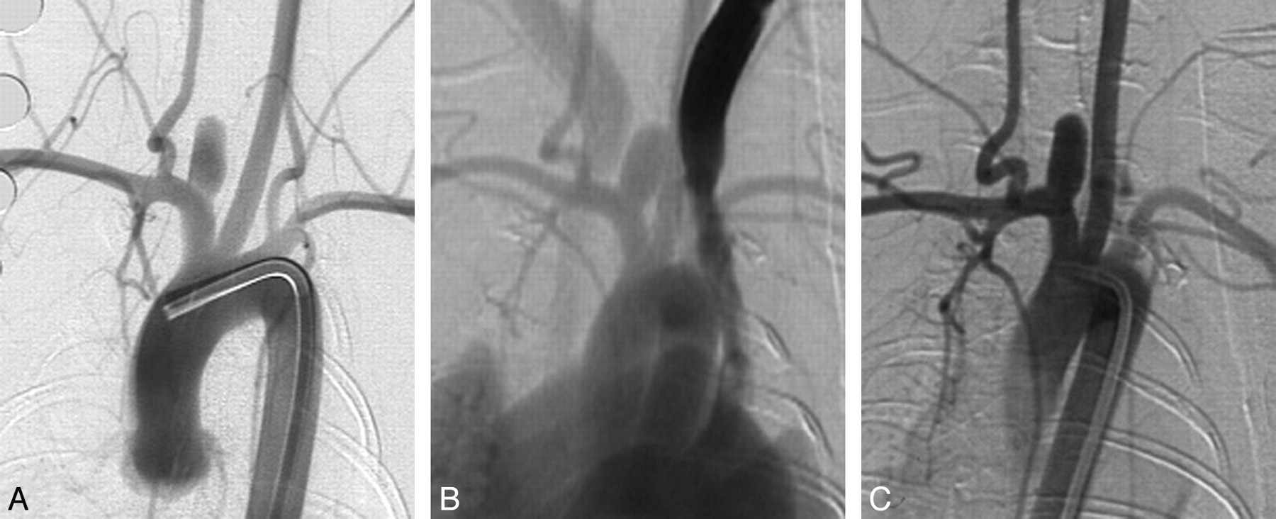
Eleven (11/12, 92%) aneurysms remained completely patent angiographically throughout the 5 years following creation (Figs 1 and 2). At 2 years, a single narrow-neck aneurysm (neck size, 1.4 mm; width, 4:5 mm; ratio of width/neck, 3:2) showed partial thrombosis (Fig 3), which remained stable at 5 years.

Fig 1.
  • Download figure
  • Open in new tab
  • Download powerpoint
Fig 1.

Serial DSA in an elastase-induced aneurysm. A, AP IADSA obtained 1 month after creation demonstrates an aneurysm cavity along the brachiocephalic artery, at the origin of the ligated RCCA. B, AP IVDSA in the same aneurysm 2 years after creation. C, AP IADSA in the same aneurysm 5 years after creation. Aneurysm dimensions remain constant throughout follow-up.

Fig 2.
  • Download figure
  • Open in new tab
  • Download powerpoint
Fig 2.

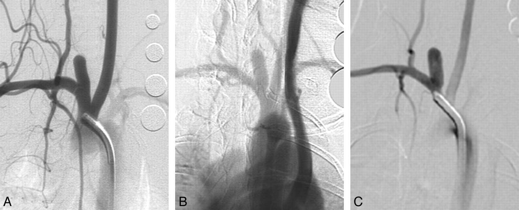
Serial DSA in another elastase-induced aneurysm. A, AP IADSA obtained 1 month after creation demonstrates an aneurysm cavity along the brachiocephalic artery, at the origin of the ligated RCCA. B, AP IVDSA in the same aneurysm 2 years after creation. C, AP IADSA in the same aneurysm 5 years after creation. Aneurysm dimensions remain stable throughout follow-up.

Fig 3.
  • Download figure
  • Open in new tab
  • Download powerpoint
Fig 3.

Serial DSA in an elastase-induced aneurysm undergoing partial thrombosis. A, AP IADSA 1 month after creation shows a patent aneurysm cavity. B, AP IVDSA at 2 years demonstrates partial thrombosis. C, AP IADSA 5 years after creation demonstrates similar partial patency compared with B.

Aneurysm Neck Dimensions

Mean neck widths at 1 month, 2 years, and 5 years after creation were 3.2 ± 1.1 mm, 3.0 ± 0.9 mm, and 3.0 ± 1.0 mm, respectively. There were no significant differences observed among these neck widths (P = .79).

Aneurysm Width

Mean aneurysm widths at 1 month, 2 years, and 5 years after creation were 3.9 ± 1.2 mm, 3.9 ± 1.2 mm, and 4.1 ± 1.0 mm, respectively. There were no significant differences among these aneurysm widths (P = .64).

Aneurysm Height

Mean aneurysm heights at 1 month and 2 and 5 years after creation were 8.9 ± 2.4 mm, 8.4 ± 2.3 mm, and 8.4 ± 2.1 mm, respectively. There were no significant differences among these aneurysm heights (P = .60).

Discussion

This study confirmed excellent long-term patency of the elastase-induced aneurysm models in rabbits. In this relatively small study of long-term patency, only a single narrow-neck aneurysm showed partial spontaneous thrombosis with time. Untreated human aneurysms also showed low rates of spontaneous thrombosis over time.21–24 Indeed, observation of spontaneous aneurysm thrombosis in humans is the subject of case reports.22–24 As such, our observed long-term patency rate of the elastase-induced aneurysms offers further support for the application of this aneurysm model, not only for evaluation of endovascular devices but also for aneurysm hemodynamics and biology.25,26

The short- and long-term patency of the elastase-induced aneurysm model has previously been reported. The model demonstrates early growth, up to 3 weeks following creation, after which aneurysm dimensions stabilize.17 This early growth may reflect either ongoing injury to the elastic lamina or ongoing interplay between aneurysm hemodynamics and mechanical integrity. Previous studies have demonstrated ongoing patency of this model for up to 2 years.18 The current study confirms ongoing patency in untreated aneurysms up to 5 years after creation.

Long-term patency rates of most types of experimental aneurysm models are rarely reported. One study reported the patency of 326 vein patch aneurysms in 310 canines during a 6-year period. Of these, 102 were sidewall (lateral) and 224 were bifurcation aneurysms. Spontaneous occlusion occurred in 9 (9/102, 9%) of the sidewall aneurysms and in only 1 (1/224, 0.4%) of the bifurcation aneurysms.2 Future model development would ideally offer such long-term patency reports.

This study had several limitations. The cohort was small, limited by the substantial financial requirements for maintaining experimental subjects for up to 5 years. Thus, the impact of aneurysm neck size or aneurysm dimension could not be determined. Second, a relatively high proportion of the rabbits in our study died from age-related pathologies, including chronic liver failure. One might argue that if we based our patency rate on the initial cohort instead of the surviving subset, our actual patency rate would have been 11 (44%) of 25 subjects. In any event, the high age-related mortality rate should be carefully considered by investigators planning very long-term rabbit aneurysm studies, with extra subjects added at the outset as needed.

Conclusions

Experimental elastase-induced aneurysms in rabbits demonstrate high rates of patency up to 5 years following creation. When planning for very long-term studies, investigators should plan for relatively high rates of mortality unrelated to aneurysm pathology.

Footnotes

  • Supported by the National Institutes of Health grant R01 NS46246.

Indicates open access to non-subscribers at www.ajnr.org

References

  1. 1.
    1. Byrne JV,
    2. Hope JK,
    3. Hubbard N,
    4. et al
    . The nature of thrombosis induced by platinum and tungsten coils in saccular aneurysms. AJNR Am J Neuroradiol 1997;18:29–33
  2. 2.
    1. Turk AS,
    2. Aagaard-Kienitz B,
    3. Niemann D,
    4. et al
    . Natural history of the canine vein pouch aneurysm model. AJNR Am J Neuroradiol 2007;28:531–32
  3. 3.
    1. Kallmes DF,
    2. Altes TA,
    3. Vincent DA,
    4. et al
    . Experimental side-wall aneurysms: a natural history study. Neuroradiology 1999;41:338–41
  4. 4.
    1. Raymond J,
    2. Salazkin I,
    3. Metcalfe A,
    4. et al
    . Lingual artery bifurcation aneurysms for training and evaluation of neurovascular devices. AJNR Am J Neuroradiol 2004;25:1387–90
  5. 5.
    1. Altes TA,
    2. Cloft HJ,
    3. Short JG,
    4. et al
    . 1999 ARRS Executive Council Award: creation of saccular aneurysms in the rabbit—a model suitable for testing endovascular devices. American Roentgen Ray Society. AJR Am J Roentgenol 2000;174:349–54
  6. 6.
    1. Cloft HJ,
    2. Altes TA,
    3. Marx WF,
    4. et al
    . Endovascular creation of an in vivo bifurcation aneurysm model in rabbits. Radiology 1999;213:223–28
  7. 7.
    1. Kallmes DF,
    2. Fujiwara NH
    . New expandable hydrogel-platinum coil hybrid device for aneurysm embolization. AJNR Am J Neuroradiol 2002;23:1580–88
  8. 8.
    1. Kallmes DF,
    2. Fujiwara NH,
    3. Yuen D,
    4. et al
    . A collagen-based coil for embolization of saccular aneurysms in a New Zealand white rabbit model. AJNR Am J Neuroradiol 2003;24:591–96
  9. 9.
    1. Kallmes DF,
    2. Williams AD,
    3. Cloft HJ,
    4. et al
    . Platinum coil-mediated implantation of growth factor-secreting endovascular tissue grafts: an in vivo study. Radiology 1998;207:519–23
  10. 10.
    1. De Gast AN,
    2. Altes TA,
    3. Marx WF,
    4. et al
    . Transforming growth factor beta-coated platinum coils for endovascular treatment of aneurysms: an animal study. Neurosurgery 2001;49:690–94
  11. 11.
    1. Aassar OS,
    2. Fujiwara NH,
    3. Marx WF,
    4. et al
    . Aneurysm growth, elastinolysis, and attempted doxycycline inhibition of elastase-induced aneurysms in rabbits. J Vasc Interv Radiol 2003;14:1427–32
  12. 12.
    1. Thiex R,
    2. Moller-Hartmann W,
    3. Hans FJ,
    4. et al
    . Are the configuration and neck morphology of experimental aneurysms predictable? A technical approach. Neuroradiology 2004;46:571–76
  13. 13.
    1. Hoh BL,
    2. Rabinov JD,
    3. Pryor JC,
    4. et al
    . A modified technique for using elastase to create saccular aneurysms in animals that histologically and hemodynamically resemble aneurysms in human. Acta Neurochir (Wien) 2004;146:705–11
  14. 14.
    1. Hans FJ,
    2. Krings T,
    3. Moller-Hartmann W,
    4. et al
    . Endovascular treatment of experimentally induced aneurysms in rabbits using stents: a feasibility study. Neuroradiology 2003;45:430–34
  15. 15.
    1. Fujiwara NH,
    2. Kallmes DF
    . Healing response in elastase-induced rabbit aneurysms after embolization with a new platinum coil system. AJNR Am J Neuroradiol 2002;23:1137–44
  16. 16.
    1. Krings T,
    2. Hans FJ,
    3. Moller-Hartmann W,
    4. et al
    . Time-of-flight, phase contrast and contrast-enhanced magnetic resonance angiography for pre-interventional determination of aneurysm size, configuration, and neck morphology in an aneurysm model in rabbits. Neurosci Lett 2002;326:46–50
  17. 17.
    1. Fujiwara NH,
    2. Cloft HJ,
    3. Marx WF,
    4. et al
    . Serial angiography in an elastase-induced aneurysm model in rabbits: evidence for progressive aneurysm enlargement after creation. AJNR Am J Neuroradiol 2001;22:698–703
  18. 18.
    1. Ding YH,
    2. Dai D,
    3. Danielson MA,
    4. et al
    . Long-term patency of elastase-induced aneurysm model in rabbits. AJNR Am J Neuroradiol 2006;27:139–41
  19. 19.
    1. Ding YH,
    2. Dai D,
    3. Lewis DA,
    4. et al
    . Intra-venous digital subtraction angiography (IVDSA): an alternative method to intra-arterial digital subtraction angiography (IADSA) for experimental aneurysm imaging. Neuroradiology 2005;47:792–95. Epub 2005 Aug 25
  20. 20.
    1. Dai D,
    2. Ding YH,
    3. Kallmes DF
    . Histopathologic and immunohistochemical comparison in human, rabbit, and swine aneurysms embolized with platinum coils. AJNR Am J Neuroradiol 2005;26:2560–68
  21. 21.
    1. Wiebers DO,
    2. Whisnant JP,
    3. Huston J 3rd.,
    4. et al.,
    5. for the International Study of Unruptured Intracranial Aneurysms Investigators
    . Natural history, clinical outcome, and risks of surgical and endovascular treatment Lancet 2003;362:103–10
  22. 22.
    1. Brownlee RD,
    2. Tranmer BI,
    3. Sevick RJ,
    4. et al
    . Spontaneous thrombosis of an unruptured anterior communicating artery aneurysm: an unusual cause of ischemic stroke. Stroke 1995;26:1945–49
  23. 23.
    1. Hamilton MG,
    2. Dold ON
    . Spontaneous disappearance of an intracranial aneurysm after subarachnoid hemorrhage. Can J Neurol Sci 1992;19:389–91
  24. 24.
    1. Tanabe M,
    2. Inoue Y,
    3. Hori T
    . Spontaneous thrombosis of an aneurysm of the middle cerebral artery with subarachnoid haemorrhage in a 6-year-old child: case report. Neurol Res 1991;13:202–04
  25. 25.
    1. Kadirve R,
    2. Ding YH,
    3. Dai D,
    4. et al
    . Gene expression profiling of experimental saccular aneurysms using deoxyribonucleic acid. AJNR Am J Neuroradiol 2008;29:1566–99
  26. 26.
    1. Kadirvel R,
    2. Ding YH,
    3. Dai D,
    4. et al
    . The influence of hemodynamic forces on biomarkers in the walls of elastase-induced aneurysms in rabbits. Neuroradiology 2007;49:1041–53
  • Received November 17, 2009.
  • Accepted after revision January 9, 2009.
  • Copyright © American Society of Neuroradiology
Advertisement

Indexed Content

  • Current Issue
  • Accepted Manuscripts
  • Article Preview
  • Past Issues
  • Editorials
  • Editor's Choice
  • Fellows' Journal Club
  • Letters to the Editor
  • Video Articles

Cases

  • Case Collection
  • Archive - Case of the Week
  • Archive - Case of the Month
  • Archive - Classic Case

More from AJNR

  • Trainee Corner
  • Imaging Protocols
  • MRI Safety Corner
  • Book Reviews

Multimedia

  • AJNR Podcasts
  • AJNR Scantastics

Resources

  • Turnaround Time
  • Submit a Manuscript
  • Submit a Video Article
  • Submit an eLetter to the Editor/Response
  • Manuscript Submission Guidelines
  • Statistical Tips
  • Fast Publishing of Accepted Manuscripts
  • Graphical Abstract Preparation
  • Imaging Protocol Submission
  • Evidence-Based Medicine Level Guide
  • Publishing Checklists
  • Author Policies
  • Become a Reviewer/Academy of Reviewers
  • News and Updates

About Us

  • About AJNR
  • Editorial Board
  • Editorial Board Alumni
  • Alerts
  • Permissions
  • Not an AJNR Subscriber? Join Now
  • Advertise with Us
  • Librarian Resources
  • Feedback
  • Terms and Conditions
  • AJNR Editorial Board Alumni

American Society of Neuroradiology

  • Not an ASNR Member? Join Now

© 2025 by the American Society of Neuroradiology All rights, including for text and data mining, AI training, and similar technologies, are reserved.
Print ISSN: 0195-6108 Online ISSN: 1936-959X

Powered by HighWire