Advertisement

Main menu

  • Home
  • Content
    • Current Issue
    • Accepted Manuscripts
    • Article Preview
    • Past Issue Archive
    • Video Articles
    • AJNR Case Collection
    • Case of the Week Archive
    • Case of the Month Archive
    • Classic Case Archive
  • Special Collections
    • AJNR Awards
    • Low-Field MRI
    • Alzheimer Disease
    • ASNR Foundation Special Collection
    • Photon-Counting CT
    • View All
  • Multimedia
    • AJNR Podcasts
    • AJNR SCANtastic
    • Trainee Corner
    • MRI Safety Corner
    • Imaging Protocols
  • For Authors
    • Submit a Manuscript
    • Submit a Video Article
    • Submit an eLetter to the Editor/Response
    • Manuscript Submission Guidelines
    • Statistical Tips
    • Fast Publishing of Accepted Manuscripts
    • Graphical Abstract Preparation
    • Imaging Protocol Submission
    • Author Policies
  • About Us
    • About AJNR
    • Editorial Board
    • Editorial Board Alumni
  • More
    • Become a Reviewer/Academy of Reviewers
    • Subscribers
    • Permissions
    • Alerts
    • Feedback
    • Advertisers
    • ASNR Home

User menu

  • Alerts
  • Log in

Search

  • Advanced search
American Journal of Neuroradiology

American Journal of Neuroradiology

ASHNR American Society of Functional Neuroradiology ASHNR American Society of Pediatric Neuroradiology ASSR
  • Alerts
  • Log in

Advanced Search

  • Home
  • Content
    • Current Issue
    • Accepted Manuscripts
    • Article Preview
    • Past Issue Archive
    • Video Articles
    • AJNR Case Collection
    • Case of the Week Archive
    • Case of the Month Archive
    • Classic Case Archive
  • Special Collections
    • AJNR Awards
    • Low-Field MRI
    • Alzheimer Disease
    • ASNR Foundation Special Collection
    • Photon-Counting CT
    • View All
  • Multimedia
    • AJNR Podcasts
    • AJNR SCANtastic
    • Trainee Corner
    • MRI Safety Corner
    • Imaging Protocols
  • For Authors
    • Submit a Manuscript
    • Submit a Video Article
    • Submit an eLetter to the Editor/Response
    • Manuscript Submission Guidelines
    • Statistical Tips
    • Fast Publishing of Accepted Manuscripts
    • Graphical Abstract Preparation
    • Imaging Protocol Submission
    • Author Policies
  • About Us
    • About AJNR
    • Editorial Board
    • Editorial Board Alumni
  • More
    • Become a Reviewer/Academy of Reviewers
    • Subscribers
    • Permissions
    • Alerts
    • Feedback
    • Advertisers
    • ASNR Home
  • Follow AJNR on Twitter
  • Visit AJNR on Facebook
  • Follow AJNR on Instagram
  • Join AJNR on LinkedIn
  • RSS Feeds

AJNR Awards, New Junior Editors, and more. Read the latest AJNR updates

LetterLetter

Solitary Plasmacytoma of the Spine: An Unusual Presentation

P.D. Afonso and A. Almeida
American Journal of Neuroradiology January 2010, 31 (1) E5; DOI: https://doi.org/10.3174/ajnr.A1948
P.D. Afonso
  • Find this author on Google Scholar
  • Find this author on PubMed
  • Search for this author on this site
A. Almeida
  • Find this author on Google Scholar
  • Find this author on PubMed
  • Search for this author on this site

Solitary plasmacytoma of bone (SBP) is a rare hematologic malignancy, which occurs with radiologic evidence of a solitary lesion in the absence of significant bone marrow plasma cell infiltration (<5%–10%), leading to absent (or low) serum and urine levels of monoclonal protein.1 This isolated lesion is most commonly found in the spine, without evidence of multiple myeloma elsewhere.2–4 Our letter will present a biopsy-proved thoracolumbar SBP with extensive involvement of the soft tissues.

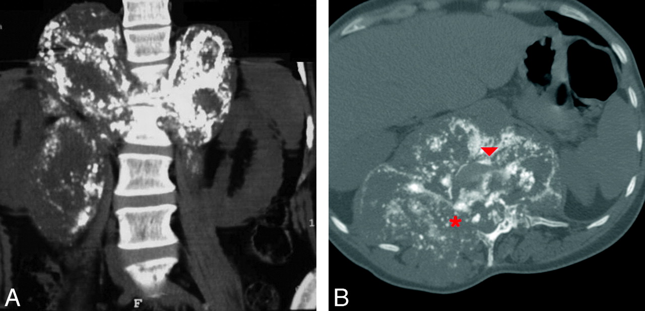
A 41-year-old woman presented with back pain, paraparesis, and hyporeflexia of the lower limbs. Spine CT revealed osteolytic areas from T12 to L2 with vertebrae and disk-space destruction, extension to the posterior elements (Fig 1A), and a perivertebral mass (Fig 1B). MR imaging showed extension of the lesion to the spinal canal, with an important mildly enhancing epidural mass and compression of the cord. CT-guided bone marrow vertebral biopsy was consistent with plasmocytoma. Laboratory tests confirmed this diagnosis.

Fig 1.
  • Download figure
  • Open in new tab
  • Download powerpoint
Fig 1.

A, Coronal nonenhanced CT image through the lumbosacral spine (soft-tissue windows) shows bone destruction (from T12 to L2), most severe at L1 and the adjacent disk space (L1-L2), and a 17 × 15 cm calcified paravertebral soft-tissue mass, involving almost the entire right psoas and the upper third of the left psoas muscle, extending up to the diaphragm and displacing the right kidney laterally. B, Axial CT image (bone windows) reveals complete destruction of the L1 vertebra with ill-defined osteolytic areas (arrowhead) and involvement of the posterior arch and pedicles (asterisk).

SBP most commonly affects the axial skeleton (25%–60%) and has a predilection for the thoracic spine.1,4 It can present for many years as an isolated lesion, but on occasion, multiple plasmacytomas can develop; therefore, an association with multiple myeloma is described.4 However, involvement of adjacent bone and tissue sites by direct extension does not constitute evidence of multiple myeloma. SBP of the spine is predominantly lytic; typically involves the vertebral body and the posterior elements with compression of the cord; but, on occasion, may present as an expansible lesion with a soft-tissue mass, fractures, or, rarely, osteosclerosis.2–4 Its MR imaging signal intensity is not specific (low on T1-weighted images and high on T2-weighted or short tau inversion recovery images). The “mini-brain” sign is the only pathognomonic finding.2 CT and MR imaging findings can be very helpful in a patient with plasma cell dyscrasia and should be kept in mind, even though biopsy is usually required. To our knowledge, the imaging findings of such an unusual presentation of SBP have not been previously described.

References

  1. 1.
    1. Jyothirmayi R,
    2. Gangadharan VP,
    3. Nair MK.
    . Radiotherapy in the treatment of solitary plasmocytoma. Br J Radiol 1997; 70: 511–16
  2. 2.
    1. Major NM,
    2. Helms CA,
    3. Richardson WJ
    . The “mini brain”: plasmocytoma in a vertebral body on MR imaging. AJR Am J Roentgenol 2000; 175: 261–63
  3. 3.
    1. Takahashi T,
    2. Koshu K,
    3. Tominaga T,
    4. et al
    . Solitary plasmacytoma in the spine: two case reports. Neurosurg Rev 1998; 21: 121–25
  4. 4.
    1. Baba H,
    2. Maezawa Y,
    3. Furusawa N,
    4. et al
    . Solitary plasmacytomas of the spine associated with neurological complications. Spinal Cord 1998; 36: 4170–75
  • Copyright © American Society of Neuroradiology
Advertisement

Indexed Content

  • Current Issue
  • Accepted Manuscripts
  • Article Preview
  • Past Issues
  • Editorials
  • Editor's Choice
  • Fellows' Journal Club
  • Letters to the Editor
  • Video Articles

Cases

  • Case Collection
  • Archive - Case of the Week
  • Archive - Case of the Month
  • Archive - Classic Case

More from AJNR

  • Trainee Corner
  • Imaging Protocols
  • MRI Safety Corner

Multimedia

  • AJNR Podcasts
  • AJNR Scantastics

Resources

  • Turnaround Time
  • Submit a Manuscript
  • Submit a Video Article
  • Submit an eLetter to the Editor/Response
  • Manuscript Submission Guidelines
  • Statistical Tips
  • Fast Publishing of Accepted Manuscripts
  • Graphical Abstract Preparation
  • Imaging Protocol Submission
  • Evidence-Based Medicine Level Guide
  • Publishing Checklists
  • Author Policies
  • Become a Reviewer/Academy of Reviewers
  • News and Updates

About Us

  • About AJNR
  • Editorial Board
  • Editorial Board Alumni
  • Alerts
  • Permissions
  • Not an AJNR Subscriber? Join Now
  • Advertise with Us
  • Librarian Resources
  • Feedback
  • Terms and Conditions
  • AJNR Editorial Board Alumni

American Society of Neuroradiology

  • Not an ASNR Member? Join Now

© 2025 by the American Society of Neuroradiology All rights, including for text and data mining, AI training, and similar technologies, are reserved.
Print ISSN: 0195-6108 Online ISSN: 1936-959X

Powered by HighWire