Advertisement

Main menu

  • Home
  • Content
    • Current Issue
    • Accepted Manuscripts
    • Article Preview
    • Past Issue Archive
    • Video Articles
    • AJNR Case Collection
    • Case of the Week Archive
    • Case of the Month Archive
    • Classic Case Archive
  • Special Collections
    • AJNR Awards
    • Low-Field MRI
    • Alzheimer Disease
    • ASNR Foundation Special Collection
    • Photon-Counting CT
    • View All
  • Multimedia
    • AJNR Podcasts
    • AJNR SCANtastic
    • Trainee Corner
    • MRI Safety Corner
    • Imaging Protocols
  • For Authors
    • Submit a Manuscript
    • Submit a Video Article
    • Submit an eLetter to the Editor/Response
    • Manuscript Submission Guidelines
    • Statistical Tips
    • Fast Publishing of Accepted Manuscripts
    • Graphical Abstract Preparation
    • Imaging Protocol Submission
    • Author Policies
  • About Us
    • About AJNR
    • Editorial Board
    • Editorial Board Alumni
  • More
    • Become a Reviewer/Academy of Reviewers
    • Subscribers
    • Permissions
    • Alerts
    • Feedback
    • Advertisers
    • ASNR Home

User menu

  • Alerts
  • Log in

Search

  • Advanced search
American Journal of Neuroradiology

American Journal of Neuroradiology

ASHNR American Society of Functional Neuroradiology ASHNR American Society of Pediatric Neuroradiology ASSR
  • Alerts
  • Log in

Advanced Search

  • Home
  • Content
    • Current Issue
    • Accepted Manuscripts
    • Article Preview
    • Past Issue Archive
    • Video Articles
    • AJNR Case Collection
    • Case of the Week Archive
    • Case of the Month Archive
    • Classic Case Archive
  • Special Collections
    • AJNR Awards
    • Low-Field MRI
    • Alzheimer Disease
    • ASNR Foundation Special Collection
    • Photon-Counting CT
    • View All
  • Multimedia
    • AJNR Podcasts
    • AJNR SCANtastic
    • Trainee Corner
    • MRI Safety Corner
    • Imaging Protocols
  • For Authors
    • Submit a Manuscript
    • Submit a Video Article
    • Submit an eLetter to the Editor/Response
    • Manuscript Submission Guidelines
    • Statistical Tips
    • Fast Publishing of Accepted Manuscripts
    • Graphical Abstract Preparation
    • Imaging Protocol Submission
    • Author Policies
  • About Us
    • About AJNR
    • Editorial Board
    • Editorial Board Alumni
  • More
    • Become a Reviewer/Academy of Reviewers
    • Subscribers
    • Permissions
    • Alerts
    • Feedback
    • Advertisers
    • ASNR Home
  • Follow AJNR on Twitter
  • Visit AJNR on Facebook
  • Follow AJNR on Instagram
  • Join AJNR on LinkedIn
  • RSS Feeds

AJNR Awards, New Junior Editors, and more. Read the latest AJNR updates

LetterLetter

Presence of Activated Microglia in a High-Signal Lesion on T1-Weighted MR Images: A Biopsy Sample Re-Examined

Din-E Shan, Hung-Chi Pan, Donald M.T. Ho, Michael M.H. Teng and Chen Chang
American Journal of Neuroradiology April 2007, 28 (4) 602;
Din-E Shan
  • Find this author on Google Scholar
  • Find this author on PubMed
  • Search for this author on this site
Hung-Chi Pan
  • Find this author on Google Scholar
  • Find this author on PubMed
  • Search for this author on this site
Donald M.T. Ho
  • Find this author on Google Scholar
  • Find this author on PubMed
  • Search for this author on this site
Michael M.H. Teng
  • Find this author on Google Scholar
  • Find this author on PubMed
  • Search for this author on this site
Chen Chang
  • Find this author on Google Scholar
  • Find this author on PubMed
  • Search for this author on this site

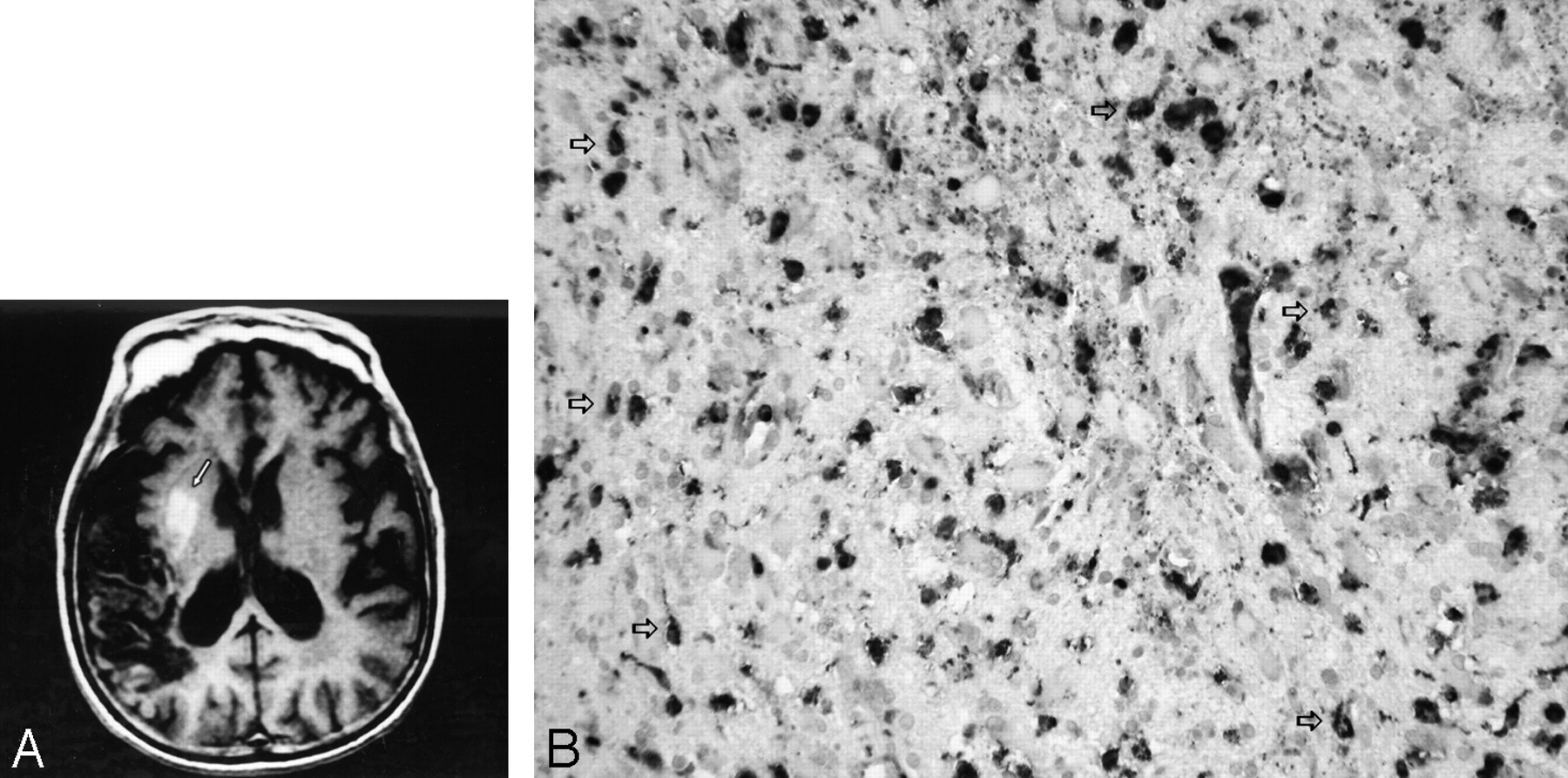
To determine the pathologic process responsible for the signal intensity changes of hyperintense putamen on T1-weighted MR images (Fig 1A), we had previously performed stereotactic biopsy in a 72-year-old man 3 months after the onset of stroke and hemiballism.1 The results revealed a fragment of gliotic brain tissue with abundant gemistocytes, which we assumed to be responsible for the signal-intensity changes.1 Aoe et al2 recently reproduced similar MR imaging findings in 29 rats by 20-minute occlusion of 4 cerebral arteries and disclosed the accumulation of fatty droplets in the activated microglia. To determine if activated microglia were present in the human tissue with similar MR imaging findings, we re-examined the previously obtained biopsy specimen with immunohistochemical staining by using a monoclonal antibody CD68. The abundant presence of microglia and their shape transformation into typical phagocytic macrophages without dendritic processes indicated that microglia activation was present in our biopsy specimen (Fig 1B). From morphologic observation, there were no discernible fat droplets in the section examined. The abundant presence of microglia per se might not be enough to explain the MR signal-intensity changes because not all lesions with microglia infiltration show hyperintense lesions on T1-weighted MR images. Fatty degeneration may be a more important factor. Our previous study with high-resolution 1H-nuclear MR spectroscopy in 1 of 3 biopsy specimens did show a marked increase of lipids.1 It is possible that the high lipid content in the brain tissues may explain the signal-intensity changes on T1-weighted MR images.

Six mechanisms may explain the high-signal-intensity changes on T1-weighted MR images,2 and publications to date have provided evidence for 5 of them. The mechanism appears to be more complex than we originally proposed, a high protein content in gemistocytes.1 A common finding in the postmortem reports confirms the presence of multiple infarcts surrounded by hypertrophic astrocytes.3 However, because of the lag between the time of MR imaging and the time of autopsy, it is difficult to confirm in humans if the appearance and disappearance of gemistocytes correlate with the appearance and disappearance of the hyperintensity on the T1-weighted MR images. Although animal experiments of Aoe et al suggested that lesions with fatty components are responsible for the signal intensity change, Fujioka et al4 did not find lipid accumulation in their animals. In contrast, they reproduced the MR imaging finding with a similar time course to that of the accumulation of tissue manganese, accompanied by Mn-superoxide dismutase induction in reactive astrocytes. Finally, an autopsy performed by Nath et al3 at 36 days after the onset demonstrated the presence of microhemorrhage, punctuate calcification, and mineralized neurons. The presence of hemosiderin-laden macrophages in our biopsy specimen indicated that microhemorrhage might have occurred earlier.1 However, Fujioka et al did not find hemorrhage in their animals.

Although a high protein content, lipid component, manganese accumulation, microhemorrhage, or punctuate calcification can all explain the high-signal-intensity changes on T1-weighted MR images, 2 observations deserve attention. The first is that the duration of MR signal-intensity changes persists much longer than that of CT signal intensity changes,1 suggesting that microhemorrhage or punctuate calcification is likely responsible for the earlier CT signal-intensity changes but unlikely responsible for the later MR signal-intensity changes. The second is that the MR signal-intensity changes extend beyond the vascular territory of ischemia,1 which makes mineralized neurons or a glial reaction secondary to neuronal changes but not to ischemic insults likely responsible. Further studies focusing on the area beyond the territory of ischemia but with the MR signal-intensity changes may help us to clarify the problem. However, because signal hyperintensity on T1-weighted MR images was reported at midbrain in primates and in a patient with manganese intoxication, manganese accumulation in reactive astrocytes remains the most likely cause.

  Fig 1.
  • Download figure
  • Open in new tab
  • Download powerpoint
Fig 1.

A, Axial section of T1-weighted (TR/TE/TI, 550/10/1 ms) MR image at the level of the basal ganglia shows hyperintensities in the right putamen and the external segment of globus pallidus (arrow), along with an infarct in the right temporoparietal cortex. B, Immunohistochemical staining of the brain biopsy specimen for CD68 revealed numerous darkly stained microglia with or without dendritic processes (white arrows) (original magnification ×100).

References

  1. Shan DE, Ho DM, Chang C, et al. Hemichorea-hemiballism: an explanation for MR signal changes. AJNR Am J Neuroradiol 1998;19:863–70
  2. Aoe H, Takeda Y, Kawahara H, et al. Clinical significance of T1-weighted MR images following transient cerebral ischemia. J Neurol Sci 2006;241:19–24
  3. Nath J, Jambhekar K, Rao C, et al. Radiological and pathological changes in hemiballism-hemichorea with striatal hyperintensity. J Magn Reson Imaging 2006;23:564–68
  4. Fujioka M, Taoka T, Matsuo Y, et al. Magnetic resonance imaging shows delayed ischemic striatal neurodegeneration. Ann Neurol 2003;54:732–47
  • Copyright © American Society of Neuroradiology
Advertisement

Indexed Content

  • Current Issue
  • Accepted Manuscripts
  • Article Preview
  • Past Issues
  • Editorials
  • Editor's Choice
  • Fellows' Journal Club
  • Letters to the Editor
  • Video Articles

Cases

  • Case Collection
  • Archive - Case of the Week
  • Archive - Case of the Month
  • Archive - Classic Case

More from AJNR

  • Trainee Corner
  • Imaging Protocols
  • MRI Safety Corner

Multimedia

  • AJNR Podcasts
  • AJNR Scantastics

Resources

  • Turnaround Time
  • Submit a Manuscript
  • Submit a Video Article
  • Submit an eLetter to the Editor/Response
  • Manuscript Submission Guidelines
  • Statistical Tips
  • Fast Publishing of Accepted Manuscripts
  • Graphical Abstract Preparation
  • Imaging Protocol Submission
  • Evidence-Based Medicine Level Guide
  • Publishing Checklists
  • Author Policies
  • Become a Reviewer/Academy of Reviewers
  • News and Updates

About Us

  • About AJNR
  • Editorial Board
  • Editorial Board Alumni
  • Alerts
  • Permissions
  • Not an AJNR Subscriber? Join Now
  • Advertise with Us
  • Librarian Resources
  • Feedback
  • Terms and Conditions
  • AJNR Editorial Board Alumni

American Society of Neuroradiology

  • Not an ASNR Member? Join Now

© 2025 by the American Society of Neuroradiology All rights, including for text and data mining, AI training, and similar technologies, are reserved.
Print ISSN: 0195-6108 Online ISSN: 1936-959X

Powered by HighWire