Advertisement

Main menu

  • Home
  • Content
    • Current Issue
    • Accepted Manuscripts
    • Article Preview
    • Past Issue Archive
    • Video Articles
    • AJNR Case Collection
    • Case of the Week Archive
    • Case of the Month Archive
    • Classic Case Archive
  • Special Collections
    • AJNR Awards
    • Low-Field MRI
    • Alzheimer Disease
    • ASNR Foundation Special Collection
    • Photon-Counting CT
    • View All
  • Multimedia
    • AJNR Podcasts
    • AJNR SCANtastic
    • Trainee Corner
    • MRI Safety Corner
    • Imaging Protocols
  • For Authors
    • Submit a Manuscript
    • Submit a Video Article
    • Submit an eLetter to the Editor/Response
    • Manuscript Submission Guidelines
    • Statistical Tips
    • Fast Publishing of Accepted Manuscripts
    • Graphical Abstract Preparation
    • Imaging Protocol Submission
    • Author Policies
  • About Us
    • About AJNR
    • Editorial Board
    • Editorial Board Alumni
  • More
    • Become a Reviewer/Academy of Reviewers
    • Subscribers
    • Permissions
    • Alerts
    • Feedback
    • Advertisers
    • ASNR Home

User menu

  • Alerts
  • Log in

Search

  • Advanced search
American Journal of Neuroradiology

American Journal of Neuroradiology

ASHNR American Society of Functional Neuroradiology ASHNR American Society of Pediatric Neuroradiology ASSR
  • Alerts
  • Log in

Advanced Search

  • Home
  • Content
    • Current Issue
    • Accepted Manuscripts
    • Article Preview
    • Past Issue Archive
    • Video Articles
    • AJNR Case Collection
    • Case of the Week Archive
    • Case of the Month Archive
    • Classic Case Archive
  • Special Collections
    • AJNR Awards
    • Low-Field MRI
    • Alzheimer Disease
    • ASNR Foundation Special Collection
    • Photon-Counting CT
    • View All
  • Multimedia
    • AJNR Podcasts
    • AJNR SCANtastic
    • Trainee Corner
    • MRI Safety Corner
    • Imaging Protocols
  • For Authors
    • Submit a Manuscript
    • Submit a Video Article
    • Submit an eLetter to the Editor/Response
    • Manuscript Submission Guidelines
    • Statistical Tips
    • Fast Publishing of Accepted Manuscripts
    • Graphical Abstract Preparation
    • Imaging Protocol Submission
    • Author Policies
  • About Us
    • About AJNR
    • Editorial Board
    • Editorial Board Alumni
  • More
    • Become a Reviewer/Academy of Reviewers
    • Subscribers
    • Permissions
    • Alerts
    • Feedback
    • Advertisers
    • ASNR Home
  • Follow AJNR on Twitter
  • Visit AJNR on Facebook
  • Follow AJNR on Instagram
  • Join AJNR on LinkedIn
  • RSS Feeds

AJNR Awards, New Junior Editors, and more. Read the latest AJNR updates

LetterLetter

Desperate Appliance

David M. Pelz, Stephen P. Lownie, Max Kole and David Ramsay
American Journal of Neuroradiology January 2005, 26 (1) 198-200;
David M. Pelz
  • Find this author on Google Scholar
  • Find this author on PubMed
  • Search for this author on this site
Stephen P. Lownie
  • Find this author on Google Scholar
  • Find this author on PubMed
  • Search for this author on this site
Max Kole
  • Find this author on Google Scholar
  • Find this author on PubMed
  • Search for this author on this site
David Ramsay
  • Find this author on Google Scholar
  • Find this author on PubMed
  • Search for this author on this site

We read with interest the recent case report by Michael Chow and the Cleveland Clinic group regarding treatment of a wide-necked basilar bifurcation aneurysm by using the Y-configuration double-stent technique (1). This report and others (2) show the enormous potential of flexible intracranial stents for therapy of complex cerebral aneurysms. As stated by the authors, however, limitations of the technique exist (3), and more will be discovered. We relate one such cautionary tale.

A 62-year-old woman was referred to our institution for treatment of a large, wide-necked basilar bifurcation aneurysm (Fig 1). Both P1 segments of the posterior cerebral arteries were incorporated in the neck. Our knowledge of the Cleveland experience and the anticipated difficulties with conventional endovascular therapies for aneurysms in this location led us to proceed with the double-stent assisted coiling approach.

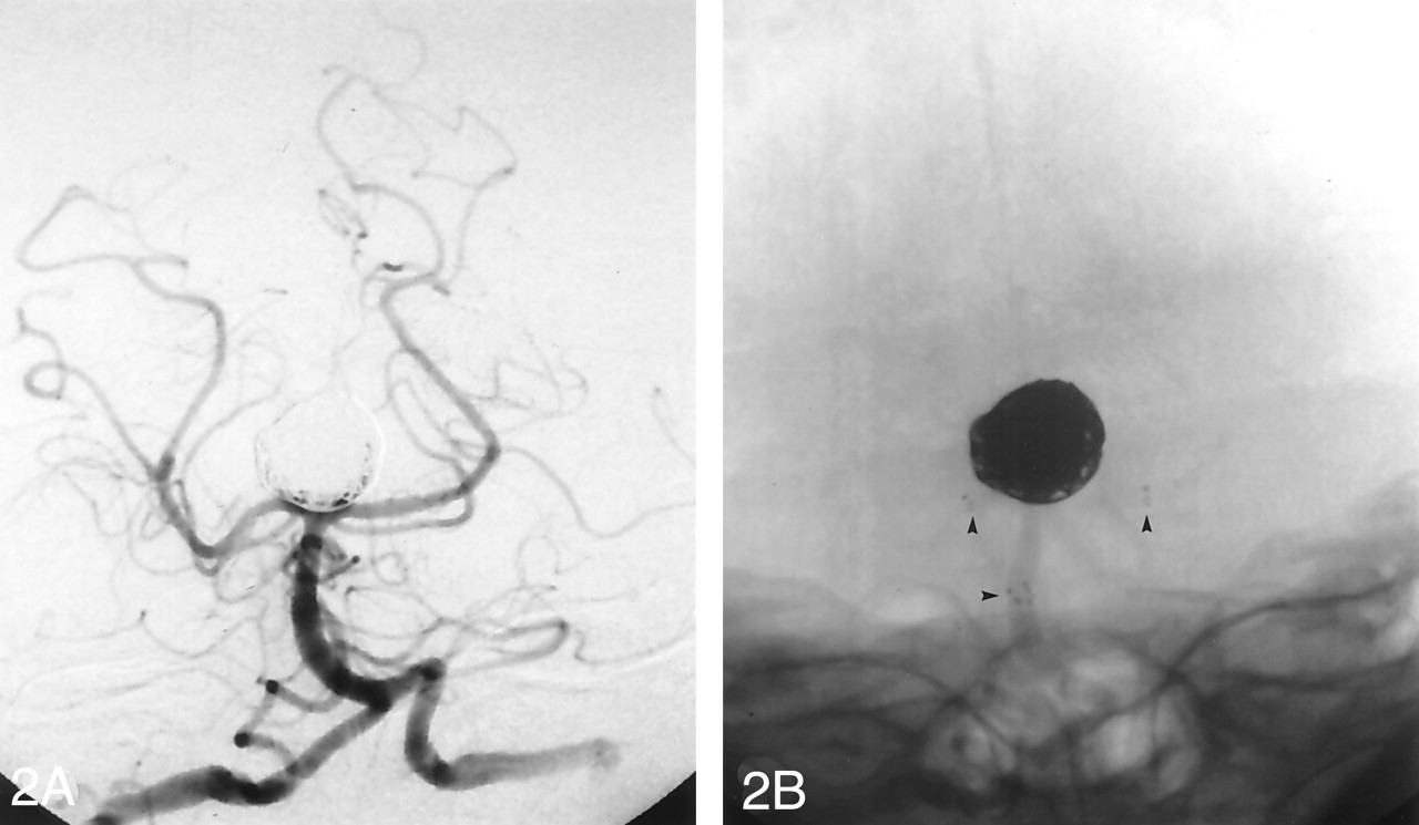
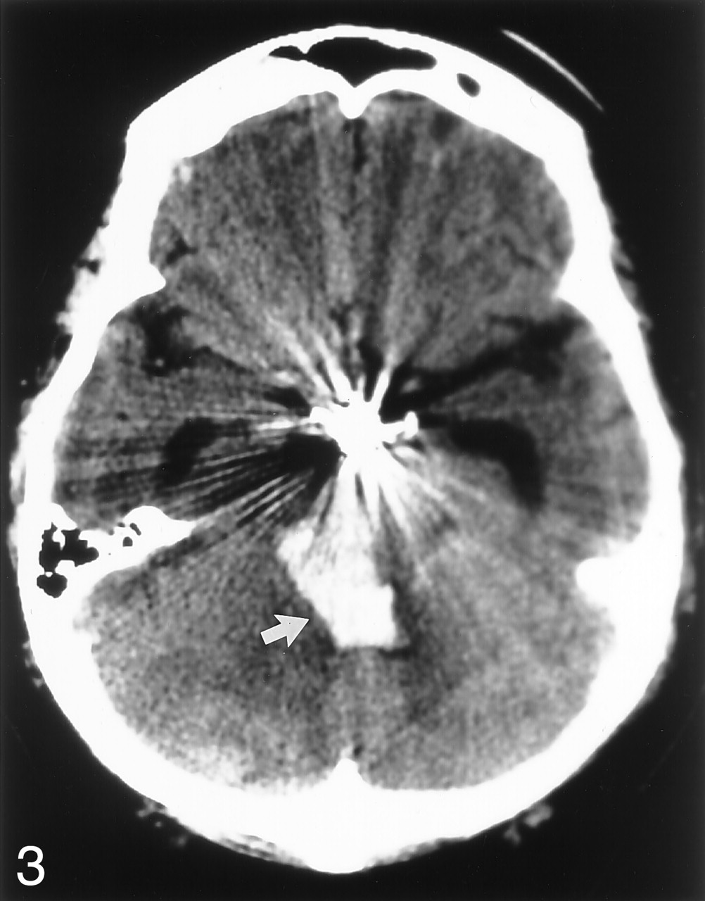
This was achieved by using two 3.5 × 20 mm Neuroform (Boston Scientific, Natick, MA) stents and 386 cm of GDC (Boston Scientific) coils in a fashion similar to the method described by Chow et al (Figs 2). The procedure was performed under general anesthesia and full heparinization, with the standard pretreatment for Neuroform cases including aspirin and clopidigrel. There was near-complete obliteration of the aneurysm with preservation of both posterior cerebral arteries and no evidence of perforation, vessel dissection, or flow-limiting stenosis. The patient awoke from anesthesia with no focal neurologic deficit; however, approximately 6 hours later, she became progressively obtunded with left-sided weakness. A CT head scan showed a midbrain and upper pontine parenchymal hemorrhage (Fig 3), and cerebral angiography showed no change from the postprocedural study. The patient had a limited neurologic recovery but eventually succumbed to chronic hydrocephalus and pneumonia.

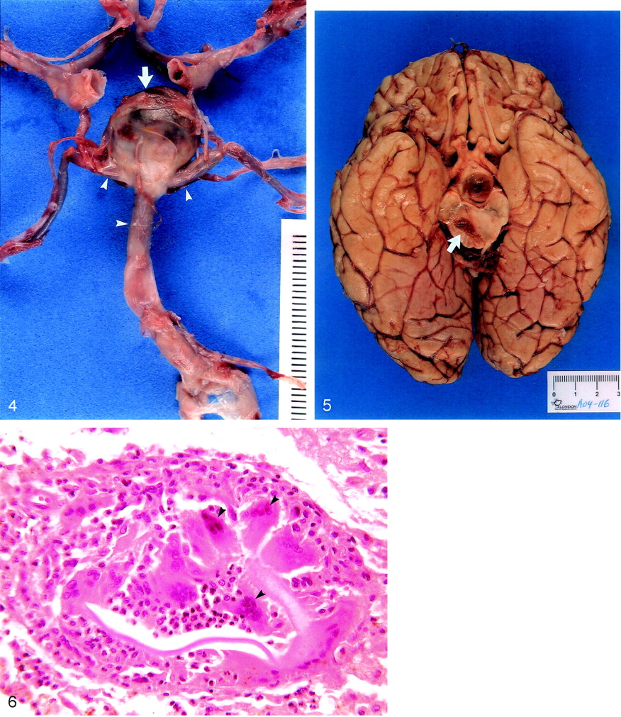
A subsequent review of the procedure showed considerable stretching of the right posterior cerebral artery during guidewire placement and positioning of the first Neuroform stent. There were no other technical problems encountered during the procedure. The postmortem neuropathologic examination showed no bleeding from the aneurysm (Fig 4) and no gross obstruction of pontine perforating vessels by the stents. There was a large right midbrain hemorrhage (Fig 5), with smaller hemorrhages in the right thalamus and right cerebellar hemisphere.

Unusual foreign body granulomas were also seen microscopically (Fig 6) associated with the hemorrhages, possibly due to microembolism from the hardware. Although the cause of the midbrain hemorrhage is unknown, it may be due to stretching of perforating vessels, infarction, and bleeding related to anticoagulation.

Despite an excellent angiographic result, this tragic clinicaloutcome demonstrates the risks inherent in novel, aggressive interventional strategies. This, however, should come as no surprise. As William Shakespeare noted long ago, Diseases desperate grownBy desperate appliance are reliev’dOr not at all (4)

Fig 1.
  • Download figure
  • Open in new tab
  • Download powerpoint
Fig 1.

Right vertebral arteriogram (anteroposterior [AP]), shows a wide-necked basilar bifurcation aneurysm.

Fig 2.
  • Download figure
  • Open in new tab
  • Download powerpoint
Fig 2.

Postoperative arteriograms.

A, Right vertebral arteriogram (AP), obtained after treatment with stents and coils, shows near complete obliteration of the aneurysm.

B, Unsubtracted image obtained from the postoperative arteriogram shows the radiopaque markers at the ends of the stents (arrowheads).

Fig 3.
  • Download figure
  • Open in new tab
  • Download powerpoint
Fig 3.

CT head scan obtained 6 hours after treatment shows the pontine component of the acute brain stem hemorrhage (arrow).

Fig 4.
  • Download figure
  • Open in new tab
  • Download powerpoint
Fig 4.

Postmortem dissection of the circle of Willis, showing the coils inside an intact basilar bifurcation aneurysm (arrow). The stents are visible through the basilar and posterior cerebral arteries (arrowheads).

Fig 5.
  • Download figure
  • Open in new tab
  • Download powerpoint
Fig 5.

Gross specimen. Section through the midbrain shows the hematoma (arrow)

Fig 6.
  • Download figure
  • Open in new tab
  • Download powerpoint
Fig 6.

Microscopic section through the midbrain hemorrhage shows unusual intravascular foreign body granulomas (arrowheads) (H&E, ×8).

References

  1. Chow MM, Woo HH, Masaryk TJ, Rasmussen PA. A novel endovascular treatment of a wide-necked basilar apex aneurysm by using a Y-configuration, double-stent technique. AJNR Am J Neuroradiol 2004;25:509–512
  2. Fiorella D, Albuquerque FC, Han P, McDougall CG. Preliminary experience using the Neuroform stent for the treatment of cerebral aneurysms. Neurosurgery 2004;54:6–16
  3. Broadbent LP, Moran CJ, Cross DT 3rd, Derdeyn CP. Management of Neuroform stent dislodgement and misplacement. AJNR Am J Neuroradiol 2003;24:1819–1822
  4. Shakespeare W. Hamlet. In: The tragedies of Shakespeare. New York: The Modern Library. 2:IV, iii, 9
  • Copyright © American Society of Neuroradiology
Advertisement

Indexed Content

  • Current Issue
  • Accepted Manuscripts
  • Article Preview
  • Past Issues
  • Editorials
  • Editor's Choice
  • Fellows' Journal Club
  • Letters to the Editor
  • Video Articles

Cases

  • Case Collection
  • Archive - Case of the Week
  • Archive - Case of the Month
  • Archive - Classic Case

More from AJNR

  • Trainee Corner
  • Imaging Protocols
  • MRI Safety Corner

Multimedia

  • AJNR Podcasts
  • AJNR Scantastics

Resources

  • Turnaround Time
  • Submit a Manuscript
  • Submit a Video Article
  • Submit an eLetter to the Editor/Response
  • Manuscript Submission Guidelines
  • Statistical Tips
  • Fast Publishing of Accepted Manuscripts
  • Graphical Abstract Preparation
  • Imaging Protocol Submission
  • Evidence-Based Medicine Level Guide
  • Publishing Checklists
  • Author Policies
  • Become a Reviewer/Academy of Reviewers
  • News and Updates

About Us

  • About AJNR
  • Editorial Board
  • Editorial Board Alumni
  • Alerts
  • Permissions
  • Not an AJNR Subscriber? Join Now
  • Advertise with Us
  • Librarian Resources
  • Feedback
  • Terms and Conditions
  • AJNR Editorial Board Alumni

American Society of Neuroradiology

  • Not an ASNR Member? Join Now

© 2025 by the American Society of Neuroradiology All rights, including for text and data mining, AI training, and similar technologies, are reserved.
Print ISSN: 0195-6108 Online ISSN: 1936-959X

Powered by HighWire