Advertisement

Main menu

  • Home
  • Content
    • Current Issue
    • Accepted Manuscripts
    • Article Preview
    • Past Issue Archive
    • Video Articles
    • AJNR Case Collection
    • Case of the Week Archive
    • Case of the Month Archive
    • Classic Case Archive
  • Special Collections
    • AJNR Awards
    • Low-Field MRI
    • Alzheimer Disease
    • ASNR Foundation Special Collection
    • Photon-Counting CT
    • View All
  • Multimedia
    • AJNR Podcasts
    • AJNR SCANtastic
    • Trainee Corner
    • MRI Safety Corner
    • Imaging Protocols
  • For Authors
    • Submit a Manuscript
    • Submit a Video Article
    • Submit an eLetter to the Editor/Response
    • Manuscript Submission Guidelines
    • Statistical Tips
    • Fast Publishing of Accepted Manuscripts
    • Graphical Abstract Preparation
    • Imaging Protocol Submission
    • Author Policies
  • About Us
    • About AJNR
    • Editorial Board
    • Editorial Board Alumni
  • More
    • Become a Reviewer/Academy of Reviewers
    • Subscribers
    • Permissions
    • Alerts
    • Feedback
    • Advertisers
    • ASNR Home

User menu

  • Alerts
  • Log in

Search

  • Advanced search
American Journal of Neuroradiology

American Journal of Neuroradiology

ASHNR American Society of Functional Neuroradiology ASHNR American Society of Pediatric Neuroradiology ASSR
  • Alerts
  • Log in

Advanced Search

  • Home
  • Content
    • Current Issue
    • Accepted Manuscripts
    • Article Preview
    • Past Issue Archive
    • Video Articles
    • AJNR Case Collection
    • Case of the Week Archive
    • Case of the Month Archive
    • Classic Case Archive
  • Special Collections
    • AJNR Awards
    • Low-Field MRI
    • Alzheimer Disease
    • ASNR Foundation Special Collection
    • Photon-Counting CT
    • View All
  • Multimedia
    • AJNR Podcasts
    • AJNR SCANtastic
    • Trainee Corner
    • MRI Safety Corner
    • Imaging Protocols
  • For Authors
    • Submit a Manuscript
    • Submit a Video Article
    • Submit an eLetter to the Editor/Response
    • Manuscript Submission Guidelines
    • Statistical Tips
    • Fast Publishing of Accepted Manuscripts
    • Graphical Abstract Preparation
    • Imaging Protocol Submission
    • Author Policies
  • About Us
    • About AJNR
    • Editorial Board
    • Editorial Board Alumni
  • More
    • Become a Reviewer/Academy of Reviewers
    • Subscribers
    • Permissions
    • Alerts
    • Feedback
    • Advertisers
    • ASNR Home
  • Follow AJNR on Twitter
  • Visit AJNR on Facebook
  • Follow AJNR on Instagram
  • Join AJNR on LinkedIn
  • RSS Feeds

AJNR Awards, New Junior Editors, and more. Read the latest AJNR updates

Third Ventricle Choroid Plexus Papilloma

  • Background:
    • Choroid plexus papillomas (CPP) are rare, benign (WHO grade 1) neuroepithelial intraventricular neoplasms that occur in both pediatric and adult populations. They are especially rare in the third ventricle.
    • Choroid plexus tumors account for less than 1% of all intracranial neoplasms and 2–4% of brain tumors in children, most commonly occurring in the first year of life. In pediatric populations, 80% arise in the lateral ventricle (atrium), 16% in the fourth ventricle, and 4–5% in the third ventricle.
    • CPPs are far more common than the aggressive choroid plexus carcinomas.
  • Clinical Presentation:
    • Presenting symptoms of choroid plexus tumors are typically related to elevated intracranial pressure and hydrocephalus, either from obstruction of CSF flow or CSF overproduction. Signs and symptoms include enlarging head circumference, bulging fontanelle, headache, vomiting, irritability, papilledema, ataxia, and seizure.
  • Key Diagnostic Features:
    • Typical CT findings are an isodense or hyperdense lobular tumor with postcontrast enhancement. Hydrocephalus is common. Calcifications have been reported to occur in 25–50% of patients.
    • CT angiography may show choroidal artery enlargement in lateral ventricle CPPs.
    • On MR imaging, CPPs are typically lobular and iso- to hyperintense on T1 and isointense on T2 sequences. T2 may also show vascular flow voids. The tumors typically show avid gadolinium enhancement, with occasional cystic foci and small areas of necrosis. FLAIR may show transependymal interstitial CSF flow due to obstructive hydrocephalus.
    • Conventional angiography may show a vascular tumor with prolonged blush and enlarged choroidal artery.
  • Differential Diagnosis:
    • Compared with the more commonly occurring colloid cyst, choroid plexus papillomas typically show homogeneous enhancement. Colloid cysts will usually not enhance but can rarely have rim enhancement. As in this case, CPPs may have irregular margins, while colloid cysts should have sharp, well-defined margins.
    • It is difficult to distinguish choroid plexus papillomas from carcinomas by imaging. Carcinomas can have more heterogeneous enhancement. Invasion of brain parenchyma may also be a clue.
    • Subependymal giant cell astrocytomas are enhancing tumors that occur near the foramen of Monro in the setting of tuberous sclerosis complex.
  • Treatment:
    • Surgical resection is the treatment of choice, with curative rates of 100%. Total resection can be achieved in the majority of cases, and the tumors rarely recur. Early case series reported operative complications and perioperative mortality due to excessive blood loss. Due to the vascular nature of the tumor, preoperative embolization has been attempted. Subdural fluid collections may occur postoperatively.
October 2015

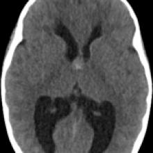
A 9-year-old girl with progressively worsening headaches over 4 months and papilledema seen at recent eye exam

View Case
  • Read more about 2015oct
  • Comments
Advertisement
Subscribe to RSS - Third Ventricle Choroid Plexus Papilloma

Indexed Content

  • Current Issue
  • Accepted Manuscripts
  • Article Preview
  • Past Issues
  • Editorials
  • Editor's Choice
  • Fellows' Journal Club
  • Letters to the Editor
  • Video Articles

Cases

  • Case Collection
  • Archive - Case of the Week
  • Archive - Case of the Month
  • Archive - Classic Case

More from AJNR

  • Trainee Corner
  • Imaging Protocols
  • MRI Safety Corner
  • Book Reviews

Multimedia

  • AJNR Podcasts
  • AJNR Scantastics

Resources

  • Turnaround Time
  • Submit a Manuscript
  • Submit a Video Article
  • Submit an eLetter to the Editor/Response
  • Manuscript Submission Guidelines
  • Statistical Tips
  • Fast Publishing of Accepted Manuscripts
  • Graphical Abstract Preparation
  • Imaging Protocol Submission
  • Evidence-Based Medicine Level Guide
  • Publishing Checklists
  • Author Policies
  • Become a Reviewer/Academy of Reviewers
  • News and Updates

About Us

  • About AJNR
  • Editorial Board
  • Editorial Board Alumni
  • Alerts
  • Permissions
  • Not an AJNR Subscriber? Join Now
  • Advertise with Us
  • Librarian Resources
  • Feedback
  • Terms and Conditions
  • AJNR Editorial Board Alumni

American Society of Neuroradiology

  • Not an ASNR Member? Join Now

© 2025 by the American Society of Neuroradiology All rights, including for text and data mining, AI training, and similar technologies, are reserved.
Print ISSN: 0195-6108 Online ISSN: 1936-959X

Powered by HighWire