Advertisement

Main menu

  • Home
  • Content
    • Current Issue
    • Accepted Manuscripts
    • Article Preview
    • Past Issue Archive
    • Video Articles
    • AJNR Case Collection
    • Case of the Week Archive
    • Case of the Month Archive
    • Classic Case Archive
  • Special Collections
    • AJNR Awards
    • Low-Field MRI
    • Alzheimer Disease
    • ASNR Foundation Special Collection
    • Photon-Counting CT
    • View All
  • Multimedia
    • AJNR Podcasts
    • AJNR SCANtastic
    • Trainee Corner
    • MRI Safety Corner
    • Imaging Protocols
  • For Authors
    • Submit a Manuscript
    • Submit a Video Article
    • Submit an eLetter to the Editor/Response
    • Manuscript Submission Guidelines
    • Statistical Tips
    • Fast Publishing of Accepted Manuscripts
    • Graphical Abstract Preparation
    • Imaging Protocol Submission
    • Author Policies
  • About Us
    • About AJNR
    • Editorial Board
    • Editorial Board Alumni
  • More
    • Become a Reviewer/Academy of Reviewers
    • Subscribers
    • Permissions
    • Alerts
    • Feedback
    • Advertisers
    • ASNR Home

User menu

  • Alerts
  • Log in

Search

  • Advanced search
American Journal of Neuroradiology

American Journal of Neuroradiology

ASHNR American Society of Functional Neuroradiology ASHNR American Society of Pediatric Neuroradiology ASSR
  • Alerts
  • Log in

Advanced Search

  • Home
  • Content
    • Current Issue
    • Accepted Manuscripts
    • Article Preview
    • Past Issue Archive
    • Video Articles
    • AJNR Case Collection
    • Case of the Week Archive
    • Case of the Month Archive
    • Classic Case Archive
  • Special Collections
    • AJNR Awards
    • Low-Field MRI
    • Alzheimer Disease
    • ASNR Foundation Special Collection
    • Photon-Counting CT
    • View All
  • Multimedia
    • AJNR Podcasts
    • AJNR SCANtastic
    • Trainee Corner
    • MRI Safety Corner
    • Imaging Protocols
  • For Authors
    • Submit a Manuscript
    • Submit a Video Article
    • Submit an eLetter to the Editor/Response
    • Manuscript Submission Guidelines
    • Statistical Tips
    • Fast Publishing of Accepted Manuscripts
    • Graphical Abstract Preparation
    • Imaging Protocol Submission
    • Author Policies
  • About Us
    • About AJNR
    • Editorial Board
    • Editorial Board Alumni
  • More
    • Become a Reviewer/Academy of Reviewers
    • Subscribers
    • Permissions
    • Alerts
    • Feedback
    • Advertisers
    • ASNR Home
  • Follow AJNR on Twitter
  • Visit AJNR on Facebook
  • Follow AJNR on Instagram
  • Join AJNR on LinkedIn
  • RSS Feeds

AJNR Awards, New Junior Editors, and more. Read the latest AJNR updates

Papillary Tumor of the Pineal Region (PTPR)

  • Background:
    • PTPR is a rare neuroepithelial tumor that arises from specialized ependymocites in the subcommissural organ, located in the posteroinferior wall of the third ventricle (pineal region).
    • Recognized neoplasm in the WHO 2007 classification; grading criterion has yet to be defined, but in the 2016 WHO, PTPR corresponds to grade II or III neoplasms.
    • Only a few PTPRs with imaging findings have been reported.
    • The immunohistochemical findings help differentiate PTPR from other lesions in the pineal region, such as pineal parenchymal tumor of intermediate differentiation (PPTID).
  • Clinical Presentation:
    • PTPR occurs in both children and adults, but most cases are seen in adults (mean age at diagnosis is 32 years).
    • PTPR can compress the tectum and cerebral aqueduct, causing hydrocephalus.
  • Key Diagnostic Features:
    • PTPRs tend to be large and lobulated, relatively not well circumscribed, and partially cystic.
    • MRI: T1WI variable signal intensity (intrinsic hyperintensity has been described); T2WI isointense or hyperintense and heterogeneous postcontrast enhancement; restricted diffusion on DWI
    • In such scenarios, it is necessary to screen the entire neuraxis to assess for CSF spread.
    • Radiologic findings are nonspecific in most patients. PTPRs are easily differentiated microscopically, showing distinct papillary architecture with pseudostratified columnar epithelium.
  • Differential Diagnoses:
    • PPTID: No radiologic features that would distinguish these tumors
    • Pineocytoma: Well-demarcated round or nodular masses; the imaging appearance is less “aggressive” than a PTPR. MRI: Iso-hypointense on T1WI, hyperintense on T2WI, and variable enhancement
    • Metastasis: Metastases to the pineal gland are rare (prevalence of 0.4%–3.8%), but may be present without metastases to brain parenchyma. Can be indistinguishable on imaging studies from primary pineal neoplasms.
    • Germinoma: More than 90% of patients are younger than 20 years at initial diagnosis. MRI: Iso-hyperintense on T2WI, with intense and homogeneous enhancement. Dissemination by CSF and invasion of the adjacent brain are also typical.
    • Brainstem astrocytoma with extension to the pineal region: Those that take place in the region of the tectum are usually low grade (WHO I–II) and occur more frequently in childhood. MRI: Bulbous enlargement of the tectal plate; isointense on T1WI and hyperintense on T2WI with no to minimal enhancement.
  • Treatment:
    • Surgical resection if it is possible
    • The clinical course of PTPR is characterized by frequent local recurrence.
    • The value of radiotherapy on disease progression in PTPR will need to be investigated in a future prospective trial, which will also provide data on long-term follow-up and may further aid the identification of histopathologic factors possibly associated with prognosis in PTPR.
November 11, 2021

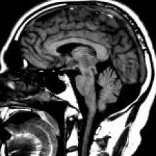
A 56-year-old man with 2 months of headaches and progressive mild left-sided weakness; MRI studies are shown below.

View Case
  • Read more about 11112021
  • Comments
Advertisement
Subscribe to RSS - Papillary Tumor of the Pineal Region (PTPR)

Indexed Content

  • Current Issue
  • Accepted Manuscripts
  • Article Preview
  • Past Issues
  • Editorials
  • Editor's Choice
  • Fellows' Journal Club
  • Letters to the Editor
  • Video Articles

Cases

  • Case Collection
  • Archive - Case of the Week
  • Archive - Case of the Month
  • Archive - Classic Case

More from AJNR

  • Trainee Corner
  • Imaging Protocols
  • MRI Safety Corner
  • Book Reviews

Multimedia

  • AJNR Podcasts
  • AJNR Scantastics

Resources

  • Turnaround Time
  • Submit a Manuscript
  • Submit a Video Article
  • Submit an eLetter to the Editor/Response
  • Manuscript Submission Guidelines
  • Statistical Tips
  • Fast Publishing of Accepted Manuscripts
  • Graphical Abstract Preparation
  • Imaging Protocol Submission
  • Evidence-Based Medicine Level Guide
  • Publishing Checklists
  • Author Policies
  • Become a Reviewer/Academy of Reviewers
  • News and Updates

About Us

  • About AJNR
  • Editorial Board
  • Editorial Board Alumni
  • Alerts
  • Permissions
  • Not an AJNR Subscriber? Join Now
  • Advertise with Us
  • Librarian Resources
  • Feedback
  • Terms and Conditions
  • AJNR Editorial Board Alumni

American Society of Neuroradiology

  • Not an ASNR Member? Join Now

© 2025 by the American Society of Neuroradiology All rights, including for text and data mining, AI training, and similar technologies, are reserved.
Print ISSN: 0195-6108 Online ISSN: 1936-959X

Powered by HighWire