Advertisement

Main menu

  • Home
  • Content
    • Current Issue
    • Accepted Manuscripts
    • Article Preview
    • Past Issue Archive
    • Video Articles
    • AJNR Case Collection
    • Case of the Week Archive
    • Case of the Month Archive
    • Classic Case Archive
  • Special Collections
    • AJNR Awards
    • Low-Field MRI
    • Alzheimer Disease
    • ASNR Foundation Special Collection
    • Photon-Counting CT
    • View All
  • Multimedia
    • AJNR Podcasts
    • AJNR SCANtastic
    • Trainee Corner
    • MRI Safety Corner
    • Imaging Protocols
  • For Authors
    • Submit a Manuscript
    • Submit a Video Article
    • Submit an eLetter to the Editor/Response
    • Manuscript Submission Guidelines
    • Statistical Tips
    • Fast Publishing of Accepted Manuscripts
    • Graphical Abstract Preparation
    • Imaging Protocol Submission
    • Author Policies
  • About Us
    • About AJNR
    • Editorial Board
    • Editorial Board Alumni
  • More
    • Become a Reviewer/Academy of Reviewers
    • Subscribers
    • Permissions
    • Alerts
    • Feedback
    • Advertisers
    • ASNR Home

User menu

  • Alerts
  • Log in

Search

  • Advanced search
American Journal of Neuroradiology

American Journal of Neuroradiology

ASHNR American Society of Functional Neuroradiology ASHNR American Society of Pediatric Neuroradiology ASSR
  • Alerts
  • Log in

Advanced Search

  • Home
  • Content
    • Current Issue
    • Accepted Manuscripts
    • Article Preview
    • Past Issue Archive
    • Video Articles
    • AJNR Case Collection
    • Case of the Week Archive
    • Case of the Month Archive
    • Classic Case Archive
  • Special Collections
    • AJNR Awards
    • Low-Field MRI
    • Alzheimer Disease
    • ASNR Foundation Special Collection
    • Photon-Counting CT
    • View All
  • Multimedia
    • AJNR Podcasts
    • AJNR SCANtastic
    • Trainee Corner
    • MRI Safety Corner
    • Imaging Protocols
  • For Authors
    • Submit a Manuscript
    • Submit a Video Article
    • Submit an eLetter to the Editor/Response
    • Manuscript Submission Guidelines
    • Statistical Tips
    • Fast Publishing of Accepted Manuscripts
    • Graphical Abstract Preparation
    • Imaging Protocol Submission
    • Author Policies
  • About Us
    • About AJNR
    • Editorial Board
    • Editorial Board Alumni
  • More
    • Become a Reviewer/Academy of Reviewers
    • Subscribers
    • Permissions
    • Alerts
    • Feedback
    • Advertisers
    • ASNR Home
  • Follow AJNR on Twitter
  • Visit AJNR on Facebook
  • Follow AJNR on Instagram
  • Join AJNR on LinkedIn
  • RSS Feeds

AJNR Awards, New Junior Editors, and more. Read the latest AJNR updates

Levamisole Leukoencephalopathy

•Background:
  • Levamisole is an adulterant that has been reported in 69% of cocaine entering the United States (Drug Enforcement Agency), and is also used for adjuvant chemotherapy or recurrent aphthous ulcer treatment.
  • Although the mechanism is unclear, it is hypothesized that levamisole may activate macrophages and lymphocytes, triggering a delayed hypersensitivity reaction that causes inflammation and demyelination.
•Clinical Presentation:
  • Variable, but may include confusion, aphasia/dysphasia, gait ataxia, hemiparesis, cognitive impairment, diplopia, facial palsy, incontinence, or parasthesia.
  • In this case, the patient presented with levamisole-induced leukoencephalopathy and cutaneous vasculopathy, along with a neuroleptic malignant syndrome-type appearance from cocaine use.
•Key Diagnostic Features:
  • Labs: Urine toxicology, CSF studies demonstrating lymphocytic pleocytosis +/- red blood cells or oligoclonal bands
  • Imaging: MRI with predominantly T2/FLAIR hyperintense, ovoid white matter lesions, often supratentorial in the periventricular or subcortical areas, along with corresponding T1 hypointensities, +/- restricted diffusion on DWI and ring enhancement. CT may initially be normal, but over time progressive hypodense lesions can be seen.
  • Brain biopsy: Demyelination with infiltration of macrophages and perivascular lymphocytes  
•Differential Diagnosis:
  • Multiple sclerosis: Periventricular and subcortical white matter lesions in perivenular distribution, with active lesions demonstrating a “leading edge” of incomplete peripheral enhancement and restricted diffusion. Optic nerve involvement is common.
  • Posterior reversible encephalopathy syndrome (PRES): Patchy subcortical white matter lesions commonly involve parietal and occipital lobes. Diffusion restriction on DWI is rare.
  • Acute disseminated encephalomyelitis: Postinfectious clinical course with self-resolving lesions. Usually deep and subcortical white matter is involved, while periventricular lesions are less common.
  • Progressive multifocal leukoencephalopathy: Seen in immunocompromised patients with JC virus reactivation. Patchy, asymmetric T2 hyperintense lesions typically in the parieto-occipital white matter with prominent subcortical U-fiber involvement. Usually non-enhancing, +/- surrounding punctate lesions (“Milky Way sign”) and corpus callosum involvement. Little to no mass effect.
  • CNS lymphoma: Hyperdense on CT  with solid enhancement on MRI and restricted diffusion.
  • Glioblastoma: Irregular mass lesion with vasogenic edema, and enhancement and restricted diffusion of solid component. Necrosis and hemorrhage are common. 
•Treatment:
  • Currently no definitive treatment.
  • Discontinuation of levamisole-containing cocaine and supportive care including steroids, plasmapheresis, and/or IVIG have been shown to improve clinical courses in case reports.
October 27, 2022

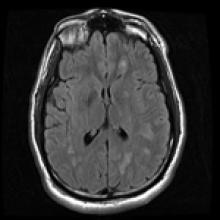
A 30-year-old man with 2 days of headaches and malaise, is found down by roommate. In the emergency department, he is found to have altered mental status, mutism, fever, tachycardia, hypertension, rigidity, hyperreflexia, and a purpuric rash.

View Case
  • Read more about 10272022
  • Comments
Advertisement
Subscribe to RSS - Levamisole Leukoencephalopathy

Indexed Content

  • Current Issue
  • Accepted Manuscripts
  • Article Preview
  • Past Issues
  • Editorials
  • Editor's Choice
  • Fellows' Journal Club
  • Letters to the Editor
  • Video Articles

Cases

  • Case Collection
  • Archive - Case of the Week
  • Archive - Case of the Month
  • Archive - Classic Case

More from AJNR

  • Trainee Corner
  • Imaging Protocols
  • MRI Safety Corner
  • Book Reviews

Multimedia

  • AJNR Podcasts
  • AJNR Scantastics

Resources

  • Turnaround Time
  • Submit a Manuscript
  • Submit a Video Article
  • Submit an eLetter to the Editor/Response
  • Manuscript Submission Guidelines
  • Statistical Tips
  • Fast Publishing of Accepted Manuscripts
  • Graphical Abstract Preparation
  • Imaging Protocol Submission
  • Evidence-Based Medicine Level Guide
  • Publishing Checklists
  • Author Policies
  • Become a Reviewer/Academy of Reviewers
  • News and Updates

About Us

  • About AJNR
  • Editorial Board
  • Editorial Board Alumni
  • Alerts
  • Permissions
  • Not an AJNR Subscriber? Join Now
  • Advertise with Us
  • Librarian Resources
  • Feedback
  • Terms and Conditions
  • AJNR Editorial Board Alumni

American Society of Neuroradiology

  • Not an ASNR Member? Join Now

© 2025 by the American Society of Neuroradiology All rights, including for text and data mining, AI training, and similar technologies, are reserved.
Print ISSN: 0195-6108 Online ISSN: 1936-959X

Powered by HighWire