Advertisement

Main menu

  • Home
  • Content
    • Current Issue
    • Accepted Manuscripts
    • Article Preview
    • Past Issue Archive
    • Video Articles
    • AJNR Case Collection
    • Case of the Week Archive
    • Case of the Month Archive
    • Classic Case Archive
  • Special Collections
    • AJNR Awards
    • Low-Field MRI
    • Alzheimer Disease
    • ASNR Foundation Special Collection
    • Photon-Counting CT
    • View All
  • Multimedia
    • AJNR Podcasts
    • AJNR SCANtastic
    • Trainee Corner
    • MRI Safety Corner
    • Imaging Protocols
  • For Authors
    • Submit a Manuscript
    • Submit a Video Article
    • Submit an eLetter to the Editor/Response
    • Manuscript Submission Guidelines
    • Statistical Tips
    • Fast Publishing of Accepted Manuscripts
    • Graphical Abstract Preparation
    • Imaging Protocol Submission
    • Author Policies
  • About Us
    • About AJNR
    • Editorial Board
    • Editorial Board Alumni
  • More
    • Become a Reviewer/Academy of Reviewers
    • Subscribers
    • Permissions
    • Alerts
    • Feedback
    • Advertisers
    • ASNR Home

User menu

  • Alerts
  • Log in

Search

  • Advanced search
American Journal of Neuroradiology

American Journal of Neuroradiology

ASHNR American Society of Functional Neuroradiology ASHNR American Society of Pediatric Neuroradiology ASSR
  • Alerts
  • Log in

Advanced Search

  • Home
  • Content
    • Current Issue
    • Accepted Manuscripts
    • Article Preview
    • Past Issue Archive
    • Video Articles
    • AJNR Case Collection
    • Case of the Week Archive
    • Case of the Month Archive
    • Classic Case Archive
  • Special Collections
    • AJNR Awards
    • Low-Field MRI
    • Alzheimer Disease
    • ASNR Foundation Special Collection
    • Photon-Counting CT
    • View All
  • Multimedia
    • AJNR Podcasts
    • AJNR SCANtastic
    • Trainee Corner
    • MRI Safety Corner
    • Imaging Protocols
  • For Authors
    • Submit a Manuscript
    • Submit a Video Article
    • Submit an eLetter to the Editor/Response
    • Manuscript Submission Guidelines
    • Statistical Tips
    • Fast Publishing of Accepted Manuscripts
    • Graphical Abstract Preparation
    • Imaging Protocol Submission
    • Author Policies
  • About Us
    • About AJNR
    • Editorial Board
    • Editorial Board Alumni
  • More
    • Become a Reviewer/Academy of Reviewers
    • Subscribers
    • Permissions
    • Alerts
    • Feedback
    • Advertisers
    • ASNR Home
  • Follow AJNR on Twitter
  • Visit AJNR on Facebook
  • Follow AJNR on Instagram
  • Join AJNR on LinkedIn
  • RSS Feeds

AJNR Awards, New Junior Editors, and more. Read the latest AJNR updates

Erdheim-Chester Disease (ECD) with Involvement of the Central Nervous System

  • Background:
    • ECD is a rare non-Langerhans cell histiocytosis that typically affects middle-aged patients and is slightly more common in men.
    • Histopathologic assessment reveals tissue infiltration by lipid-laden histiocytes and an immunohistochemical profile distinct from Langerhans cell histiocytosis.
    • Multiple somatic mutations are implicated in ECD, most commonly BRAF V600E.
    • It is a multisystem disease with common involvement of the musculoskeletal system, CNS, orbits, retroperitoneum, pericardium, periaortic region, and pulmonary interstitium.
  • Clinical Presentation:
    • Symptoms vary according to the organ and extent of involvement.
    • CNS involvement often affects the hypothalamic-pituitary axis (HPA) and may present with diabetes insipidus.
    • Additionally, ataxia is a common symptom owing to involvement of posterior fossa structures, particularly the cerebellar dentate nuclei.
    • Orbital involvement is seen in about one-fourth of patients and can present with exophthalmos, retroorbital pain, extraocular muscle palsies, and progressive vision loss.
    • Additional common symptoms include bone pain, weakness and fatigue, cough and progressive respiratory failure, ureteral obstruction, renal impairment, cardiac dysfunction and tamponade, and xanthelasmas and xanthomas.
  • Key Diagnostic Features:
    • Intracranial disease may be intra-axial and/or extra-axial and is commonly multifocal.
    • Extra-axial lesions may take a plaquelike morphology.
    • Intra-axial lesions are often T2 hyperintense, but extra-axial and intraorbital lesions are more variable and may be T2 hypointense.
    • Avid postcontrast enhancement is typical.
    • CNS findings are generally nonspecific, but the additional findings of sclerotic bone lesions and pericardial, pulmonary interstitial, and retroperitoneal infiltrative soft tissue thickening should raise suspicion for this disease.
  • Differential Diagnoses:
    • Langerhans cell histiocytosis (LCH): CNS disease in LCH may be identical to ECD and also has a propensity for involvement of the HPA. Unlike ECD, LCH typically results in lytic bone lesions, rarely results in bilateral orbital involvement, and more commonly affects children.
    • Rosai-Dorfman disease (RDD): CNS and orbital manifestations of RDD may be identical to ECD. Unlike ECD, RDD characteristically presents with cervical lymphadenopathy and more commonly affects children and young adults.
    • Listeria rhomboencephalitis: Like intra-axial disease in ECD, Listeria rhomboencephalitis may present with T2-hyperintense lesions with patchy postcontrast enhancement involving the brainstem and cerebellum. Unlike ECD, Listeria rhomboencephalitis may result in subcortical abscesses and has a more acutely progressive clinical course.
    • Behçet disease: CNS involvement of Behçet disease typically manifests as T2-hyperintense, enhancing lesions involving the brainstem, basal ganglia, and thalami. Unlike ECD, neuro-Behçet disease is typically preceded by the findings of oral and genital ulcers and has an association with cerebral vein thrombosis.
    • Chronic lymphocytic inflammation with pontine perivascular enhancement responsive to steroids (CLIPPERS): Stippled and patchy enhancement is seen involving the pons but may also involve the cerebellar peduncles, cerebellar hemispheres, and cervical spinal cord. Unlike ECD, there is no orbital or systemic involvement.
    • Myelin oligodendrocyte glycoprotein​ antibody-associated disease (MOGAD): Intracranial findings are variable, but characteristically include T2-hyperintense lesions with variable enhancement and restricted diffusion involving the thalami, pons, and cerebellar peduncles. Coexistent optic neuritis and longitudinally extensive spinal cord lesions are common.
    • Neurosarcoidosis and lymphoma: Multifocal intra-axial and extra-axial enhancing lesions as well as orbital involvement may be seen, similar to ECD. Intra-axial lesions in lymphoma are more typically T2 hypointense and demonstrate restricted diffusion. Multisystem involvement is common to all 3 diseases, often requiring tissue sampling for differentiation. 
  • Treatment:
    • Management is reserved for symptomatic disease.
    • Current treatment approaches target underlying mutations when possible. For example, the BRAF inhibitor vemurafenib is an initial management option for patients with BRAF V600E mutations.
    • Interferon alpha may also be used when genetic mutation–targeted therapy is not possible.
June 30, 2022

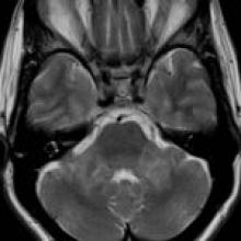
A 56-year-old woman with a several-year history of progressive dizziness, hearing loss, diplopia, gait instability, emotional lability, and fatigue

View Case
  • Read more about 06302022
  • Comments
Advertisement
Subscribe to RSS - Erdheim-Chester Disease (ECD) with Involvement of the Central Nervous System

Indexed Content

  • Current Issue
  • Accepted Manuscripts
  • Article Preview
  • Past Issues
  • Editorials
  • Editor's Choice
  • Fellows' Journal Club
  • Letters to the Editor
  • Video Articles

Cases

  • Case Collection
  • Archive - Case of the Week
  • Archive - Case of the Month
  • Archive - Classic Case

More from AJNR

  • Trainee Corner
  • Imaging Protocols
  • MRI Safety Corner
  • Book Reviews

Multimedia

  • AJNR Podcasts
  • AJNR Scantastics

Resources

  • Turnaround Time
  • Submit a Manuscript
  • Submit a Video Article
  • Submit an eLetter to the Editor/Response
  • Manuscript Submission Guidelines
  • Statistical Tips
  • Fast Publishing of Accepted Manuscripts
  • Graphical Abstract Preparation
  • Imaging Protocol Submission
  • Evidence-Based Medicine Level Guide
  • Publishing Checklists
  • Author Policies
  • Become a Reviewer/Academy of Reviewers
  • News and Updates

About Us

  • About AJNR
  • Editorial Board
  • Editorial Board Alumni
  • Alerts
  • Permissions
  • Not an AJNR Subscriber? Join Now
  • Advertise with Us
  • Librarian Resources
  • Feedback
  • Terms and Conditions
  • AJNR Editorial Board Alumni

American Society of Neuroradiology

  • Not an ASNR Member? Join Now

© 2025 by the American Society of Neuroradiology All rights, including for text and data mining, AI training, and similar technologies, are reserved.
Print ISSN: 0195-6108 Online ISSN: 1936-959X

Powered by HighWire