Advertisement

Main menu

  • Home
  • Content
    • Current Issue
    • Accepted Manuscripts
    • Article Preview
    • Past Issue Archive
    • Video Articles
    • AJNR Case Collection
    • Case of the Week Archive
    • Case of the Month Archive
    • Classic Case Archive
  • Special Collections
    • AJNR Awards
    • Low-Field MRI
    • Alzheimer Disease
    • ASNR Foundation Special Collection
    • Photon-Counting CT
    • View All
  • Multimedia
    • AJNR Podcasts
    • AJNR SCANtastic
    • Trainee Corner
    • MRI Safety Corner
    • Imaging Protocols
  • For Authors
    • Submit a Manuscript
    • Submit a Video Article
    • Submit an eLetter to the Editor/Response
    • Manuscript Submission Guidelines
    • Statistical Tips
    • Fast Publishing of Accepted Manuscripts
    • Graphical Abstract Preparation
    • Imaging Protocol Submission
    • Author Policies
  • About Us
    • About AJNR
    • Editorial Board
    • Editorial Board Alumni
  • More
    • Become a Reviewer/Academy of Reviewers
    • Subscribers
    • Permissions
    • Alerts
    • Feedback
    • Advertisers
    • ASNR Home

User menu

  • Alerts
  • Log in

Search

  • Advanced search
American Journal of Neuroradiology

American Journal of Neuroradiology

ASHNR American Society of Functional Neuroradiology ASHNR American Society of Pediatric Neuroradiology ASSR
  • Alerts
  • Log in

Advanced Search

  • Home
  • Content
    • Current Issue
    • Accepted Manuscripts
    • Article Preview
    • Past Issue Archive
    • Video Articles
    • AJNR Case Collection
    • Case of the Week Archive
    • Case of the Month Archive
    • Classic Case Archive
  • Special Collections
    • AJNR Awards
    • Low-Field MRI
    • Alzheimer Disease
    • ASNR Foundation Special Collection
    • Photon-Counting CT
    • View All
  • Multimedia
    • AJNR Podcasts
    • AJNR SCANtastic
    • Trainee Corner
    • MRI Safety Corner
    • Imaging Protocols
  • For Authors
    • Submit a Manuscript
    • Submit a Video Article
    • Submit an eLetter to the Editor/Response
    • Manuscript Submission Guidelines
    • Statistical Tips
    • Fast Publishing of Accepted Manuscripts
    • Graphical Abstract Preparation
    • Imaging Protocol Submission
    • Author Policies
  • About Us
    • About AJNR
    • Editorial Board
    • Editorial Board Alumni
  • More
    • Become a Reviewer/Academy of Reviewers
    • Subscribers
    • Permissions
    • Alerts
    • Feedback
    • Advertisers
    • ASNR Home
  • Follow AJNR on Twitter
  • Visit AJNR on Facebook
  • Follow AJNR on Instagram
  • Join AJNR on LinkedIn
  • RSS Feeds

AJNR Awards, New Junior Editors, and more. Read the latest AJNR updates

Adult-Onset Leigh Syndrome Presenting with Encephalopathy and Peripheral Neuropathy Caused by a Homoplasmic MT-ATP6 Gene Mutation

  • Background:
    • Leigh syndrome (LS), also known as subacute necrotizing encephalomyelopathy, is a rare (1 in 40,000 births) neurodegenerative mitochondrial disorder characterized by symmetric spongiform lesions in the brain.
    • LS is considered to be a disease of infancy and early childhood, typically reported to present within the first 2 years of life and characteristically presenting during an episode of metabolic stress following a period of normal development. Juvenile or adult forms are uncommon.
    • LS is generally attributed to mutations in mitochondrial or nuclear genes, and most mitochondrial (mt) DNA defects are associated with mutations in the mitochondrial ATPase 6 gene. Frequently reported mutations in the MT-ATP6 gene are m.8993T>C and m.8993T>G, followed by m.9176T>C. Genetic etiology is confirmed in approximately 50% of patients.
  • Clinical Presentation:
    • Clinical features include psychomotor regression with a wide clinical spectrum of neurologic involvement associated with feeding difficulties, intermittent abnormalities of the respiratory rhythm, cranial nerve palsies, dystonia, and ataxia.
    • The age of onset has important implications for disease manifestation. The late-onset LS is generally associated with disorders of movement and coordination, including dystonia, ataxia, and dysarthria. The other atypical signs and symptoms may also occur in late-onset LS, such as intellectual decline, encephalopathy, headache, memory loss, vertical gaze paralysis, visual hallucinations, and peripheral neuropathy.
  • Key Diagnostic Features:
    • Imaging features are heterogeneous and differ depending on the underlying genetic abnormality.
    • The most common characteristic finding on T2-weighted MR images is bilateral, symmetric hyperintensity in the basal ganglia, midbrain and medulla oblongata, periaqueductal gray matter, thalami, subthalamic nuclei, substantia nigra, dentate nuclei, and, rarely, cerebral/cerebellar white matter.
    • The involvement of the mammillary bodies is very unusual.
    • DWI is highly sensitive and may demonstrate transient abnormalities in the bilateral thalamus, internal segments of the globus pallidus, substantia nigra, and pontine tegmentum, even before apparent clinical onset.
    • The "double panda" sign refers to the combination of the "face of the giant panda" in the midbrain and "face of the miniature panda" in the pons, seen as hyperintense signals on T2-weighted images. Although this sign is traditionally considered to be characteristic of Wilson disease, it has also been reported in other metabolic disorders. LS should also be considered in the differential diagnosis of this rare imaging finding.
  • Differential Diagnoses:
    • In our patient, bilateral symmetric T2-hyperintense signal in the basal ganglia, thalami, midbrain, periaqueductal gray matter, and central tegmental tract, as well as symmetric diffusion restriction and mild enhancement in the affected areas, suggested diffuse systemic toxic or metabolic conditions. Causes include LS, Wernicke encephalopathy, methanol poisoning, Creutzfeldt–Jakob disease, CO poisoning, central pontine and extrapontine myelinolysis, biotin-thiamine–responsive basal ganglia disease, and Wilson disease.
    • Muscle biopsy exhibited atrophic muscle fibers and scattered amorphous cytoplasmic aggregates but no vacuoles. Many muscle fibers demonstrated glycogen depletion or abnormal increase in lipid droplets, suggesting a metabolic myopathy.
    • Genetic testing revealed a known pathogenic, 100% homoplasmic missense variant (m.9176T>C, p.Leu217Pro) in the MT-ATP6 gene.
  • Treatment
    • LS is a devastating disease with a very poor prognosis. Death usually occurs in childhood, most frequently due to respiratory failure.
    • The onset age of LS is important in predicting clinical severity or prognosis. Patients with early-onset LS have a poor prognosis compared with those with late-onset LS.
    • Treatment options are limited, with the most common being thiamine or vitamin B1. Ubiquinol, L-carnitine, and coenzyme Q10 may also be used.
    • Diagnostic delay affects the clinical outcome by delaying the possibility to start a treatment and provide genetic counseling.
January 2023

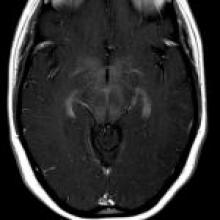
A 21-year-old woman presents with gait abnormality, recurrent falls, hand and lower extremity numbness, and subsequently altered mental status, encephalopathy, and fever. EMG reveals a sensory axonal polyneuropathy affecting mostly the upper limbs, while EEG is normal.

View Case
  • Read more about 2023jan
  • Comments
Advertisement
Subscribe to RSS - Adult-Onset Leigh Syndrome Presenting with Encephalopathy and Peripheral Neuropathy Caused by a Homoplasmic MT-ATP6 Gene Mutation

Indexed Content

  • Current Issue
  • Accepted Manuscripts
  • Article Preview
  • Past Issues
  • Editorials
  • Editor's Choice
  • Fellows' Journal Club
  • Letters to the Editor
  • Video Articles

Cases

  • Case Collection
  • Archive - Case of the Week
  • Archive - Case of the Month
  • Archive - Classic Case

More from AJNR

  • Trainee Corner
  • Imaging Protocols
  • MRI Safety Corner
  • Book Reviews

Multimedia

  • AJNR Podcasts
  • AJNR Scantastics

Resources

  • Turnaround Time
  • Submit a Manuscript
  • Submit a Video Article
  • Submit an eLetter to the Editor/Response
  • Manuscript Submission Guidelines
  • Statistical Tips
  • Fast Publishing of Accepted Manuscripts
  • Graphical Abstract Preparation
  • Imaging Protocol Submission
  • Evidence-Based Medicine Level Guide
  • Publishing Checklists
  • Author Policies
  • Become a Reviewer/Academy of Reviewers
  • News and Updates

About Us

  • About AJNR
  • Editorial Board
  • Editorial Board Alumni
  • Alerts
  • Permissions
  • Not an AJNR Subscriber? Join Now
  • Advertise with Us
  • Librarian Resources
  • Feedback
  • Terms and Conditions
  • AJNR Editorial Board Alumni

American Society of Neuroradiology

  • Not an ASNR Member? Join Now

© 2025 by the American Society of Neuroradiology All rights, including for text and data mining, AI training, and similar technologies, are reserved.
Print ISSN: 0195-6108 Online ISSN: 1936-959X

Powered by HighWire