
Article Figures & Data
Figures
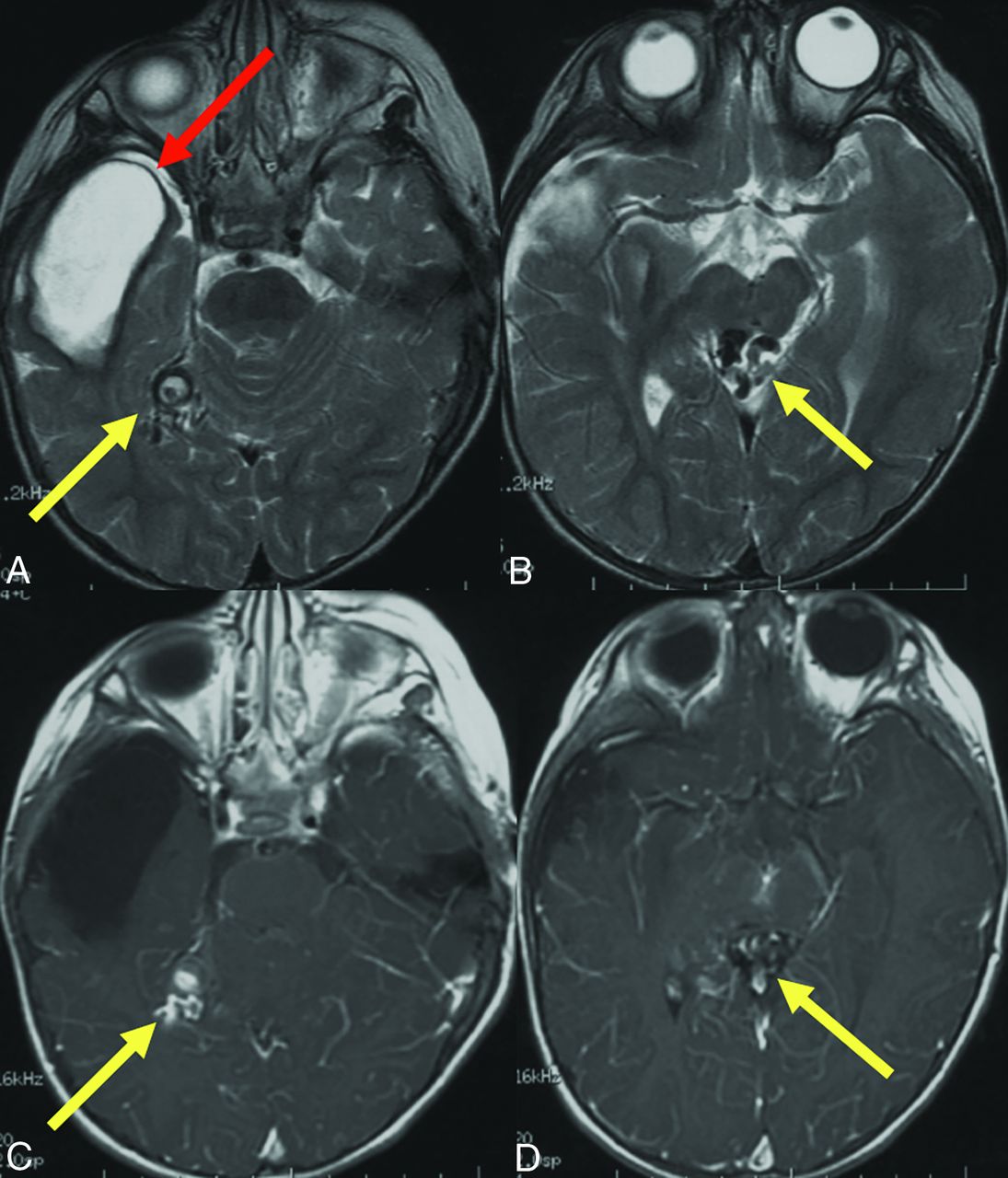
- FIG 1.
Prior brain hemorrhage in an infant with HHT. T2-weighted MR imaging (A) demonstrates a hemosiderin-lined cavity (red arrow) in the right temporal lobe of a 15-month-old girl with HHT. Additional AVFs (yellow arrows) are evident on T2 (A and B) and contrast-enhanced T1-weighted images (C and D).
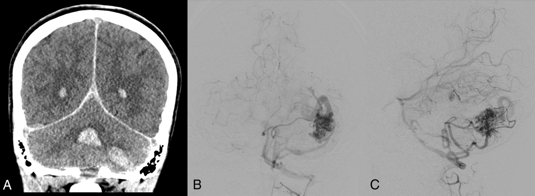
- FIG 2.
A 13-year-old boy with HHT who presented with severe headache and altered mental status. A, Coronal noncontrast head CT shows an acute left cerebellar hemisphere hemorrhage and intraventricular hemorrhage in the bilateral lateral ventricles and fourth ventricle. Anterior-posterior (B) and lateral (C) views on digital DSA depict a left cerebellar AVM with arterial supply from the left anterior and posterior inferior cerebellar arteries, left superior cerebellar artery, and deep venous drainage through the transverse sinus.
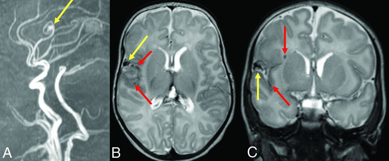
- FIG 3.
An infant diagnosed with HHT due to a familial ENG variant who had screening MR imaging of the brain and MRA of the head at 2 months of life had a pial AVF with hemosiderin deposition. Sagittal reformat (A) TOF-MRA demonstrates the right fontal AVF (yellow arrow). Axial (B) and coronal (C) T2-weighted MR images confirm the location of the AVF (yellow arrows) as well as adjacent hemosiderin deposition (red arrows) suggestive of prior hemorrhage.
- FIG 4.
A girl diagnosed with HHT due to a familial ENG variant had screening MR imaging that revealed a punctate area of enhancement in the left posterior frontal lobe surrounded by a subtle halo of enhancement (yellow arrow) on a high-resolution 3D T1 postgadolinium image (A). This finding corresponds to a subcentimeter AVM nidus (yellow arrow) on lateral (B) and anterior-posterior (C) DSA. An additional subcentimeter AVM nidus (blue arrow) supplied by the contralateral anterior cerebral artery is also identified on DSA.


Tables
∼Ten percent of patients with HHT have brain VMs (up to 20% of those with ENG variants) Risk of hemorrhagic stroke or SAH in children with HHT is 60 times the risk in children without HHT In cohorts of children with HHT, 3%–5% have intracerebral hemorrhage Rupture risk of brain AVMs in children with HHT is ∼0.7% per lesion per year (5.5 hemorrhages per 1000 child-years) Many brain VMs in children with HHT are asymptomatic until rupture Pros Cons Detect brain VM before rupture, allowing experts to determine whether treatment may be beneficial before a child has neurologic disability or even death Uncertain benefit of treatment of unruptured brain AVMs based on adult data from patients with sporadic AVMs Allow VMs that do not require treatment upon identification to be monitored with serial imaging to detect changes that may cause experts to decide to treat the VM Need for sedation in some children MR images that detect blood products can evaluate signs of asymptomatic bleeding, which may assist in risk stratification Causes anxiety for some parents and children if a brain VM is identified - Table 3:
Sample MR imaging–based protocol for brain VM screening in children with HHT (see also Vella et al17)
MR imaging brain with and without contrast DWI and ADC: assess for acute ischemia ASL: evaluate for asymmetric perfusion that can identify arteriovenous shunting FLAIR: sensitive for edema or gliosis related to VMs SWI: most sensitive for evidence of hemorrhage or microhemorrhage T1-weighted pre and postcontrast: differentiate intrinsic T1 shortening from enhancement High-resolution 3D postcontrast: most sensitive sequence for small HHT-related shunting AVMs and nonshunting capillary malformations High-resolution 3D T2-weighted: sensitive for edema or gliosis related to VMs MRA head Obtain TOF images from skull base to vertex (“whole head” MRA) because VMs can occur anywhere in brain parenchyma, not just near the circle of Willis


