
Article Figures & Data
Figures
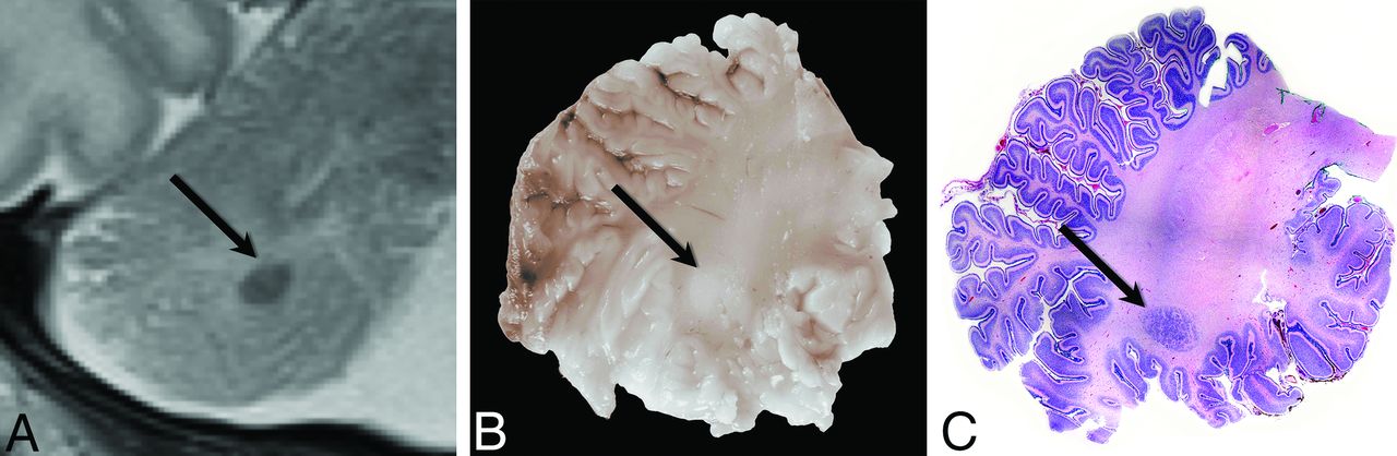
- Fig 1.
Radiologic-pathologic correlation in patient 7. Cropped coronal T2-weighted MR image of the right cerebellar hemisphere (A) obtained at 5 days of age shows the subcortical heterotopia (black arrow). Corresponding gross pathologic specimen (B) demonstrates the lesion as a white nodule (black arrow). Whole-mount hematoxylin-eosin–stained microscopic pathologic specimen (C) demonstrates the corresponding neuronal heterotopia (black arrow) (original magnification ×1).
- Fig 2.
Photomicrographs demonstrate the histopathology of the heterotopia in patient 7. A, A cluster of disorganized neurons (black arrows) is juxtaposed to an adjacent normal-layered foliar cortex (bracketed by black arrowheads) (hematoxylin-eosin stain, original magnification ×4). B, Heterotopia comprises a dominant population of morphologically normal granular cell neurons in a background of mixed glial cells (hematoxylin-eosin stain, original magnification ×20). C, Purkinje cells (darkly staining cells) are scattered within the granular cell neurons as well as within the adjacent cortex (HuC stain, original magnification ×4). D, Nerve fibers (darkly staining material) surround and traverse the cluster of heterotopic neurons (calretinin stain, original magnification ×4).
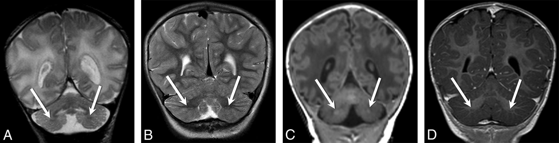
- Fig 3.
Coronal (A) and axial (B) T2-weighted images from patient 7 demonstrate bilateral cerebellar heterotopias (white arrows, A and B) with characteristic ellipsoid morphology and subcortical location in the bilateral inferior cerebellar hemispheres.
- Fig 4.
Coronal T2-weighted images from patient 15 at 3 days (A) and again at 3 years (B) of age demonstrate the expected evolution of the signal intensity of the heterotopias—hypointense relative to surrounding unmyelinated white matter (white arrows, A), becoming hyperintense relative to surrounding myelinated white matter (white arrows, B). Coronal T1-weighted images from patient 7 at 3 days of age (C) and patient 17 at 13 months of age (D) demonstrate similar isointensity of the heterotopias (white arrows, C and D) to gray matter.
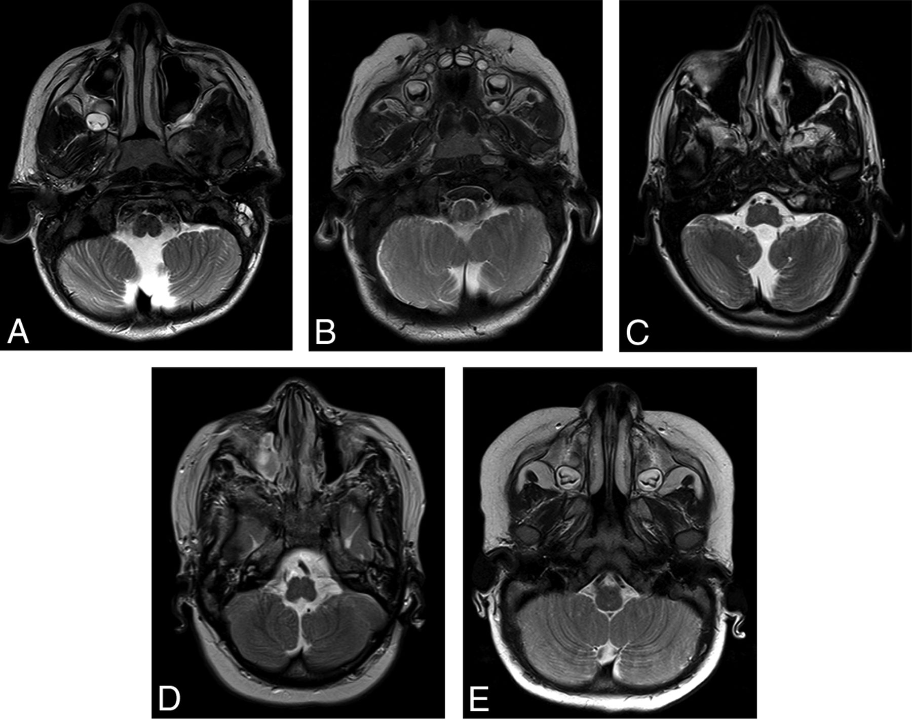
- Fig 5.
Axial T2-weighted images from patient 22 (A), patient 33 (B), patient 13 (C), and patient 1 (D) demonstrate anteromedial rotation of the inferior cerebellar tonsils. Axial T2-weighted image from a 10-month-old control patient is included for comparison (E).
- Fig 6.
Sagittal T1-weighted images in patient 25 (A), patient 19 (B), patient 16 (C), and patient 12 (D) demonstrate a spectrum of inferior vermian hypoplasia. Axial T2-weighted image from a 10-month-old control patient is included for comparison (E).
- Fig 7.
Coronal and axial T2-weighted images from patient 11 (A and B), patient 30 (C and D), and patient 22 (E and F) demonstrate a diffusely disorganized pattern of cerebellar foliation of the bilateral cerebellar hemispheres. Coronal (G) and axial (H) T2-weighted images from a 10-month-old control patient are included for comparison.


Tables
Diagnostic criteria for CHARGE syndrome
Diagnostic Characteristics Manifestations Major criteria Ocular coloboma Coloboma of the iris, retina, choroid, disc; microphthalmos Choanal atresia or stenosis Unilateral or bilateral, bony or membranous, atresia or stenosis Cranial nerve dysfunction or anomaly I, Hyposmia or anosmia VII, Facial palsy (unilateral or bilateral) VIII, Hypoplasia of auditory nerve IX/X, Swallowing problems with aspiration CHARGE syndrome ear Outer ear: short, wide ear with little or no lobe, “snipped off” helix, prominent antihelix that is often discontinuous with tragus, triangular concha, decreased cartilage; often protruding and usually asymmetric Middle ear: ossicular malformations Cochlea: incomplete partition Vestibular apparatus: absent or hypoplastic semicircular canals Minor criteria Genital hypoplasia Males: micropenis, cryptorchidism Females: hypoplastic labia Developmental delay Delayed milestones, hypotonia Cardiovascular malformation Conotruncal defects (eg, tetralogy of Fallot), atrioventricular canal defects, aortic arch anomalies Growth deficiency Short stature, usually postnatal with or without growth hormone deficiency Orofacial cleft Cleft lip and/or palate Tracheoesophageal fistula Tracheoesophageal defects of all types Distinctive facial features Square face with broad prominent forehead, prominent nasal bridge and columella, flat midface


