Advertisement

Main menu

  • Home
  • Content
    • Current Issue
    • Accepted Manuscripts
    • Article Preview
    • Past Issue Archive
    • Video Articles
    • AJNR Case Collection
    • Case of the Week Archive
    • Case of the Month Archive
    • Classic Case Archive
  • Special Collections
    • AJNR Awards
    • Low-Field MRI
    • Alzheimer Disease
    • ASNR Foundation Special Collection
    • Photon-Counting CT
    • View All
  • Multimedia
    • AJNR Podcasts
    • AJNR SCANtastic
    • Trainee Corner
    • MRI Safety Corner
    • Imaging Protocols
  • For Authors
    • Submit a Manuscript
    • Submit a Video Article
    • Submit an eLetter to the Editor/Response
    • Manuscript Submission Guidelines
    • Statistical Tips
    • Fast Publishing of Accepted Manuscripts
    • Graphical Abstract Preparation
    • Imaging Protocol Submission
    • Author Policies
  • About Us
    • About AJNR
    • Editorial Board
    • Editorial Board Alumni
  • More
    • Become a Reviewer/Academy of Reviewers
    • Subscribers
    • Permissions
    • Alerts
    • Feedback
    • Advertisers
    • ASNR Home

User menu

  • Alerts
  • Log in

Search

  • Advanced search
American Journal of Neuroradiology

American Journal of Neuroradiology

ASHNR American Society of Functional Neuroradiology ASHNR American Society of Pediatric Neuroradiology ASSR
  • Alerts
  • Log in

Advanced Search

  • Home
  • Content
    • Current Issue
    • Accepted Manuscripts
    • Article Preview
    • Past Issue Archive
    • Video Articles
    • AJNR Case Collection
    • Case of the Week Archive
    • Case of the Month Archive
    • Classic Case Archive
  • Special Collections
    • AJNR Awards
    • Low-Field MRI
    • Alzheimer Disease
    • ASNR Foundation Special Collection
    • Photon-Counting CT
    • View All
  • Multimedia
    • AJNR Podcasts
    • AJNR SCANtastic
    • Trainee Corner
    • MRI Safety Corner
    • Imaging Protocols
  • For Authors
    • Submit a Manuscript
    • Submit a Video Article
    • Submit an eLetter to the Editor/Response
    • Manuscript Submission Guidelines
    • Statistical Tips
    • Fast Publishing of Accepted Manuscripts
    • Graphical Abstract Preparation
    • Imaging Protocol Submission
    • Author Policies
  • About Us
    • About AJNR
    • Editorial Board
    • Editorial Board Alumni
  • More
    • Become a Reviewer/Academy of Reviewers
    • Subscribers
    • Permissions
    • Alerts
    • Feedback
    • Advertisers
    • ASNR Home
  • Follow AJNR on Twitter
  • Visit AJNR on Facebook
  • Follow AJNR on Instagram
  • Join AJNR on LinkedIn
  • RSS Feeds

AJNR Awards, New Junior Editors, and more. Read the latest AJNR updates

Research ArticleSpine Imaging and Spine Image-Guided Interventions

MR Imaging Features of Critical Spinal Demyelinating Lesions Associated with Progressive Motor Impairment

B. Mark Keegan, Steven A. Messina, Dennis Hanson, David Holmes, Jon Camp, Elia Sechi, Shreya Nayak, Benan Barakat, Rowaid Ahmad, Jay Mandrekar, W. Scott Harmsen, Orhun Kantarci, Brian G. Weinshenker and Eoin P. Flanagan
American Journal of Neuroradiology July 2024, 45 (7) 943-950; DOI: https://doi.org/10.3174/ajnr.A8304
B. Mark Keegan
aFrom the Department of Neurology (B.M.K., E.S., S.N., B.B., R.A., J.M., O.K., B.G.W., E.P.F.), Center for Multiple Sclerosis and Autoimmune Neurology, Mayo Clinic, Rochester, Minnesota
  • Find this author on Google Scholar
  • Find this author on PubMed
  • Search for this author on this site
  • ORCID record for B. Mark Keegan
Steven A. Messina
bDepartment of Radiology (S.A.M.), Mayo Clinic, Rochester, Minnesota
  • Find this author on Google Scholar
  • Find this author on PubMed
  • Search for this author on this site
  • ORCID record for Steven A. Messina
Dennis Hanson
cBiomedical Imaging Resource (D. Hanson, D. Holmes, J.C.), Mayo Clinic, Rochester, Minnesota
  • Find this author on Google Scholar
  • Find this author on PubMed
  • Search for this author on this site
  • ORCID record for Dennis Hanson
David Holmes
cBiomedical Imaging Resource (D. Hanson, D. Holmes, J.C.), Mayo Clinic, Rochester, Minnesota
  • Find this author on Google Scholar
  • Find this author on PubMed
  • Search for this author on this site
  • ORCID record for David Holmes
Jon Camp
cBiomedical Imaging Resource (D. Hanson, D. Holmes, J.C.), Mayo Clinic, Rochester, Minnesota
  • Find this author on Google Scholar
  • Find this author on PubMed
  • Search for this author on this site
Elia Sechi
aFrom the Department of Neurology (B.M.K., E.S., S.N., B.B., R.A., J.M., O.K., B.G.W., E.P.F.), Center for Multiple Sclerosis and Autoimmune Neurology, Mayo Clinic, Rochester, Minnesota
dUniversità degli Studi di Sassari (E.S.), Sassari, Italy
  • Find this author on Google Scholar
  • Find this author on PubMed
  • Search for this author on this site
Shreya Nayak
aFrom the Department of Neurology (B.M.K., E.S., S.N., B.B., R.A., J.M., O.K., B.G.W., E.P.F.), Center for Multiple Sclerosis and Autoimmune Neurology, Mayo Clinic, Rochester, Minnesota
eSt. Elizabeth Dearborn Hospital (S.N.), Lawrenceburg, Indiana
  • Find this author on Google Scholar
  • Find this author on PubMed
  • Search for this author on this site
Benan Barakat
aFrom the Department of Neurology (B.M.K., E.S., S.N., B.B., R.A., J.M., O.K., B.G.W., E.P.F.), Center for Multiple Sclerosis and Autoimmune Neurology, Mayo Clinic, Rochester, Minnesota
fMercy St. Vincent Medical Center (B.B.), Toledo, Ohio
  • Find this author on Google Scholar
  • Find this author on PubMed
  • Search for this author on this site
  • ORCID record for Benan Barakat
Rowaid Ahmad
aFrom the Department of Neurology (B.M.K., E.S., S.N., B.B., R.A., J.M., O.K., B.G.W., E.P.F.), Center for Multiple Sclerosis and Autoimmune Neurology, Mayo Clinic, Rochester, Minnesota
gUniversity of Texas Medical Branch (R.A.), Galveston, Texas
  • Find this author on Google Scholar
  • Find this author on PubMed
  • Search for this author on this site
Jay Mandrekar
aFrom the Department of Neurology (B.M.K., E.S., S.N., B.B., R.A., J.M., O.K., B.G.W., E.P.F.), Center for Multiple Sclerosis and Autoimmune Neurology, Mayo Clinic, Rochester, Minnesota
hQuantitative Health Services (J.M., W.S.H,), Mayo Clinic, Rochester, Minnesota
  • Find this author on Google Scholar
  • Find this author on PubMed
  • Search for this author on this site
W. Scott Harmsen
hQuantitative Health Services (J.M., W.S.H,), Mayo Clinic, Rochester, Minnesota
  • Find this author on Google Scholar
  • Find this author on PubMed
  • Search for this author on this site
Orhun Kantarci
aFrom the Department of Neurology (B.M.K., E.S., S.N., B.B., R.A., J.M., O.K., B.G.W., E.P.F.), Center for Multiple Sclerosis and Autoimmune Neurology, Mayo Clinic, Rochester, Minnesota
  • Find this author on Google Scholar
  • Find this author on PubMed
  • Search for this author on this site
  • ORCID record for Orhun Kantarci
Brian G. Weinshenker
aFrom the Department of Neurology (B.M.K., E.S., S.N., B.B., R.A., J.M., O.K., B.G.W., E.P.F.), Center for Multiple Sclerosis and Autoimmune Neurology, Mayo Clinic, Rochester, Minnesota
iDepartment of Neurology (B.G.W.), University of Virginia Health, Charlottesville, Virginia
  • Find this author on Google Scholar
  • Find this author on PubMed
  • Search for this author on this site
Eoin P. Flanagan
aFrom the Department of Neurology (B.M.K., E.S., S.N., B.B., R.A., J.M., O.K., B.G.W., E.P.F.), Center for Multiple Sclerosis and Autoimmune Neurology, Mayo Clinic, Rochester, Minnesota
  • Find this author on Google Scholar
  • Find this author on PubMed
  • Search for this author on this site
  • ORCID record for Eoin P. Flanagan
  • Article
  • Figures & Data
  • Info & Metrics
  • Responses
  • References
  • PDF
Loading

Article Figures & Data

Figures

  • Tables
  • FIG 1.
    • Download figure
    • Open in new tab
    • Download powerpoint
    FIG 1.

    Patient ascertainment algorithm.
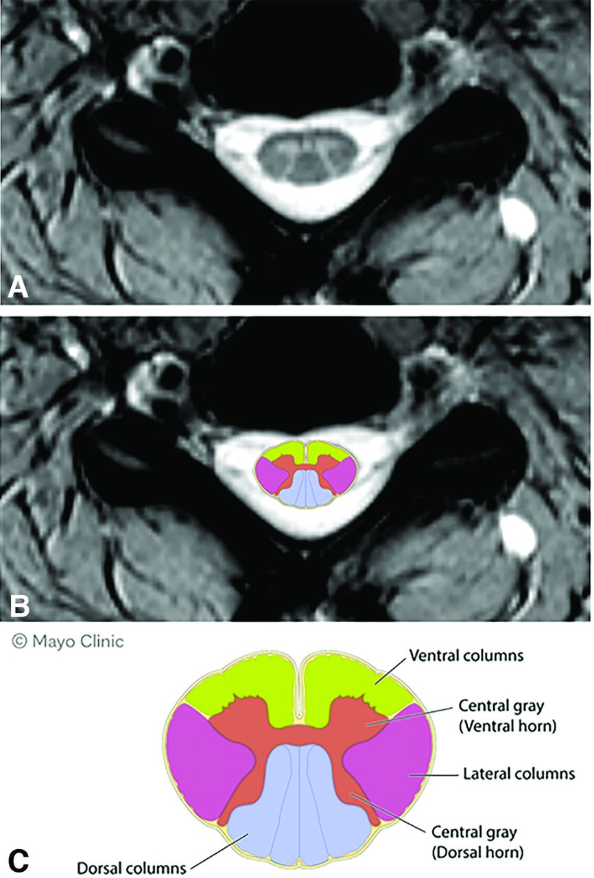
  • FIG 2.
    • Download figure
    • Open in new tab
    • Download powerpoint
    FIG 2.

    Axial spinal cord columns. A, Axial T2 spinal cord imaging. B, Superimposed schematic of axial columns on axial MR image. C, Schematic diagram of axial columns of spinal cord.
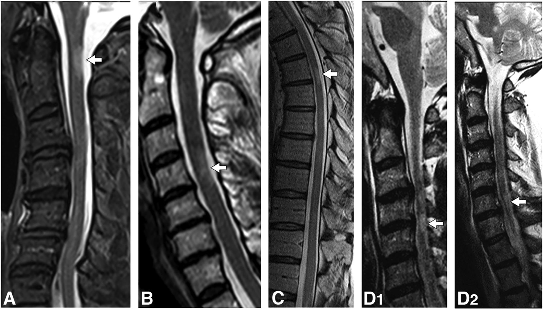
  • FIG 3.
    • Download figure
    • Open in new tab
    • Download powerpoint
    FIG 3.

    Examples of critical T2-hyperintense demyelinating lesions on sagittal T2-weighted images. Imaging composite of sagittal T2-weighted images in patients with critical lesions with corresponding axial images. A right C1 T2-hyperintense lesion with focal atrophy (A, arrow). A left C4 T2-hyperintense lesion with focal atrophy (B, arrow). A right-sided upper thoracic spine; T2-hyperintense lesion with focal atrophy (C, arrow). A left-sided C4-C5 T2-hyperintense lesion with focal atrophy (D1, arrow). An additional noncritical lesion is noted at the C6 level (D2, arrow).
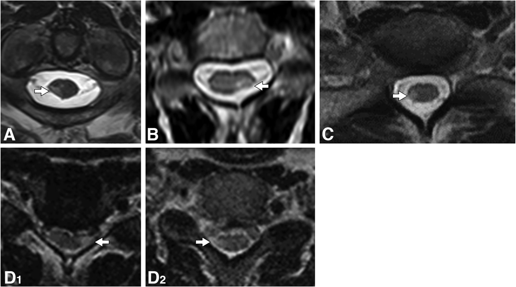
  • FIG 4.
    • Download figure
    • Open in new tab
    • Download powerpoint
    FIG 4.

    Examples of critical T2-hyperintense demyelinating lesions on axial T2-weighted images. Imaging composite of axial T2-weighted images in patients with critical lesions, corresponding to sagittal images and clinical details. The corresponding axial images reveal a T2-hyperintense lesion in the right lateral column with focal atrophy (A, arrow), a T2-hyperintense lesion in the left lateral column with focal atrophy (B, arrow), a T2-hyperintense lesion in the right lateral column with focal atrophy (C, arrow), a T2-hyperintense lesion in the left lateral column with focal atrophy (D1, arrow), and a T2-hyperintense lesion in the right lateral column without focal atrophy (D2, arrow).

Tables

  • Figures
    • View popup
    Table 1:

    Clinical characteristics of 91 individuals with critical demyelinating lesions

    Clinical Characteristics
    Demographics
     Female sex (No.) (%)51 (56%)
     White ethnicity (No.) (%)88 (97%)
     Median age at first symptom (range) (yr)50 (28–65)
     Median age at last MR imaging (range) (yr)58 (36–80)
     Median age at progression (range) (yr)52 (30–73)
    Clinical course (No.) (%)
     Progression from onset (primary-progressive)56/91 (62%)
     Relapse onset (secondary-progressive)25/91 (28%)
     Clinical cohort (No.) (%)
     PUHMS37 (41%)
     PPS35 (38%)
     PSS19 (21%)
    • View popup
    Table 2:

    MR imaging characteristics in 91 critical and 88 noncritical spinal cord demyelinating lesions

    Critical Spinal Cord Demyelinating Lesion (n = 91)Noncritical Spinal Cord Demyelinating Lesion (n = 88)OR 95% CIP Value
    Sagittal location (No.) (%)
     Cervical74 (81)63 (64)
     Thoracic17 (19)25 (26)
    Moderate/severe atrophy (No.) (%)41 (45)0 (0%)161.91 (9.43 to >999.9).0005
    Axial column location (No.) (%)
     Lateral86 (95)61 (62)10.43 (3.88–28.07)<.0001
     Central62 (68)39 (40)3.23 (1.78–5.88) .0001
     Ventral38 (42)19 (19)2.98 (1.55–5.72).001
     Dorsal21 (23)39 (40)0.45 (0.24–0.86).0145
    Axial column lateral involvement<.0001
     Lateral with additional columns63 (69)34 (35)
     Lateral only23 (25)27 (28)
     No lateral involved5 (6)37 (38)
    Median lesion size (mm)
     Axial A/P width4.13.32.01 (1.49–2.72)<.0001
     Transverse axial (mm3)5.23.51.66 (1.36–2.01)<.0001
     Area lesion14.07.61.14 (1.08–1.2)<.0001
    • Note:—A/P indicates anterior-posterior.

PreviousNext
Back to top

In this issue

American Journal of Neuroradiology: 45 (7)
American Journal of Neuroradiology
Vol. 45, Issue 7
1 Jul 2024
  • Table of Contents
  • Index by author
  • Complete Issue (PDF)
Advertisement
Print
Download PDF
Email Article

Thank you for your interest in spreading the word on American Journal of Neuroradiology.

NOTE: We only request your email address so that the person you are recommending the page to knows that you wanted them to see it, and that it is not junk mail. We do not capture any email address.

Enter multiple addresses on separate lines or separate them with commas.
MR Imaging Features of Critical Spinal Demyelinating Lesions Associated with Progressive Motor Impairment
(Your Name) has sent you a message from American Journal of Neuroradiology
(Your Name) thought you would like to see the American Journal of Neuroradiology web site.
CAPTCHA
This question is for testing whether or not you are a human visitor and to prevent automated spam submissions.
Cite this article
B. Mark Keegan, Steven A. Messina, Dennis Hanson, David Holmes, Jon Camp, Elia Sechi, Shreya Nayak, Benan Barakat, Rowaid Ahmad, Jay Mandrekar, W. Scott Harmsen, Orhun Kantarci, Brian G. Weinshenker, Eoin P. Flanagan
MR Imaging Features of Critical Spinal Demyelinating Lesions Associated with Progressive Motor Impairment
American Journal of Neuroradiology Jul 2024, 45 (7) 943-950; DOI: 10.3174/ajnr.A8304

Citation Manager Formats

  • BibTeX
  • Bookends
  • EasyBib
  • EndNote (tagged)
  • EndNote 8 (xml)
  • Medlars
  • Mendeley
  • Papers
  • RefWorks Tagged
  • Ref Manager
  • RIS
  • Zotero
0 Responses
Respond to this article
Share
Bookmark this article
MRI of Spinal Demyelinating Lesions
B. Mark Keegan, Steven A. Messina, Dennis Hanson, David Holmes, Jon Camp, Elia Sechi, Shreya Nayak, Benan Barakat, Rowaid Ahmad, Jay Mandrekar, W. Scott Harmsen, Orhun Kantarci, Brian G. Weinshenker, Eoin P. Flanagan
American Journal of Neuroradiology Jul 2024, 45 (7) 943-950; DOI: 10.3174/ajnr.A8304
del.icio.us logo Twitter logo Facebook logo Mendeley logo
  • Tweet Widget
  • Facebook Like
  • Google Plus One
Purchase

Jump to section

  • Article
    • Abstract
    • ABBREVIATIONS:
    • MATERIALS AND METHODS
    • RESULTS
    • DISCUSSION
    • CONCLUSIONS
    • Acknowledgments
    • Footnotes
    • References
  • Figures & Data
  • Info & Metrics
  • Responses
  • References
  • PDF

Related Articles

  • No related articles found.
  • PubMed
  • Google Scholar

Cited By...

  • No citing articles found.
  • Crossref
  • Google Scholar

This article has not yet been cited by articles in journals that are participating in Crossref Cited-by Linking.

More in this TOC Section

  • Dynamic CT Myelography: Patient Positioning
  • Evaluation of SIH MR Scoring Systems in Normals
  • Management Outcomes For VO Spine Biopsy
Show more SPINE IMAGING AND SPINE IMAGE-GUIDED INTERVENTIONS

Similar Articles

Advertisement

Indexed Content

  • Current Issue
  • Accepted Manuscripts
  • Article Preview
  • Past Issues
  • Editorials
  • Editor's Choice
  • Fellows' Journal Club
  • Letters to the Editor
  • Video Articles

Cases

  • Case Collection
  • Archive - Case of the Week
  • Archive - Case of the Month
  • Archive - Classic Case

More from AJNR

  • Trainee Corner
  • Imaging Protocols
  • MRI Safety Corner
  • Book Reviews

Multimedia

  • AJNR Podcasts
  • AJNR Scantastics

Resources

  • Turnaround Time
  • Submit a Manuscript
  • Submit a Video Article
  • Submit an eLetter to the Editor/Response
  • Manuscript Submission Guidelines
  • Statistical Tips
  • Fast Publishing of Accepted Manuscripts
  • Graphical Abstract Preparation
  • Imaging Protocol Submission
  • Evidence-Based Medicine Level Guide
  • Publishing Checklists
  • Author Policies
  • Become a Reviewer/Academy of Reviewers
  • News and Updates

About Us

  • About AJNR
  • Editorial Board
  • Editorial Board Alumni
  • Alerts
  • Permissions
  • Not an AJNR Subscriber? Join Now
  • Advertise with Us
  • Librarian Resources
  • Feedback
  • Terms and Conditions
  • AJNR Editorial Board Alumni

American Society of Neuroradiology

  • Not an ASNR Member? Join Now

© 2025 by the American Society of Neuroradiology All rights, including for text and data mining, AI training, and similar technologies, are reserved.
Print ISSN: 0195-6108 Online ISSN: 1936-959X

Powered by HighWire