Advertisement

Main menu

  • Home
  • Content
    • Current Issue
    • Accepted Manuscripts
    • Article Preview
    • Past Issue Archive
    • Video Articles
    • AJNR Case Collection
    • Case of the Week Archive
    • Case of the Month Archive
    • Classic Case Archive
  • Special Collections
    • AJNR Awards
    • Low-Field MRI
    • Alzheimer Disease
    • ASNR Foundation Special Collection
    • Photon-Counting CT
    • View All
  • Multimedia
    • AJNR Podcasts
    • AJNR SCANtastic
    • Trainee Corner
    • MRI Safety Corner
    • Imaging Protocols
  • For Authors
    • Submit a Manuscript
    • Submit a Video Article
    • Submit an eLetter to the Editor/Response
    • Manuscript Submission Guidelines
    • Statistical Tips
    • Fast Publishing of Accepted Manuscripts
    • Graphical Abstract Preparation
    • Imaging Protocol Submission
    • Author Policies
  • About Us
    • About AJNR
    • Editorial Board
    • Editorial Board Alumni
  • More
    • Become a Reviewer/Academy of Reviewers
    • Subscribers
    • Permissions
    • Alerts
    • Feedback
    • Advertisers
    • ASNR Home

User menu

  • Alerts
  • Log in

Search

  • Advanced search
American Journal of Neuroradiology

American Journal of Neuroradiology

ASHNR American Society of Functional Neuroradiology ASHNR American Society of Pediatric Neuroradiology ASSR
  • Alerts
  • Log in

Advanced Search

  • Home
  • Content
    • Current Issue
    • Accepted Manuscripts
    • Article Preview
    • Past Issue Archive
    • Video Articles
    • AJNR Case Collection
    • Case of the Week Archive
    • Case of the Month Archive
    • Classic Case Archive
  • Special Collections
    • AJNR Awards
    • Low-Field MRI
    • Alzheimer Disease
    • ASNR Foundation Special Collection
    • Photon-Counting CT
    • View All
  • Multimedia
    • AJNR Podcasts
    • AJNR SCANtastic
    • Trainee Corner
    • MRI Safety Corner
    • Imaging Protocols
  • For Authors
    • Submit a Manuscript
    • Submit a Video Article
    • Submit an eLetter to the Editor/Response
    • Manuscript Submission Guidelines
    • Statistical Tips
    • Fast Publishing of Accepted Manuscripts
    • Graphical Abstract Preparation
    • Imaging Protocol Submission
    • Author Policies
  • About Us
    • About AJNR
    • Editorial Board
    • Editorial Board Alumni
  • More
    • Become a Reviewer/Academy of Reviewers
    • Subscribers
    • Permissions
    • Alerts
    • Feedback
    • Advertisers
    • ASNR Home
  • Follow AJNR on Twitter
  • Visit AJNR on Facebook
  • Follow AJNR on Instagram
  • Join AJNR on LinkedIn
  • RSS Feeds

AJNR Awards, New Junior Editors, and more. Read the latest AJNR updates

Research ArticleAdult Brain

Tumefactive Primary Central Nervous System Vasculitis: Imaging Findings of a Rare and Underrecognized Neuroinflammatory Disease

S. Suthiphosuwan, A. Bharatha, C.C.-T. Hsu, A.W. Lin, J.A. Maloney, D.G. Munoz, C.A. Palmer and A.G. Osborn
American Journal of Neuroradiology November 2020, 41 (11) 2075-2081; DOI: https://doi.org/10.3174/ajnr.A6736
S. Suthiphosuwan
aFrom the Department of Medical Imaging (S.S., A.B., CC-T.H., A.W.L.)
  • Find this author on Google Scholar
  • Find this author on PubMed
  • Search for this author on this site
  • ORCID record for S. Suthiphosuwan
A. Bharatha
aFrom the Department of Medical Imaging (S.S., A.B., CC-T.H., A.W.L.)
bSurgery (A.B.)
  • Find this author on Google Scholar
  • Find this author on PubMed
  • Search for this author on this site
  • ORCID record for A. Bharatha
C.C.-T. Hsu
aFrom the Department of Medical Imaging (S.S., A.B., CC-T.H., A.W.L.)
dDepartment of Medical Imaging (CC-T.H.), Gold Coast University Hospital, Queensland, Australia
  • Find this author on Google Scholar
  • Find this author on PubMed
  • Search for this author on this site
  • ORCID record for C.C.-T. Hsu
A.W. Lin
aFrom the Department of Medical Imaging (S.S., A.B., CC-T.H., A.W.L.)
  • Find this author on Google Scholar
  • Find this author on PubMed
  • Search for this author on this site
  • ORCID record for A.W. Lin
J.A. Maloney
eDepartment of Radiology (J.A.M.), University of Colorado, Denver, Colorado
  • Find this author on Google Scholar
  • Find this author on PubMed
  • Search for this author on this site
  • ORCID record for J.A. Maloney
D.G. Munoz
cLaboratory Medicine and Pathobiology (D.G.M.), University of Toronto, Toronto, Canada
  • Find this author on Google Scholar
  • Find this author on PubMed
  • Search for this author on this site
  • ORCID record for D.G. Munoz
C.A. Palmer
fDepartment of Pathology (C.A.P.)
  • Find this author on Google Scholar
  • Find this author on PubMed
  • Search for this author on this site
  • ORCID record for C.A. Palmer
A.G. Osborn
gRadiology and Imaging Sciences (A.G.O.), University of Utah, Salt Lake City, Utah.
  • Find this author on Google Scholar
  • Find this author on PubMed
  • Search for this author on this site
  • ORCID record for A.G. Osborn
  • Article
  • Figures & Data
  • Supplemental
  • Info & Metrics
  • Responses
  • References
  • PDF
Loading

Article Figures & Data

Figures

  • Tables
  • FIG 1.
    • Download figure
    • Open in new tab
    • Download powerpoint
    FIG 1.

    Common MR imaging features of t-PCNSV in 4 different patients. A and E: 68-year-old man (case 5), B and F: 16-year-old male adolescent (case 6), C and G: 42-year-old man (case 8); and D and H: 55-year-old woman (case 7). Axial postcontrast T1-weighted images (A–D) demonstrate lobar masses. The masses have patchy heterogeneous enhancement (mottled appearance) on A–C (open arrow) and nodular and linear (perivascular enhancement) on D (open arrowhead). Leptomeningeal enhancement is noted on A, B, and D (black arrows). Subependymal enhancement is observed on C (white arrow). Axial susceptibility-weighted imaging (E, F, and H) and axial T2*-weighted gradient-echo imaging (G) demonstrate microhemorrhages. Punctate microhemorrhages are shown in E–H (white arrowheads). Linear microhemorrhages are demonstrated on G and H (black arrowheads).

  • FIG 2.
    • Download figure
    • Open in new tab
    • Download powerpoint
    FIG 2.

    MR imaging findings with histopathologic correlation of t-PCNSV. A 42-year-old man (case 1) presented with first episode seizure. Axial fluid-attenuated inversion recovery (A) shows a hyperintense lesion involving the cortex and subcortical white matter (white arrow). Axial post gadolinium T1-weighted imaging (B) demonstrates that the lesion has small nodular enhancement (black arrow). The initial imaging diagnosis was glioma. This patient underwent a gross total resection of the mass. Hematoxylin and eosin-stained (C: 2 mm and D: 200 µm magnifications) demonstrated vasculocentric transmural lymphocytic infiltration involving the leptomeningeal artery (C, white arrowhead) and parenchymal cortical arteries (C and D, black arrowheads). Immunohistochemically, the most infiltrating lymphocytic cell is positive for CD20 marker for B-cell lymphocytes (E) and CD3 marker for T-cell lymphocytes (F). The minority of inflammatory cells are macrophages that are positive for CD163 (G).
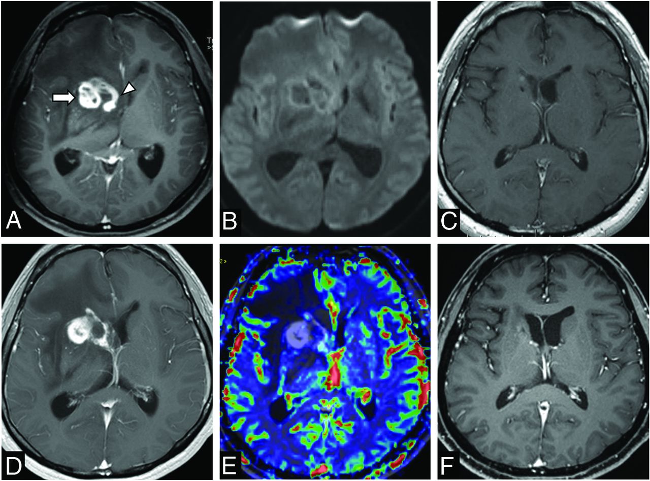
  • FIG 3.
    • Download figure
    • Open in new tab
    • Download powerpoint
    FIG 3.

    Recurrent granulomatous t-PCNSV. A 30-year-old man presented with headaches (case 4). A and B: Initial brain MR imaging shows conglomerate ring-enhancing mass centered in the right basal ganglia (arrow) with subependymal enhancement (arrowhead) on axial post gadolinium T1-weighted images (Gd-T1WI) (A). This mass does not show diffusion restriction on DWI (B). He underwent stereotactic-guided biopsy and histopathology showed granulomatous t-PCNSV. He was treated with corticosteroid and immunosuppressive agents. C: Follow-up MR imaging at 12 months shows the resolution of the disease. D and E: Follow-up MR imaging at 48 months demonstrates recurrent disease in the same region on Gd-T1WI (D). This mass does not show an increase in relative cerebral blood volume on dynamic susceptibility contrast MR perfusion (E). He was again treated with corticosteroids and an immunosuppressive agent. F: Follow-up Gd-T1WI at 77 months confirms the remission of the disease.

Tables

  • Figures
    • View popup
    Table 1:

    Patient clinical and radiologic demographics

    Patient Demographics 
    Age, median; range (years)42 (16–68)
    Sex, male (%)7 (70%)
    Clinical presentations, n (%) 
     Headache4 (40%)
     Seizure3 (30%)
     Hemiparesis3 (30%)
     Confusion2 (20%)
     Visual disturbance2 (20%)
     Cognitive impairment1 (10%)
     Word-finding difficulty1 (10%)
    Location of involvement, n (%) 
     Cortex/subcortical white matter7 (70%)
     Deep white matter6 (60%)
     Basal ganglia4 (40%)
     Brain stem1 (10%)
    Enhancement patterns, n (%)
     Patchy enhancement5 (50%)
     Small nodular enhancement5 (50%)
     Ring enhancement2 (20%)
     Linear/perivascular enhancement1 (10%)
     Leptomeningeal enhancement4 (40%)
     Subependymal enhancement3 (30%)
    Microhemorrhages, n (%)8 (80%)
    Patients with available CSF analysis, n (%)5 (50%)
     Abnormal CSF protein (>0.5 g/L)3 (60%)
     CSF leukocytosis (>5 cells/mm3)4 (80%)
    Patients with available serum inflammatory marker, n (%)8 (80%)
     High ESR (>20 mL/h)2 (25%)
     High CRP (>5 mg/L)2 (25%)
    Surgical procedure 
     Stereotactic-guided biopsy5 (50%)
     Tumor resection4 (40%)
     Open wedge biopsy1 (10%)
    Histopathology 
     Lymphocytic vasculitis7 (70%)
     Lymphocytic plus necrotizing vasculitis2 (20%)
     Granulomatous vasculitis1 (10%)
    Treatment 
     Steroid alone5 (50%)
     Steroid plus immunosuppressive agents5 (50%)
     Total duration follow-upa, median (range), months18 (4–77)a
    • ↵a Three patients did not have available clinical follow-up detail.

PreviousNext
Back to top

In this issue

American Journal of Neuroradiology: 41 (11)
American Journal of Neuroradiology
Vol. 41, Issue 11
1 Nov 2020
  • Table of Contents
  • Index by author
  • Complete Issue (PDF)
Advertisement
Print
Download PDF
Email Article

Thank you for your interest in spreading the word on American Journal of Neuroradiology.

NOTE: We only request your email address so that the person you are recommending the page to knows that you wanted them to see it, and that it is not junk mail. We do not capture any email address.

Enter multiple addresses on separate lines or separate them with commas.
Tumefactive Primary Central Nervous System Vasculitis: Imaging Findings of a Rare and Underrecognized Neuroinflammatory Disease
(Your Name) has sent you a message from American Journal of Neuroradiology
(Your Name) thought you would like to see the American Journal of Neuroradiology web site.
CAPTCHA
This question is for testing whether or not you are a human visitor and to prevent automated spam submissions.
Cite this article
S. Suthiphosuwan, A. Bharatha, C.C.-T. Hsu, A.W. Lin, J.A. Maloney, D.G. Munoz, C.A. Palmer, A.G. Osborn
Tumefactive Primary Central Nervous System Vasculitis: Imaging Findings of a Rare and Underrecognized Neuroinflammatory Disease
American Journal of Neuroradiology Nov 2020, 41 (11) 2075-2081; DOI: 10.3174/ajnr.A6736

Citation Manager Formats

  • BibTeX
  • Bookends
  • EasyBib
  • EndNote (tagged)
  • EndNote 8 (xml)
  • Medlars
  • Mendeley
  • Papers
  • RefWorks Tagged
  • Ref Manager
  • RIS
  • Zotero
0 Responses
Respond to this article
Share
Bookmark this article
Tumefactive Primary Central Nervous System Vasculitis: Imaging Findings of a Rare and Underrecognized Neuroinflammatory Disease
S. Suthiphosuwan, A. Bharatha, C.C.-T. Hsu, A.W. Lin, J.A. Maloney, D.G. Munoz, C.A. Palmer, A.G. Osborn
American Journal of Neuroradiology Nov 2020, 41 (11) 2075-2081; DOI: 10.3174/ajnr.A6736
del.icio.us logo Twitter logo Facebook logo Mendeley logo
  • Tweet Widget
  • Facebook Like
  • Google Plus One
Purchase

Jump to section

  • Article
    • Abstract
    • ABBREVIATIONS:
    • CASE SERIES
    • RESULTS
    • DISCUSSION
    • CONCLUSIONS
    • Footnotes
    • References
  • Figures & Data
  • Supplemental
  • Info & Metrics
  • Responses
  • References
  • PDF

Related Articles

  • PubMed
  • Google Scholar

Cited By...

  • More on Exploiting the T1 Shinethrough and T2* Effects Using Multiecho Susceptibility-Weighted Imaging
  • Primary Angiitis of the CNS with Unremarkable Vessel Wall MR Imaging: How the "T1 Shinethrough" Effect on SWI Adds to the Detection of Gadolinium Enhancement of Small Intraparenchymal Brain Vessels
  • Crossref
  • Google Scholar

This article has not yet been cited by articles in journals that are participating in Crossref Cited-by Linking.

More in this TOC Section

  • Diagnostic Neuroradiology of Monoclonal Antibodies
  • Cerebral ADC Changes in Fabry Disease
  • ML for Glioma Molecular Subtype Prediction
Show more Adult Brain

Similar Articles

Advertisement

Indexed Content

  • Current Issue
  • Accepted Manuscripts
  • Article Preview
  • Past Issues
  • Editorials
  • Editor's Choice
  • Fellows' Journal Club
  • Letters to the Editor
  • Video Articles

Cases

  • Case Collection
  • Archive - Case of the Week
  • Archive - Case of the Month
  • Archive - Classic Case

More from AJNR

  • Trainee Corner
  • Imaging Protocols
  • MRI Safety Corner
  • Book Reviews

Multimedia

  • AJNR Podcasts
  • AJNR Scantastics

Resources

  • Turnaround Time
  • Submit a Manuscript
  • Submit a Video Article
  • Submit an eLetter to the Editor/Response
  • Manuscript Submission Guidelines
  • Statistical Tips
  • Fast Publishing of Accepted Manuscripts
  • Graphical Abstract Preparation
  • Imaging Protocol Submission
  • Evidence-Based Medicine Level Guide
  • Publishing Checklists
  • Author Policies
  • Become a Reviewer/Academy of Reviewers
  • News and Updates

About Us

  • About AJNR
  • Editorial Board
  • Editorial Board Alumni
  • Alerts
  • Permissions
  • Not an AJNR Subscriber? Join Now
  • Advertise with Us
  • Librarian Resources
  • Feedback
  • Terms and Conditions
  • AJNR Editorial Board Alumni

American Society of Neuroradiology

  • Not an ASNR Member? Join Now

© 2025 by the American Society of Neuroradiology All rights, including for text and data mining, AI training, and similar technologies, are reserved.
Print ISSN: 0195-6108 Online ISSN: 1936-959X

Powered by HighWire