
Article Figures & Data
Figures
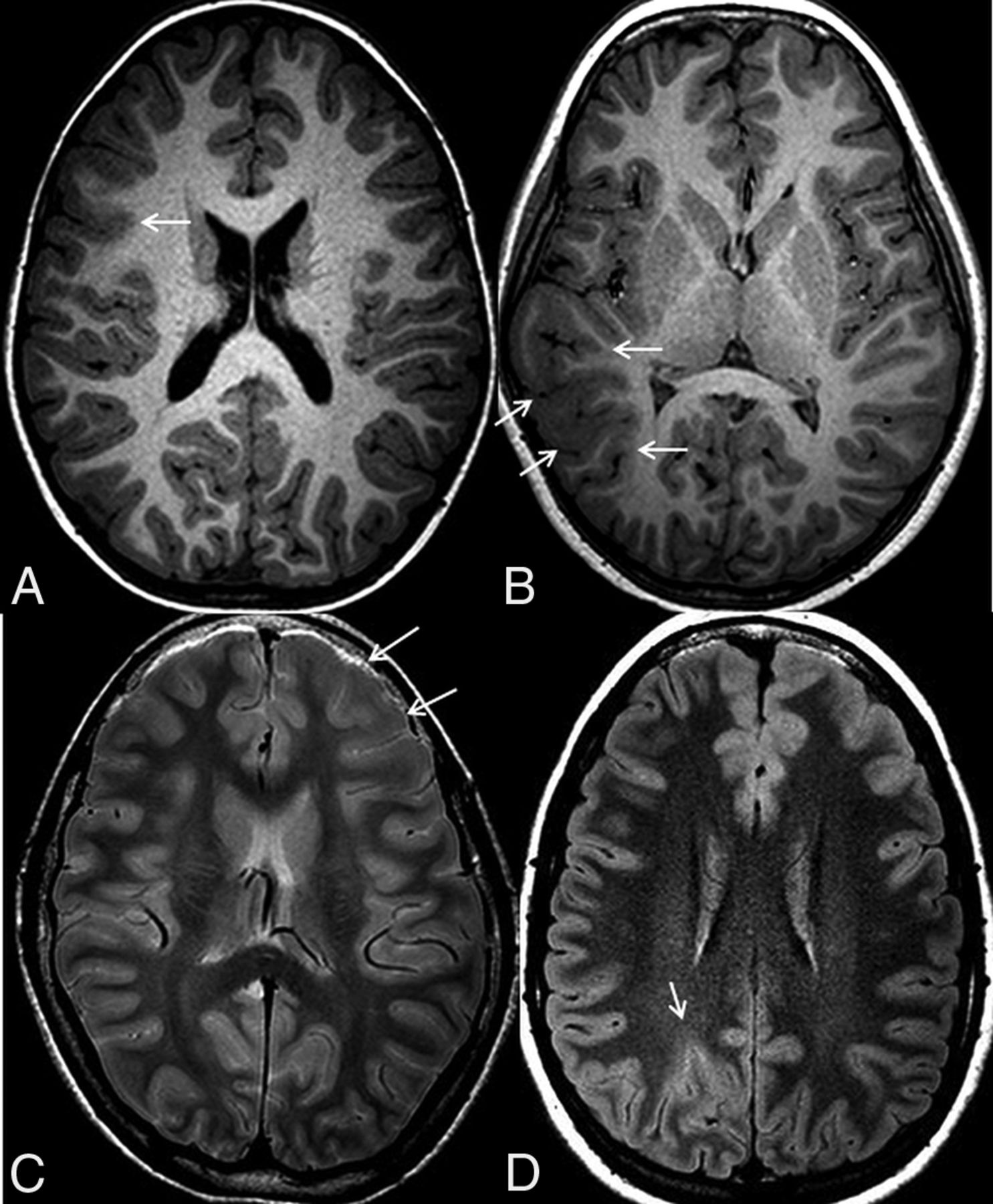
- Fig 1.
MR imaging features. Axial volumetric T1 shows high T1 signal in the cortex and blurring of the gray-white matter junction in the right inferior frontal gyrus (arrow, A) and thickening of the cortex, high T1 signal in the cortex, and an abnormal sulcation and gyration pattern (arrows, B) in the right temporal lobe. C, Axial proton-density sequence demonstrates high signal in the subcortical white matter (arrows) of the left frontal lobe. D, Axial FLAIR image shows high signal in the cortex (arrow) of the right parietal lobe.
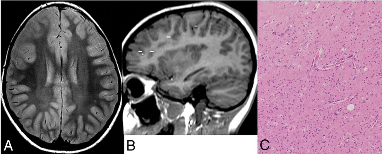
- Fig 2.
Case with oligodendrocytosis only. A, Axial proton density shows increased signal in the subcortical white matter of right frontal lobe. B, MEG demonstrates dipole scatter in the right frontal lobe. C, Hematoxylin-eosin stain magnification 10X demonstrates hypercellularity of oligodendroglial-like cells, with round nuclei and a scant amount of cytoplasm, in keeping with oligodendrocytosis.
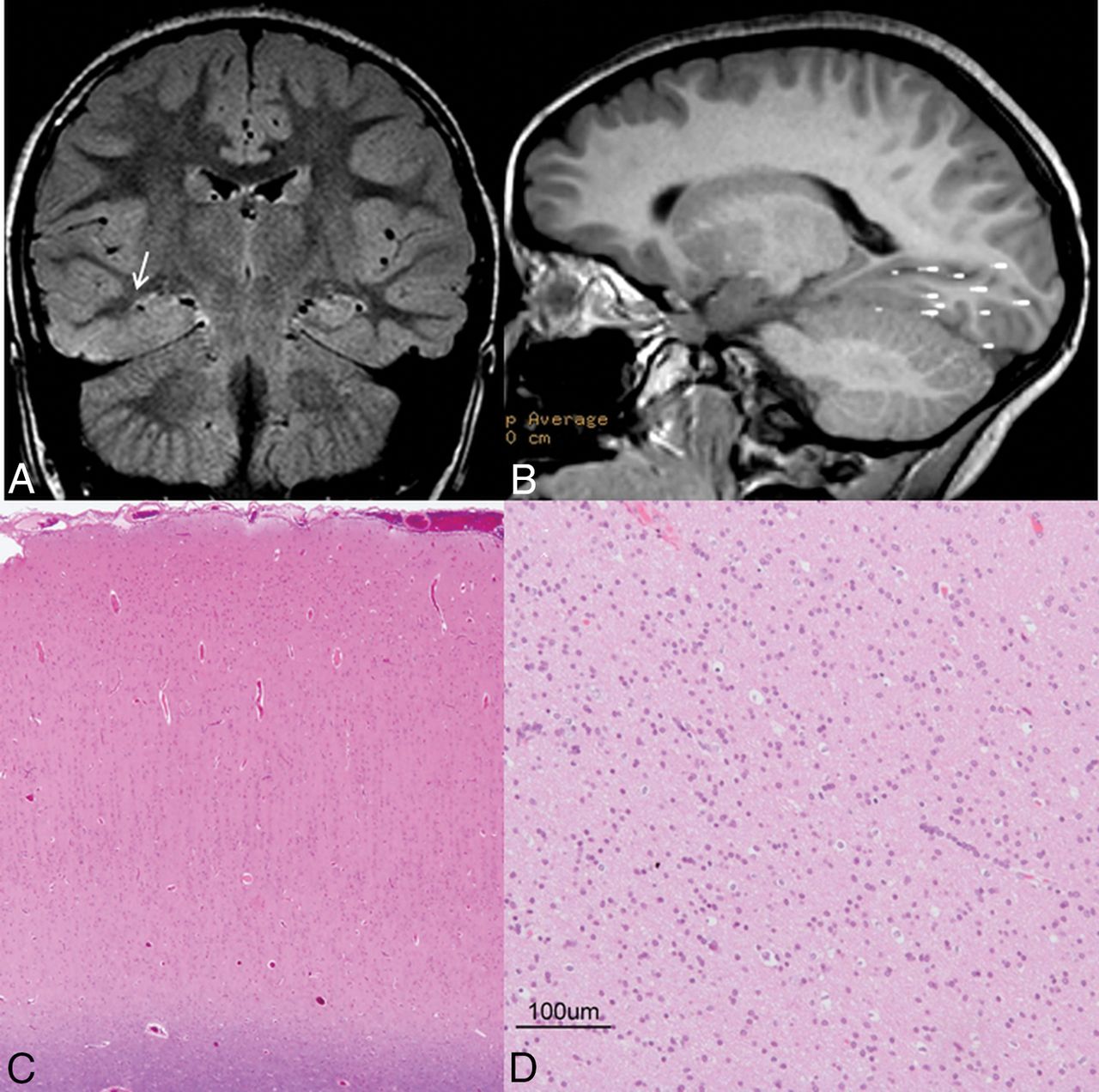
- Fig 3.
Case with oligodendrocytosis + focal cortical dysplasia type I. A, Coronal FLAIR shows increased FLAIR signal in the white matter of the right parahippocampal and fusiform gyri. B, MEG shows a dipole cluster in the right temporal and occipital lobes. Hematoxylin-eosin stain magnification 10X demonstrates abnormal radial cortical lamination, in keeping with FCD I (C), and hypercellularity of oligodendroglial-like cells with round nuclei and scant amount of cytoplasm (D), in keeping with oligodendrocytosis.


Tables
Oligodendrocytosis (n = 18) FCD I (n = 21) Oligodendrocytosis + FCD I (n = 7) P Value Sex .78 Male (%) 9 (50%) 12 (57.14%) 3 (42.9%) Female (%) 9 (50%) 9 (42.9%) 4 (57.1%) Age at operation (mean) (SD) (yr) 11.2 (4.6) 9.9 (6.0) 11.1 (5.6) .71 Age at seizure onset (mean) (SD) (yr) 6.2 (4.8) 4.6 (4.9) 2.44 (1.7) .18 Duration of epilepsy (mean) (SD) (yr) 6.3 (3.9) 6.5 (4.1) 10.4 (5.6) .11 Frequency of seizures .26 Daily (%) 8 (44.4%) 16 (76.2%) 5 (71.4%) Monthly (%) 3 (16.7%) 1 (4.8%) 0 (0%) Weekly (%) 7 (38.9%) 4 (19%) 2 (28.6%) Types of seizures .60 FOWIA/FOWIA-BTCZ (%) 12 (66.7%) 10 (47.6%) 3 (42.9%) FOWA/ FOWA-BTCZ (%) 2 (11.1%) 4 (19.0%) 1 (14.3%) Epileptic spasms (%) 4 (22.2%) 5 (23.8%) 3 (42.9%) Generalized (%) 0 (0%) 2 (9.5%) 0 (0%) Epileptiform discharges on video-EEG .95 Focal (%) 15 (83.3%) 17(81.0%) 6 (85.7%) No focal (%) 3 (16.7%) 4 (19.0%) 1 (14.3%) No. of AEDs (mean) SD) 2 (0.8) 2.2 (0.7) 2.7 (0.8) .13 Note:—AED indicates Anti-epileptic drugs; FOWIA, focal onset with impaired awareness; FOWIA-BTCZ, focal onset with impaired awareness of bilateral tonic clonic seizures; FOWA, focal onset with awareness; FOWA-BTCZ, focal onset with awareness of bilateral tonic clonic seizures.
Oligodendrocytosis (n = 18) FCD I (n = 21) Oligodendrocytosis + FCD I (n = 7) P Value MRI features High T1 in cortex (%) 5 (27.8) 4 (19.0) 3 (42.9) .44 High T2/FLAIR in cortex (%) 2 (11.1) 6 (28.6) 1 (14.3) .40 High T2/PD/FLAIR in subcortical white matter (%) 13 (72.2) 14 (66.7) 6 (85.7) .57 Increased cortical thickness (%) 0 (0) 3 (14.3) 2 (28.6) .10 Blurring of gray-white matter junction (%) 3 (16.7) 3 (14.3) 3 (42.9) .24 Abnormal sulcation and gyration pattern (%) 1 (5.6) 0 (0) 2 (28.6) .03 MEG features MEGSS cluster (%) 12 (66.7) 18 (85.7) 5 (71.4) .19 Concordance of MEGSS cluster with surgical resection (%) 12 (100) 17 (94.4) 5 (100) .30 Peak of MEGSS leading peak of EEG spike (%) 9 (50.0) 14 (66.7) 5 (71.4) .19 Spike and wave (%) 10 (55.6) 14 (66.7) 4 (57.1) .65 Oligodendrocytosis (n = 18) FCD I (n = 21) Oligodendrocytosis + FCD I (n = 7) P Value Invasive monitoring .09 Yes (%) 10 (55.6) 15 (71.4) 7 (100) No (%) 8 (44.4) 6 (28.6) 0 (0) Extension of resection .37 Unilobar (%) 10 (55.6) 7 (33.3) 3 (42.9) Multilobar (%) 8 (44.4) 12 (57.2) 4 (57.1) Hemispherectomy (%) 0 (0) 2 (9.5) 0 (0) Location of operation .10 Temporal (%) 8 (44.4) 3 (14.3) 0 (0) Temporal and extratemporal (%) 7 (38.9) 11 (52.4) 4 (57.1) Extratemporal (%) 3 (16.7) 7 (33.3) 3 (42.9) Seizure outcome at 1 yr .58 Seizure-free (ILAE) I (%) 13 (72.2) 12 (57.1) 4 (57.1) Persistent seizures (ILAE II–VI) (%) 5 (27.8) 9 (42.9) 3 (42.9) Seizure outcome at 1 yr .35 ILAE (I–IV) 18 (100) 16 (76.2) 7 (100) ILAE (V–VI) 0 (0) 5 (23.8) 0 (0)


