
Article Figures & Data
Figures
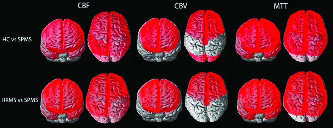
- FIGURE.
Whole-brain depiction of perfusion differences in cortical gray matter among HCs and patients with RRMS and SPMS. Units for CBF are milliliter/100 gram/minute; CBV, milliliter/100 gram; MTT, second.

Tables
- Table 1:
Comparison of demographic and clinical data for HCs and subjects with RRMS and SPMSa
Parameter HC (n = 19) RRMS (n = 19) SPMS (n = 20) RRMS vs HC (P Value) SPMS vs HC (P Value) SPMS vs RRMS (P Value) Age (yr) 49.0 ± 7.1 46.4 ± 7.2 55.2 ± 6.5 .27 .0168b .0041b Sex (F/M) 14:5 15:4 11:9 .70 .23 .12 Education (yr) 16.9 ± 2.9 16.1 ± 1.3 15.1 ± 2.6 .22 .051 .15 Disease duration (yr) 0.00 11.8 ± 5.4 16.7 ± 6.5 NA NA .0234 EDSS (median) (IQR) 0.00 1.5 (1–2) 6 (6–6.5) NA NA .0006b - Table 2:
Comparison of perfusion and volumetric data for cGM, NAWM, and T2h-l between HCs and subjects with RRMS and SPMS after adjusting for confounding factorsa
Parameter Median (Interquartiles) P Value HC (n = 19) RRMS (n = 19) SPMS (n = 20) RRMS vs HC SPMS vs HC SPMS vs RRMS cGM Global volume 890 (869–914) 864 (827–895) 673 (646–697) .10 .0026b .0020b CBF 43.20 (33.91–54.49) 41.07 (29.85–55.68) 34.05 (27.19–43.68) <.0001b <.0001b .0011b CBV 2.64 (2.02–3.29) 2.53 (1.94–3.37) 2.40 (1.92–3.07) .078 .046 .0062b MTT 3.77 (3.20–4.31) 3.90 (3.32–4.41) 4.30 (3.66–4.99) .0001b <.0001b <.0001b NAWM Global volume 878 (812–894) 818 (738–850) 697 (669–729) .0115b .0011b .0049b CBF 23.49 (18.69–27.78) 21.55 (16.05–29.50) 19.54 (16.74–24.92) .68 .58 .0080b CBV 1.59 (1.28–2.00) 1.57 (1.17–2.15) 1.51 (1.30–1.98) .89 .16 .0103b MTT 4.51 (4.09–5.10) 4.62 (4.08–5.20) 4.86 (4.28–5.41) .25 .0005b .0094b T2h-l Global volume 0.00 5.4 (1.2–9.3) 6.4 (1.0–10.9) NA NA .82 CBF 0.00 12.77 (8.23–17.24) 11.15 (8.95–15.71) NA NA .049 CBV 0.00 0.99 (0–1.32) 1.08 (0.83–1.47) NA NA .0026b MTT 0.00 4.91 (4.00–5.63) 5.64 (4.82–6.30) NA NA .43 Note:—NA, not applicable.
↵a Bonferroni-corrected P < .017 is considered statistically significant. Age was considered a confounding factor for comparing RRMS vs HC and SPMS vs HC; age, EDSS, and disease duration were considered confounding factors for comparing SPMS vs RRMS. All volumes are in cubic centimeters; CBF, milliliter/100 gram/meter; CBV, milliliter/100 gram; MTT, second.
↵b Significant.
- Table 3:
Lobar GM and WM volume differences among HCs and subjects with RRMS and SPMS after adjusting for confounding factorsa
Parameter Median (Interquartiles) P Value HC (n = 19) RRMS (n = 19) SPMS (n = 19) RRMS vs HC (n = 19) SPMS vs HC (n = 19) SPMS vs RRMS (n = 19) Gray matter Frontal lobe 200.040 (190.352–167.944) 199.248 (192.480–207.896) 200.352 (193.500–207.540) .69 .60 .22 Parietal lobe 90.296 (83.992–94.512) 89.824 (85.048–95.176) 93.368 (88.956–96.352) .72 .03 .095 Temporal lobe 122.740 (110.640–130.560) 121.336 (114.672–128.544) 106.892 (99.432–112.928) .86 .0044b .0073b Occipital lobe 101.088 (98.536–106.272) 97.160 (95.728–102.960) 72.792 (69.868–75.196) .10 .0030b .0033b White matter Frontal lobe 190.568 (170.368–201.528) 170.316 (159.520–189.112) 146.260 (134.908–157.136) <.0001b .0006b <.0001b Parietal lobe 101.200 (94.536–109.152) 93.272 (84.944–97.208) 81.952 (77.736–87.532) .0001b .0018b <.0001b Temporal lobe 70.132 (56.648–79.352) 68.512 (64.116–74.016) 61.208 (51.576–71.720) .026 .98 .0026b Occipital lobe 32.024 (30.248–35.092) 29.092 (24.136–33.768) 26.864 (23.552–31.832) .65 .0098b .0003b ↵a Bonferroni-corrected P < .017 is considered statistically significant. Age was considered a confounding factor for comparing RRMS vs HC and SPMS vs HC; age, EDSS, and disease duration were considered confounding factors for comparing SPMS vs RRMS. All volumes are in cubic centimeters.
↵b Significant.


Jump to section
Related Articles
Cited By...
- No citing articles found.