Advertisement

Main menu

  • Home
  • Content
    • Current Issue
    • Accepted Manuscripts
    • Article Preview
    • Past Issue Archive
    • Video Articles
    • AJNR Case Collection
    • Case of the Week Archive
    • Case of the Month Archive
    • Classic Case Archive
  • Special Collections
    • AJNR Awards
    • Low-Field MRI
    • Alzheimer Disease
    • ASNR Foundation Special Collection
    • Photon-Counting CT
    • View All
  • Multimedia
    • AJNR Podcasts
    • AJNR SCANtastic
    • Trainee Corner
    • MRI Safety Corner
    • Imaging Protocols
  • For Authors
    • Submit a Manuscript
    • Submit a Video Article
    • Submit an eLetter to the Editor/Response
    • Manuscript Submission Guidelines
    • Statistical Tips
    • Fast Publishing of Accepted Manuscripts
    • Graphical Abstract Preparation
    • Imaging Protocol Submission
    • Author Policies
  • About Us
    • About AJNR
    • Editorial Board
    • Editorial Board Alumni
  • More
    • Become a Reviewer/Academy of Reviewers
    • Subscribers
    • Permissions
    • Alerts
    • Feedback
    • Advertisers
    • ASNR Home

User menu

  • Alerts
  • Log in

Search

  • Advanced search
American Journal of Neuroradiology

American Journal of Neuroradiology

ASHNR American Society of Functional Neuroradiology ASHNR American Society of Pediatric Neuroradiology ASSR
  • Alerts
  • Log in

Advanced Search

  • Home
  • Content
    • Current Issue
    • Accepted Manuscripts
    • Article Preview
    • Past Issue Archive
    • Video Articles
    • AJNR Case Collection
    • Case of the Week Archive
    • Case of the Month Archive
    • Classic Case Archive
  • Special Collections
    • AJNR Awards
    • Low-Field MRI
    • Alzheimer Disease
    • ASNR Foundation Special Collection
    • Photon-Counting CT
    • View All
  • Multimedia
    • AJNR Podcasts
    • AJNR SCANtastic
    • Trainee Corner
    • MRI Safety Corner
    • Imaging Protocols
  • For Authors
    • Submit a Manuscript
    • Submit a Video Article
    • Submit an eLetter to the Editor/Response
    • Manuscript Submission Guidelines
    • Statistical Tips
    • Fast Publishing of Accepted Manuscripts
    • Graphical Abstract Preparation
    • Imaging Protocol Submission
    • Author Policies
  • About Us
    • About AJNR
    • Editorial Board
    • Editorial Board Alumni
  • More
    • Become a Reviewer/Academy of Reviewers
    • Subscribers
    • Permissions
    • Alerts
    • Feedback
    • Advertisers
    • ASNR Home
  • Follow AJNR on Twitter
  • Visit AJNR on Facebook
  • Follow AJNR on Instagram
  • Join AJNR on LinkedIn
  • RSS Feeds

AJNR Awards, New Junior Editors, and more. Read the latest AJNR updates

Research ArticleHead and Neck Imaging

Initial Performance of NI-RADS to Predict Residual or Recurrent Head and Neck Squamous Cell Carcinoma

D.A. Krieger, P.A. Hudgins, G.K. Nayak, K.L. Baugnon, A.S. Corey, M.R. Patel, J.J. Beitler, N.F. Saba, Y. Liu and A.H. Aiken
American Journal of Neuroradiology June 2017, 38 (6) 1193-1199; DOI: https://doi.org/10.3174/ajnr.A5157
D.A. Krieger
aFrom the Department of Radiology, Hackensack University Medical Center (D.A.K.), Hackensack, New Jersey
  • Find this author on Google Scholar
  • Find this author on PubMed
  • Search for this author on this site
  • ORCID record for D.A. Krieger
P.A. Hudgins
bDepartments of Radiology and Imaging Sciences (P.A.H., A.H.A., K.L.B., A.S.C.)
  • Find this author on Google Scholar
  • Find this author on PubMed
  • Search for this author on this site
  • ORCID record for P.A. Hudgins
G.K. Nayak
gDepartment of Radiology (G.K.N.), New York University School of Medicine, New York, New York.
  • Find this author on Google Scholar
  • Find this author on PubMed
  • Search for this author on this site
  • ORCID record for G.K. Nayak
K.L. Baugnon
bDepartments of Radiology and Imaging Sciences (P.A.H., A.H.A., K.L.B., A.S.C.)
  • Find this author on Google Scholar
  • Find this author on PubMed
  • Search for this author on this site
  • ORCID record for K.L. Baugnon
A.S. Corey
bDepartments of Radiology and Imaging Sciences (P.A.H., A.H.A., K.L.B., A.S.C.)
  • Find this author on Google Scholar
  • Find this author on PubMed
  • Search for this author on this site
  • ORCID record for A.S. Corey
M.R. Patel
cOtolaryngology Head and Neck Surgery (M.R.P., J.J.B.), Emory University School of Medicine, Atlanta, Georgia
  • Find this author on Google Scholar
  • Find this author on PubMed
  • Search for this author on this site
  • ORCID record for M.R. Patel
J.J. Beitler
cOtolaryngology Head and Neck Surgery (M.R.P., J.J.B.), Emory University School of Medicine, Atlanta, Georgia
dDepartments of Radiation Oncology (J.J.B.)
eHematology and Medical Oncology (J.J.B., N.F.S.)
  • Find this author on Google Scholar
  • Find this author on PubMed
  • Search for this author on this site
  • ORCID record for J.J. Beitler
N.F. Saba
eHematology and Medical Oncology (J.J.B., N.F.S.)
  • Find this author on Google Scholar
  • Find this author on PubMed
  • Search for this author on this site
  • ORCID record for N.F. Saba
Y. Liu
fBiostatistics (Y.L.), Winship Cancer Institute, Emory University School of Medicine, Atlanta, Georgia
  • Find this author on Google Scholar
  • Find this author on PubMed
  • Search for this author on this site
  • ORCID record for Y. Liu
A.H. Aiken
bDepartments of Radiology and Imaging Sciences (P.A.H., A.H.A., K.L.B., A.S.C.)
  • Find this author on Google Scholar
  • Find this author on PubMed
  • Search for this author on this site
  • ORCID record for A.H. Aiken
  • Article
  • Figures & Data
  • Info & Metrics
  • Responses
  • References
  • PDF
Loading

Article Figures & Data

Figures

  • Tables
  • Fig 1.
    • Download figure
    • Open in new tab
    • Download powerpoint
    Fig 1.

    ROC curve for NI-RADS at the primary site with AUC = 0.786 (95% CI, 0.691–0.881).

  • Fig 2.
    • Download figure
    • Open in new tab
    • Download powerpoint
    Fig 2.

    ROC curve for NI-RADS at the lymph nodes with AUC = 0.71 (95% CI, 0.597–0.826).

  • Fig 3.
    • Download figure
    • Open in new tab
    • Download powerpoint
    Fig 3.

    ROC curve for NI-RADS for primary site and lymph nodes combined, with AUC = 0.756 (95% CI, 0.682–0.8).

  • Fig 4.
    • Download figure
    • Open in new tab
    • Download powerpoint
    Fig 4.

    NI-RADS primary site category 2a: superficial mucosal abnormality. Primary T4a N2c base of tongue squamous cell carcinoma, status post chemoradiotherapy. A, CECT showed only subtle/questionable asymmetric enhancement in the right vallecula (arrow) retrospectively after review of PET. B, Fused PET image shows asymmetric uptake in the right vallecula (arrow). Direct visualization did show ulcerated mucosa, but the biopsy was negative for tumor. Clinically, this was deemed a radiation-related injury.
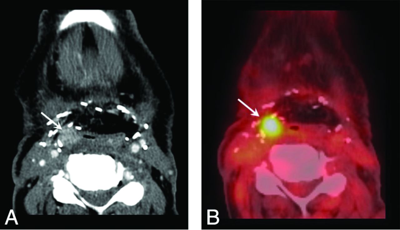
  • Fig 5.
    • Download figure
    • Open in new tab
    • Download powerpoint
    Fig 5.

    NI-RADS primary site category 2a: superficial mucosal abnormality. Primary T2 larynx squamous cell carcinoma status post chemoradiotherapy. A, CECT showed subtle irregularity of the anterior commissure and anterior true vocal cords bilaterally (arrow). B, Corresponding fused PET image shows focal mucosal uptake (arrow). After direct visualization revealed suspicious mucosal findings, the biopsy showed persistent disease. Although this lesion does demonstrate focal avid FDG uptake, it is in a special category of mucosal abnormality. In the published NI-RADS 1.0 by Aiken et al,5 these are scored as 2a because the linked management recommendation is direct visualization.

  • Fig 6.
    • Download figure
    • Open in new tab
    • Download powerpoint
    Fig 6.

    NI-RADS primary site category 2b: ill-defined asymmetric soft tissue. T4N0 oral cavity squamous cell carcinoma. CECT shows asymmetric full soft tissue around fibular reconstruction of the mandible (arrow). The linked management recommendation is shorter interval surveillance. Repeat CECT at 3 months showed no interval change (not shown). Subsequent clinical follow-up also demonstrated improvement and no disease recurrence.

  • Fig 7.
    • Download figure
    • Open in new tab
    • Download powerpoint
    Fig 7.

    NI-RADS primary site category 3: discrete enhancing lesion. T4a larynx squamous cell carcinoma, status post total laryngectomy, bilateral neck dissection, and chemoradiotherapy. A, CECT shows a 1-cm discrete rounded hyperenhancing nodule along the lateral border of neopharynx, deep to the flap (arrow). B, Fused PET images show focal high FDG uptake (arrow). This was given a category 3 score, and endoscopic biopsy demonstrated recurrence.

  • Fig 8.
    • Download figure
    • Open in new tab
    • Download powerpoint
    Fig 8.

    NI-RADS neck category 3: new or enlarged lymph node. T2N0 oral cavity squamous cell carcinoma status post resection, neck dissection, and adjuvant radiation therapy. A, CECT at 6-month intervals shows enlarging left level 1B lymph node with necrosis (arrows). B, Fused PET images show marked focal FDG uptake (arrow). Revision neck dissection was positive for disease recurrence.

Tables

  • Figures
    • View popup
    Table 1:

    Tumor site and initial stage (patient level)

    Site/Stage% (No.)
    Primary site
        Oropharynx43.2% (124)
        Larynx22.3% (64)
        Oral cavity25.4% (73)
        Hypopharynx4.2% (12)
        Skin2.1% (6)
        Unknown2.8% (8)
    Primary stage
        Tx7.7% (22)
        Tis0.3% (1)
        T116.4% (47)
        T225.4% (73)
        T312.2% (35)
        T4a32.8% (94)
        T4b4.2% (12)
        T40.8% (3)
    Nodal stage
        Nx4.5% (13)
        N028.9% (83)
        N111.8% (34)
        N2a3.1% (9)
        N2b33.8% (97)
        N2c16.4% (47)
        N31.4% (4)
    Distant stage
        M097.9% (281)
        M12.1% (6)
    • View popup
    Table 2:

    Recurrence rates among the NI-RADS categories

    NI-RADS CategoriesTotalRecurrence Rate (No.)
    Primary site
        NI-RADS 12543.5% (9)
        NI-RADS 23818.4% (7)
        NI-RADS 32254.6% (12)
        All primary site categories3148.9% (28)
    Lymph nodes
        NI-RADS 12744.0% (11)
        NI-RADS 22015.0% (3)
        NI-RADS 31070.0% (7)
        All nodal categories3046.9% (21)
    Combined primary and nodes
        NI-RADS 15283.8% (20)
        NI-RADS 25817.2% (10)
        NI-RADS 33259.4% (19)
        Combined, all categories6187.9% (49)
    • View popup
    Table 3:

    CECT alone versus CECT with PET/CT

    CECTCECT + PET/CT
    Combined primary and nodes
        NI-RADS 13.1% (12/385)5.6% (8/143)
        NI-RADS 221.9% (7/32)11.5% (3/26)
        NI-RADS 391.7% (11/12)40.0% (8/20)
    Combined, all categories7.0% (30/429)10.1% (19/189)
    • View popup
    Table 4:

    Initial posttreatment versus subsequent follow-up

    Combined Primary and NodesPosttreatmentFollow-Up
    NI-RADS 15.7% (5/88)3.4% (15/440)
    NI-RADS 220.0% (4/20)15.8% (6/38)
    NI-RADS 350.0% (4/8)62.5% (15/24)
    Combined, all categories11.2% (13/116)7.2% (36/502)
PreviousNext
Back to top

In this issue

American Journal of Neuroradiology: 38 (6)
American Journal of Neuroradiology
Vol. 38, Issue 6
1 Jun 2017
  • Table of Contents
  • Index by author
  • Complete Issue (PDF)
Advertisement
Print
Download PDF
Email Article

Thank you for your interest in spreading the word on American Journal of Neuroradiology.

NOTE: We only request your email address so that the person you are recommending the page to knows that you wanted them to see it, and that it is not junk mail. We do not capture any email address.

Enter multiple addresses on separate lines or separate them with commas.
Initial Performance of NI-RADS to Predict Residual or Recurrent Head and Neck Squamous Cell Carcinoma
(Your Name) has sent you a message from American Journal of Neuroradiology
(Your Name) thought you would like to see the American Journal of Neuroradiology web site.
CAPTCHA
This question is for testing whether or not you are a human visitor and to prevent automated spam submissions.
Cite this article
D.A. Krieger, P.A. Hudgins, G.K. Nayak, K.L. Baugnon, A.S. Corey, M.R. Patel, J.J. Beitler, N.F. Saba, Y. Liu, A.H. Aiken
Initial Performance of NI-RADS to Predict Residual or Recurrent Head and Neck Squamous Cell Carcinoma
American Journal of Neuroradiology Jun 2017, 38 (6) 1193-1199; DOI: 10.3174/ajnr.A5157

Citation Manager Formats

  • BibTeX
  • Bookends
  • EasyBib
  • EndNote (tagged)
  • EndNote 8 (xml)
  • Medlars
  • Mendeley
  • Papers
  • RefWorks Tagged
  • Ref Manager
  • RIS
  • Zotero
0 Responses
Respond to this article
Share
Bookmark this article
Initial Performance of NI-RADS to Predict Residual or Recurrent Head and Neck Squamous Cell Carcinoma
D.A. Krieger, P.A. Hudgins, G.K. Nayak, K.L. Baugnon, A.S. Corey, M.R. Patel, J.J. Beitler, N.F. Saba, Y. Liu, A.H. Aiken
American Journal of Neuroradiology Jun 2017, 38 (6) 1193-1199; DOI: 10.3174/ajnr.A5157
del.icio.us logo Twitter logo Facebook logo Mendeley logo
  • Tweet Widget
  • Facebook Like
  • Google Plus One
Purchase

Jump to section

  • Article
    • Abstract
    • ABBREVIATIONS:
    • Materials and Methods
    • Results
    • Discussion
    • Conclusions
    • Footnotes
    • References
  • Figures & Data
  • Info & Metrics
  • Responses
  • References
  • PDF

Related Articles

  • No related articles found.
  • PubMed
  • Google Scholar

Cited By...

  • Diagnostic Performance of Ultrasound in Neck Node NI-RADS Category 2
  • Adding MR Diffusion Imaging and T2 Signal Intensity to Neck Imaging Reporting and Data System Categories 2 and 3 in Primary Sites of Postsurgical Oral Cavity Carcinoma Provides Incremental Diagnostic Value
  • ADC for Differentiation between Posttreatment Changes and Recurrence in Head and Neck Cancer: A Systematic Review and Meta-analysis
  • PET/MR Imaging in Evaluating Treatment Failure of Head and Neck Malignancies: A Neck Imaging Reporting and Data System-Based Study
  • MRI Posttreatment Surveillance for Head and Neck Squamous Cell Carcinoma: Proposed MR NI-RADS Criteria
  • Posttreatment Imaging in Patients with Head and Neck Cancer without Clinical Evidence of Recurrence: Should Surveillance Imaging Extend Beyond 6 Months?
  • Positive Predictive Value of Neck Imaging Reporting and Data System Categories 3 and 4 Posttreatment FDG-PET/CT in Head and Neck Squamous Cell Carcinoma
  • Inter- and Intrareader Agreement of NI-RADS in the Interpretation of Surveillance Contrast-Enhanced CT after Treatment of Oral Cavity and Oropharyngeal Squamous Cell Carcinoma
  • RESISTing the Need to Quantify: Putting Qualitative FDG-PET/CT Tumor Response Assessment Criteria into Daily Practice
  • Negative Predictive Value of NI-RADS Category 2 in the First Posttreatment FDG-PET/CT in Head and Neck Squamous Cell Carcinoma
  • Crossref
  • Google Scholar

This article has not yet been cited by articles in journals that are participating in Crossref Cited-by Linking.

More in this TOC Section

  • Diagnostic Performance of US in Neck Node NIRADS 2
  • Hydrops Herniation into the Semicircular Canals
  • MRI of the Parasellar Ligaments
Show more HEAD AND NECK IMAGING

Similar Articles

Advertisement

Indexed Content

  • Current Issue
  • Accepted Manuscripts
  • Article Preview
  • Past Issues
  • Editorials
  • Editor's Choice
  • Fellows' Journal Club
  • Letters to the Editor
  • Video Articles

Cases

  • Case Collection
  • Archive - Case of the Week
  • Archive - Case of the Month
  • Archive - Classic Case

More from AJNR

  • Trainee Corner
  • Imaging Protocols
  • MRI Safety Corner
  • Book Reviews

Multimedia

  • AJNR Podcasts
  • AJNR Scantastics

Resources

  • Turnaround Time
  • Submit a Manuscript
  • Submit a Video Article
  • Submit an eLetter to the Editor/Response
  • Manuscript Submission Guidelines
  • Statistical Tips
  • Fast Publishing of Accepted Manuscripts
  • Graphical Abstract Preparation
  • Imaging Protocol Submission
  • Evidence-Based Medicine Level Guide
  • Publishing Checklists
  • Author Policies
  • Become a Reviewer/Academy of Reviewers
  • News and Updates

About Us

  • About AJNR
  • Editorial Board
  • Editorial Board Alumni
  • Alerts
  • Permissions
  • Not an AJNR Subscriber? Join Now
  • Advertise with Us
  • Librarian Resources
  • Feedback
  • Terms and Conditions
  • AJNR Editorial Board Alumni

American Society of Neuroradiology

  • Not an ASNR Member? Join Now

© 2025 by the American Society of Neuroradiology All rights, including for text and data mining, AI training, and similar technologies, are reserved.
Print ISSN: 0195-6108 Online ISSN: 1936-959X

Powered by HighWire