
Article Figures & Data
Figures
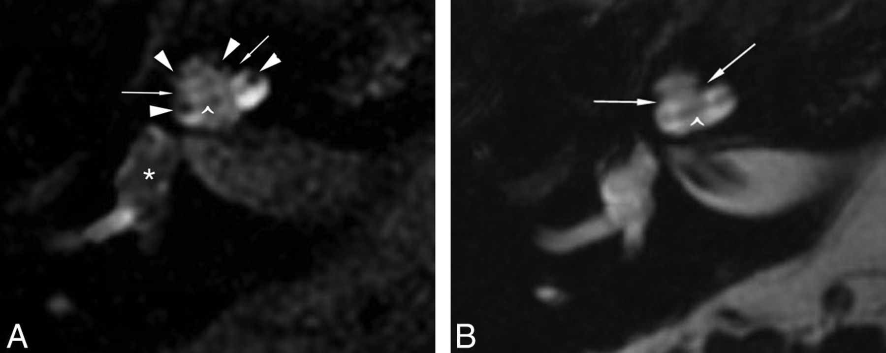
- Fig 1.
Differentiation of the perilymphatic-versus-endolymphatic spaces is evident when comparing FLAIR (A) and FIESTA images (B) obtained 21 hours after IT injection of 1:7 volume/volume diluted Magnevist contrast with a 3-inch surface coil. The nonenhancing fibro-osseous structures are evident on both sequences: the interscalar septa, separating the basal and middle turns and middle and apical turns (arrows), and the spiral lamina apparatus within each cochlear turn (caret). The FLAIR sequence shows central nonenhancement of the endolymphatic space of the vestibule (A, asterisk) and suggestion of distention of the scala media (A, arrowheads) into the scala vestibuli, whereas on FIESTA image (B), the endolymph and perilymph are both hyperintense and indistinguishable (patient 2).
- Fig 2.
The nonenhancing endolymphatic space (A, arrow) occupies >33% of the area of the vestibule on axial FLAIR image (A) obtained 28 hours after IT GBCA injection, suggesting endolymphatic distention. Coronal FLAIR obtained concurrently (B) demonstrates that the extent of distention of the endolymphatic space is overestimated on the axial view, due to partial volume averaging and section prescription through the membranous utricle (B, arrow). Partial volume averaging also likely contributes to signal heterogeneity within the semicircular canals (A). Arrowheads (B) correspond to the endolymphatic ductal ampullae of the superior and lateral semicircular canals (patient 6).
- Fig 3.
FLAIR imaging performed 28 hours after left IT contrast injection reveals a hyperintense structure (A, arrowhead) extending parallel to the expected course of the vestibular aqueduct (not seen). Comparison with positive-contrast T1-weighted images obtained before IT injection confirms that this structure is an enhancing dural vessel (B and C, arrowheads) coursing parallel to the posterior semicircular canal, extending from the middle cranial fossa to the sigmoid sinus. Correlation with anatomic imaging is imperative to avoid the misinterpretation of dilated endolymphatic space in the vestibular aqueduct (patient 4).
- Fig 4.
The impact of variable fluid suppression is visually evident with direct comparison of FLAIR sequences with TIs of 2000 ms (A and D), 2500 ms (B and E), and 2800 ms (C and F), with all other parameters remaining fixed. The nonenhancing scala media (arrowheads) becomes less conspicuous during this short range of TI (A–C), which may result in altered perception of endolymphatic space distention. Variation corresponding to the larger endolymphatic space in the vestibule (arrows, D–F) is less perceptible with changes in TI (patient 4).
- Fig 5.
Enhancement is visible at the fundus of the IAC (arrow) on delayed imaging after IT GBCA, conspicuous on FLAIR (A) and T1-weighted imaging (B) in this patient who had profound distention of the endolymphatic space in the basal and middle cochlear turns (arrowheads) (patient 5).


Tables
No. Age (yr) Sex, Ethnicity Side Years since Onset Prior Therapy Symptoms SRT/SDSa Right Left 1 62 Female Left 6 HCTZ ∼2 Vertigo episodes/year 20 dB 90 dB 96% at 60 dB 4% at 100 dB 2 45 Male Right 3 HCTZ, oral steroids Waxing/waning roaring tinnitus, occasional vertigo 15 dB 5 dB 96% at 75 dB 100% at 45 dB 3 55 Male Right 1 HCTZ Tinnitus ×1 year, 15 dB 20 dB episodic vertigo ×2 mo 96% at 55 dB 96% at 80 dB 4 63 Female Right >10 Bilateral IT steroid injection, oral steroid, HCTZ 8 Vertigo attacks within 2 mo 65 dB 25 dB 56% at 80 dB 96% at 50 dB 5 53 Female Left >10 Endolymphatic shunt, IT gentamicin ×9, IT steroids, gent/dex impregnated Gelfoamb sponge in round window niche Tinnitus and pressure 10 dB 30 dB 96% at 45 dB 92% at 65 dB 6 35 Female Right 3 HCTZ Episodic vertigo within prior 2 weeks 65 dB 10 dB 44% at 80 dB 96% at 50 dB Note:—HCTZ indicates hydrochlorothiazide; SRT, speech reception threshold; SDS, speech discrimination score (%) at supra-SRT level (decibel); gent/dex, gentamicin/dexamethasone.
↵a Normal = Less than 25, mild = 26–40, moderate = 41–55, moderate/severe = 56–70, severe = 71–90, profound hearing loss >90.
↵b Phadia, Uppsala, Sweden.
Coil Sequence TE (ms) TI (ms) TR (ms) Matrix FOV (cm) In-Plane Resolution (mm) Thickness (mm) Flip Angle Bandwidth (Hz) Time to Acquire (min:s) 8-Channel Cisternography (FIESTA) 3 6 320 × 320 18 0.56 × 0.56 1 55° 163 4:27 2D FLAIR 122 2500 9454 320 × 320 18 0.56 × 0.56 2 90° 122 4:06 T1 spin-echob 9 400 320 × 320 18 0.56 × 0.56 2 90° 163 4:19 3-Inch surface Cisternography (FIESTA) 4 8 320 × 320 12 0.38 × 0.38 1 55° 163 4:35 2D FLAIR (3 pt) 124 2000c 10,000 320 × 320 12 0.38 × 0.38 2 90° 122 4:12 2D FLAIR (3 pt) 123 2000 9000 320 × 320 12 0.38 × 0.38 2 90° 122 4:24 T1 spin-echob 11 500 320 × 320 12 0.38 × 0.38 2 90° 163 5:10 Note:—pt indicates patients.
↵a Protocol 1 (n = 3), 1 scan session: 20–28 hours post-IT contrast, includes pre- and post-IV contrast images. Protocol 2 (n = 2), 3 scan sessions: 1) pre- and post-IV contrast images (no IT), 2) 2 hours post-IT contrast/4 hours post-IV contrast, 3) 20–28 hours post-IT contrast/30 hours post-IV contrast. Protocol 2B (n = 1; scheduling conflict precluded pre-IT injection imaging as above in protocol 2 number 1), 2 scan sessions: 1) 20 hours post-IT contrast with pre-and post-IV contrast images, 2) 25 hours post-IT contrast/4 hours post-IV contrast.
↵b Parameters for T1 pre- and post-IV contrast scans did not change.
↵c FLAIR TI was varied from 1800 to 2800 ms for 2 patient scans, with all other parameters fixed.


Jump to section
Related Articles
Cited By...
- No citing articles found.