
Article Figures & Data
Figures
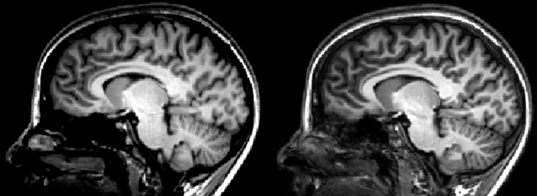
- Fig 1.
Comparison of the normal MPRAGE protocol (left and middle image, different windowing) with the optimized MPRAGE protocol (right).
- Fig 2.
Comparison of the normal MPRAGE protocol (top row) with the optimized protocol (bottom row) in different section orientations. Contrast in both images was chosen for an optimal depiction of thalamus substructures. Adjacent anatomic structures have been labeled. cc indicates corpus callosum; fx, fornix; R, red nucleus; LV, lateral ventricles; C, caudate nucleus; Pu, putamen; IC, internal capsule; GP, globus pallidus.
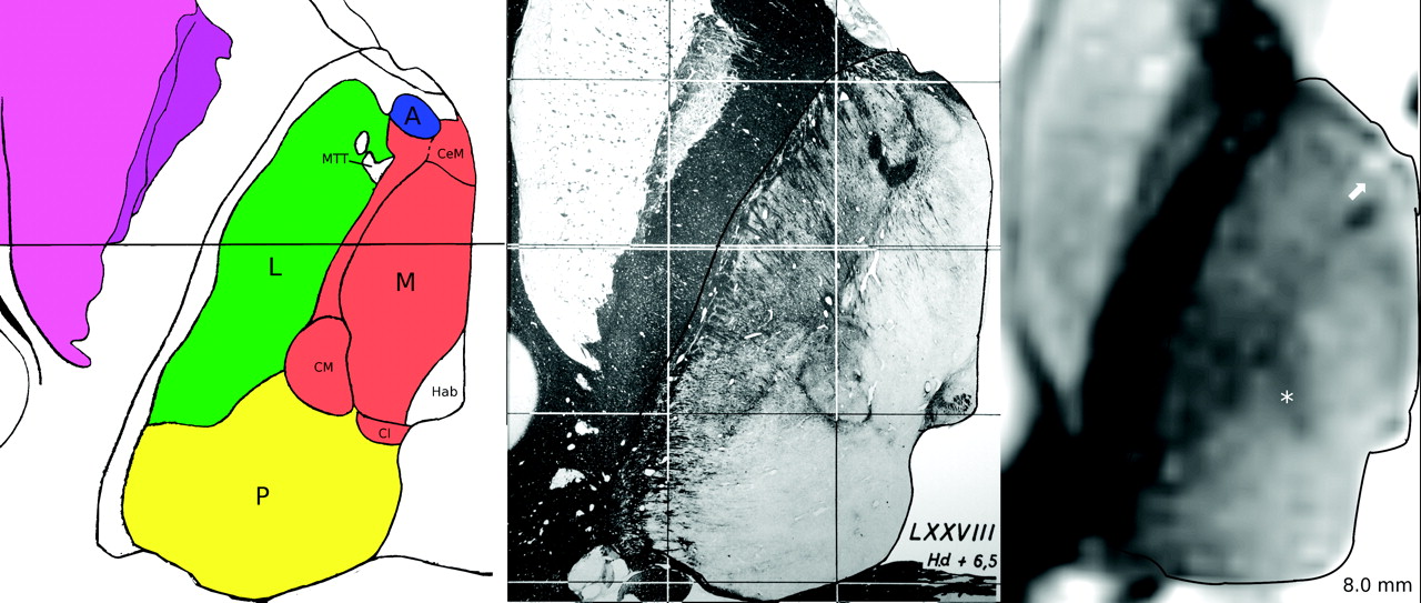
- Fig 3.
Axial myelin stain (middle) from the Schaltenbrand and Wahren atlas,17 taken it from plate 54, located 6.5 mm above Reil's plane, and a corresponding inverted gray-scale image from one of the subjects located 8 mm above Reil's plane (right). For anatomic reference, a schematic drawing adapted from the overlay of the Schaltenbrand and Wahren atlas is shown on the left. The boundaries of the thalamus have been delineated in all images with a thin black line. The white arrow marks the anteroventral nucleus, which can be clearly delineated as a hyperintense structure. The CM (marked by an asterisk) is clearly distinguishable from the surrounding structures. The anatomic structures belonging to the 4 thalamic nuclei groups used in this work have been colored (blue, anterior; red, medial; green, lateral; yellow, posterior).
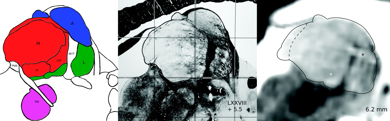
- Fig 4.
Parasagittal myelin stain (middle) from the Schaltenbrand and Wahren atlas,17 taken from plate 44, located 5.5 mm lateral to the midline and a corresponding inverted gray-scale image from one of the subjects, located 6.2 mm lateral to the midline (right). The boundaries of the thalamus have been delineated in all images with a thin black line. The white star marks the CM, which can be clearly localized. The bright structure (black star) close to the MTT is a vessel. For anatomic reference, a schematic drawing adapted from the overlay of the Schaltenbrand and Wahren atlas is shown on the left. The anatomic structures belonging to the 4 thalamic nuclei groups used in this work have been colored (blue, anterior; red, medial; green, lateral).
- Fig 5.
An axial section of the optimized MPRAGE. Note that not only the 4 large nucleus groups (middle image) but also the further substructures can be identified. Medial to the MD, a hyperintense structure is also visible (dashed line depicts border), which is most likely the superficial medial nucleus9 not described in the Morel atlas.15 In the lateral nucleus group, only thin bands of white matter can be used to distinguish the different parts. The MTT is clearly visible as a hyperintense spot between the anteroventral nucleus (AV), central medial nucleus (CeM), and ventral anterior nucleus (VA). CL indicates central lateral nucleus; VLa, ventral lateral anterior nucleus; VLp, ventral lateral posterior nucleus; VPL, ventral posterior lateral nucleus; LP, lateral posterior nucleus; PuM, medial pulvinar.
- Fig 6.
Optimized MPRAGE (bottom row) and nucleus groups in axial sections parallel to the ACPC line (from left to right: apical to caudal sections; position given in millimeters relative to the ACPC line) selected by the first and second readers (top row, light and dark gray lines).


Tables
Summary of the volume size of the thalamus and substructures of the thalamus in all subjectsa
Subject No., Age (yr), Sex Size (mm2) Anterior Medial Lateral Posterior Thalamus B.B. C.M. B.B. C.M. B.B. C.M. B.B. C.M. B.B. C.M. 1) 27, F 218 203 1608 1639 2193 2152 2273 2261 6292 6255 2) 26, M 227 205 1562 1476 2123 2134 2277 2291 6189 6106 3) 21, F 221 202 1567 1520 1969 1970 2186 2187 5943 5879 4) 23, F 200 186 1211 1184 1699 1740 1811 1934 4921 5044 5) 34, F 293 286 1555 1499 2216 2287 2008 2099 6072 6171 6) 28, M 197 209 1328 1380 1728 1757 2057 2174 5310 5520 Mean ± Std 226 ± 35 215 ± 32 1471 ± 162 1450 ± 141 1988 ± 230 2007 ± 204 2102 ± 180 2158 ± 118 5788 ± 548 5829 ± 426 Atlas 249 1380 1727 1813 5169 ↵a For both readers (B.B., C.M.) and in the 3D mean thalamus atlas from Krauth et al.10


