Advertisement

Main menu

  • Home
  • Content
    • Current Issue
    • Accepted Manuscripts
    • Article Preview
    • Past Issue Archive
    • Video Articles
    • AJNR Case Collection
    • Case of the Week Archive
    • Case of the Month Archive
    • Classic Case Archive
  • Special Collections
    • AJNR Awards
    • Low-Field MRI
    • Alzheimer Disease
    • ASNR Foundation Special Collection
    • Photon-Counting CT
    • View All
  • Multimedia
    • AJNR Podcasts
    • AJNR SCANtastic
    • Trainee Corner
    • MRI Safety Corner
    • Imaging Protocols
  • For Authors
    • Submit a Manuscript
    • Submit a Video Article
    • Submit an eLetter to the Editor/Response
    • Manuscript Submission Guidelines
    • Statistical Tips
    • Fast Publishing of Accepted Manuscripts
    • Graphical Abstract Preparation
    • Imaging Protocol Submission
    • Author Policies
  • About Us
    • About AJNR
    • Editorial Board
    • Editorial Board Alumni
  • More
    • Become a Reviewer/Academy of Reviewers
    • Subscribers
    • Permissions
    • Alerts
    • Feedback
    • Advertisers
    • ASNR Home

User menu

  • Alerts
  • Log in

Search

  • Advanced search
American Journal of Neuroradiology

American Journal of Neuroradiology

ASHNR American Society of Functional Neuroradiology ASHNR American Society of Pediatric Neuroradiology ASSR
  • Alerts
  • Log in

Advanced Search

  • Home
  • Content
    • Current Issue
    • Accepted Manuscripts
    • Article Preview
    • Past Issue Archive
    • Video Articles
    • AJNR Case Collection
    • Case of the Week Archive
    • Case of the Month Archive
    • Classic Case Archive
  • Special Collections
    • AJNR Awards
    • Low-Field MRI
    • Alzheimer Disease
    • ASNR Foundation Special Collection
    • Photon-Counting CT
    • View All
  • Multimedia
    • AJNR Podcasts
    • AJNR SCANtastic
    • Trainee Corner
    • MRI Safety Corner
    • Imaging Protocols
  • For Authors
    • Submit a Manuscript
    • Submit a Video Article
    • Submit an eLetter to the Editor/Response
    • Manuscript Submission Guidelines
    • Statistical Tips
    • Fast Publishing of Accepted Manuscripts
    • Graphical Abstract Preparation
    • Imaging Protocol Submission
    • Author Policies
  • About Us
    • About AJNR
    • Editorial Board
    • Editorial Board Alumni
  • More
    • Become a Reviewer/Academy of Reviewers
    • Subscribers
    • Permissions
    • Alerts
    • Feedback
    • Advertisers
    • ASNR Home
  • Follow AJNR on Twitter
  • Visit AJNR on Facebook
  • Follow AJNR on Instagram
  • Join AJNR on LinkedIn
  • RSS Feeds

AJNR Awards, New Junior Editors, and more. Read the latest AJNR updates

LetterLetter

Premortem Diagnosis of Sporadic Creutzfeldt-Jakob Disease Aided by Positron-Emission Tomography Imaging

W.-J. Zhang, M.B. Westover and C.J. Keary
American Journal of Neuroradiology January 2011, 32 (1) E18; DOI: https://doi.org/10.3174/ajnr.A2292
W.-J. Zhang
  • Find this author on Google Scholar
  • Find this author on PubMed
  • Search for this author on this site
M.B. Westover
  • Find this author on Google Scholar
  • Find this author on PubMed
  • Search for this author on this site
C.J. Keary
  • Find this author on Google Scholar
  • Find this author on PubMed
  • Search for this author on this site
  • Article
  • Figures & Data
  • Info & Metrics
  • Responses
  • References
  • PDF
Loading

A 72-year-old woman presented 6 months after the onset of falls and ataxia, which had progressed to wheelchair dependence, followed by the onset of bizarre behavior and rapid cognitive deterioration, for which she was hospitalized. Neurologic examination revealed marked disorientation, impairments in recent memory and attention, paranoia, disorganization of thought, dysarthria, symmetric waxy limb rigidity, ataxia of all limbs, and profound gait ataxia. Her medical history was notable only for an implanted cardiac pacemaker, precluding brain MR imaging.

Creutzfeldt-Jakob disease (CJD) was suspected clinically. A thorough search for alternative causes of rapidly progressive dementia was negative.1 CSF was noninflammatory but showed elevation of neuron-specific enolase and 14–3–3 protein. Electroencephalography (EEG) showed generalized slowing.

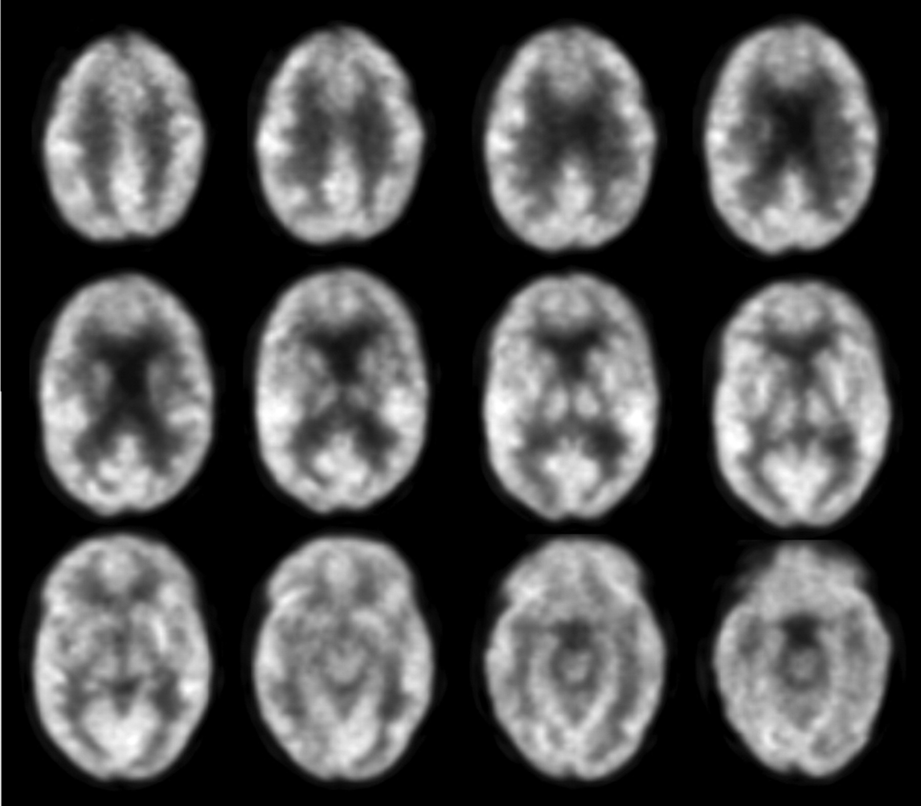
A brain [18F] fluorodeoxyglucose–positron-emission tomography (PET) scan revealed multiple scattered areas of focal cortical hypometabolism in the frontal, parietal, temporal, and occipital lobes. Additionally, there was marked hypometabolism in the bilateral caudate nuclei and heterogeneously in the cerebellum, midbrain, and upper pons (Fig 1). This pattern of basal ganglia and patchy cortical hypometabolism without white matter involvement is analogous to the anatomic distribution of MR imaging findings in CJD1,2 and thus is strongly supportive of the clinical diagnosis.

Fig 1.
  • Download figure
  • Open in new tab
  • Download powerpoint
Fig 1.

PET scans show basal ganglia and patchy cortical hypometabolism, supportive of the clinical diagnosis of CJD.

During 2 weeks of hospitalization, the patient developed hallucinations, worsened limb ataxia, swallowing apraxia, and ultimately akinetic mutism. There was no response to a brief trial of high-dose corticosteroids. She was discharged to hospice care with a diagnosis of probable CJD, and she died 1 month later.

The terminal prognosis of CJD necessitates being reasonably certain before rendering a premortem diagnosis. Our patient's clinical presentation was typical for CJD, in the absence of alternative explanations, making the pretest probability for CJD high. The sensitivities and specificities (SE, Sp) for the usual ancillary tests are as follows: Periodic sharp wave complexes on EEG are specific (Sp ∼ 90%), but EEG is insensitive (SE ∼ 50%); the nonspecific “generalized slowing” seen in our case is typical.1 CSF 14–3–3 testing in recent studies shows modest statistical utility (SE ∼ 48%, Sp ∼ 65%), though other CSF markers such as neuron-specific enolase may be more reliable.1,3 Specific MR imaging fluid-attenuated inversion recovery and diffusion-weighted imaging patterns have emerged as reliable key diagnostic indicators (SE ∼ 92%; Sp, 94%), in which key findings include cortical hyperintensity restricted to the cortex (“cortical ribbon sign”), striatum, or both.1,2

The diagnostic utility of PET imaging is not well-established for CJD. However, we suspect that cases like ours, in which MR imaging is not feasible, are fairly common. The PET findings in our case are in agreement with those in a handful of other reported CJD cases in which PET imaging was performed.4 These imaging findings are distinct from findings in other common neurodegenerative diseases such as Alzheimer disease and Lewy body dementia3 but are anatomically analogous to the distribution of abnormalities seen in CJD on MR imaging. This case presents a clinical situation in which PET imaging provided strong supporting evidence for a diagnosis of probable CJD in the absence of MR imaging and suggests a future use for PET in the evaluation of rapidly progressive dementia.

References

  1. 1.↵
    1. Geschwind MD,
    2. Shu H,
    3. Haman A,
    4. et al
    . Rapidly progressive dementia. Ann Neurol 2008;64:97–108
    CrossRefPubMed
  2. 2.↵
    1. Shiga Y,
    2. Miyazawa K,
    3. Sato S,
    4. et al
    . Diffusion-weighted MRI abnormalities as an early diagnostic marker for Creutzfeldt-Jakob disease. Neurology 2004;63:443–49
    Abstract/FREE Full Text
  3. 3.↵
    1. Geschwind MD,
    2. Martindale J,
    3. Miller D,
    4. et al
    . Challenging the clinical utility of the 14–3-3 protein for the diagnosis of sporadic Creutzfeldt-Jakob disease. Arch Neurol 2003;60:813–16
    CrossRefPubMed
  4. 4.↵
    1. Henkel K,
    2. Zerr I,
    3. Hertel A,
    4. et al
    . Positron emission tomography with [(18)F]FDG in the diagnosis of Creutzfeldt-Jakob disease (CJD). J Neurol 2002;249:699–705
    CrossRefPubMed
  • Copyright © American Society of Neuroradiology
PreviousNext
Back to top

In this issue

American Journal of Neuroradiology: 32 (1)
American Journal of Neuroradiology
Vol. 32, Issue 1
1 Jan 2011
  • Table of Contents
  • Index by author
Advertisement
Print
Download PDF
Email Article

Thank you for your interest in spreading the word on American Journal of Neuroradiology.

NOTE: We only request your email address so that the person you are recommending the page to knows that you wanted them to see it, and that it is not junk mail. We do not capture any email address.

Enter multiple addresses on separate lines or separate them with commas.
Premortem Diagnosis of Sporadic Creutzfeldt-Jakob Disease Aided by Positron-Emission Tomography Imaging
(Your Name) has sent you a message from American Journal of Neuroradiology
(Your Name) thought you would like to see the American Journal of Neuroradiology web site.
CAPTCHA
This question is for testing whether or not you are a human visitor and to prevent automated spam submissions.
Cite this article
W.-J. Zhang, M.B. Westover, C.J. Keary
Premortem Diagnosis of Sporadic Creutzfeldt-Jakob Disease Aided by Positron-Emission Tomography Imaging
American Journal of Neuroradiology Jan 2011, 32 (1) E18; DOI: 10.3174/ajnr.A2292

Citation Manager Formats

  • BibTeX
  • Bookends
  • EasyBib
  • EndNote (tagged)
  • EndNote 8 (xml)
  • Medlars
  • Mendeley
  • Papers
  • RefWorks Tagged
  • Ref Manager
  • RIS
  • Zotero
0 Responses
Respond to this article
Share
Bookmark this article
Premortem Diagnosis of Sporadic Creutzfeldt-Jakob Disease Aided by Positron-Emission Tomography Imaging
W.-J. Zhang, M.B. Westover, C.J. Keary
American Journal of Neuroradiology Jan 2011, 32 (1) E18; DOI: 10.3174/ajnr.A2292
del.icio.us logo Twitter logo Facebook logo Mendeley logo
  • Tweet Widget
  • Facebook Like
  • Google Plus One
Purchase

Jump to section

  • Article
    • References
  • Figures & Data
  • Info & Metrics
  • Responses
  • References
  • PDF

Related Articles

  • No related articles found.
  • PubMed
  • Google Scholar

Cited By...

  • No citing articles found.
  • Crossref
  • Google Scholar

This article has not yet been cited by articles in journals that are participating in Crossref Cited-by Linking.

More in this TOC Section

  • Letter to the Editor regarding “Automated Volumetric Software in Dementia: Help or Hindrance to the Neuroradiologist?”
  • Reply:
  • Brain AVM’s Nidus: What if We Hadn’t Understood Anything?
Show more LETTERS

Similar Articles

Advertisement

Indexed Content

  • Current Issue
  • Accepted Manuscripts
  • Article Preview
  • Past Issues
  • Editorials
  • Editor's Choice
  • Fellows' Journal Club
  • Letters to the Editor
  • Video Articles

Cases

  • Case Collection
  • Archive - Case of the Week
  • Archive - Case of the Month
  • Archive - Classic Case

More from AJNR

  • Trainee Corner
  • Imaging Protocols
  • MRI Safety Corner
  • Book Reviews

Multimedia

  • AJNR Podcasts
  • AJNR Scantastics

Resources

  • Turnaround Time
  • Submit a Manuscript
  • Submit a Video Article
  • Submit an eLetter to the Editor/Response
  • Manuscript Submission Guidelines
  • Statistical Tips
  • Fast Publishing of Accepted Manuscripts
  • Graphical Abstract Preparation
  • Imaging Protocol Submission
  • Evidence-Based Medicine Level Guide
  • Publishing Checklists
  • Author Policies
  • Become a Reviewer/Academy of Reviewers
  • News and Updates

About Us

  • About AJNR
  • Editorial Board
  • Editorial Board Alumni
  • Alerts
  • Permissions
  • Not an AJNR Subscriber? Join Now
  • Advertise with Us
  • Librarian Resources
  • Feedback
  • Terms and Conditions
  • AJNR Editorial Board Alumni

American Society of Neuroradiology

  • Not an ASNR Member? Join Now

© 2025 by the American Society of Neuroradiology All rights, including for text and data mining, AI training, and similar technologies, are reserved.
Print ISSN: 0195-6108 Online ISSN: 1936-959X

Powered by HighWire