Advertisement

Main menu

  • Home
  • Content
    • Current Issue
    • Accepted Manuscripts
    • Article Preview
    • Past Issue Archive
    • Video Articles
    • AJNR Case Collection
    • Case of the Week Archive
    • Case of the Month Archive
    • Classic Case Archive
  • Special Collections
    • AJNR Awards
    • Low-Field MRI
    • Alzheimer Disease
    • ASNR Foundation Special Collection
    • Photon-Counting CT
    • View All
  • Multimedia
    • AJNR Podcasts
    • AJNR SCANtastic
    • Trainee Corner
    • MRI Safety Corner
    • Imaging Protocols
  • For Authors
    • Submit a Manuscript
    • Submit a Video Article
    • Submit an eLetter to the Editor/Response
    • Manuscript Submission Guidelines
    • Statistical Tips
    • Fast Publishing of Accepted Manuscripts
    • Graphical Abstract Preparation
    • Imaging Protocol Submission
    • Author Policies
  • About Us
    • About AJNR
    • Editorial Board
    • Editorial Board Alumni
  • More
    • Become a Reviewer/Academy of Reviewers
    • Subscribers
    • Permissions
    • Alerts
    • Feedback
    • Advertisers
    • ASNR Home

User menu

  • Alerts
  • Log in

Search

  • Advanced search
American Journal of Neuroradiology

American Journal of Neuroradiology

ASHNR American Society of Functional Neuroradiology ASHNR American Society of Pediatric Neuroradiology ASSR
  • Alerts
  • Log in

Advanced Search

  • Home
  • Content
    • Current Issue
    • Accepted Manuscripts
    • Article Preview
    • Past Issue Archive
    • Video Articles
    • AJNR Case Collection
    • Case of the Week Archive
    • Case of the Month Archive
    • Classic Case Archive
  • Special Collections
    • AJNR Awards
    • Low-Field MRI
    • Alzheimer Disease
    • ASNR Foundation Special Collection
    • Photon-Counting CT
    • View All
  • Multimedia
    • AJNR Podcasts
    • AJNR SCANtastic
    • Trainee Corner
    • MRI Safety Corner
    • Imaging Protocols
  • For Authors
    • Submit a Manuscript
    • Submit a Video Article
    • Submit an eLetter to the Editor/Response
    • Manuscript Submission Guidelines
    • Statistical Tips
    • Fast Publishing of Accepted Manuscripts
    • Graphical Abstract Preparation
    • Imaging Protocol Submission
    • Author Policies
  • About Us
    • About AJNR
    • Editorial Board
    • Editorial Board Alumni
  • More
    • Become a Reviewer/Academy of Reviewers
    • Subscribers
    • Permissions
    • Alerts
    • Feedback
    • Advertisers
    • ASNR Home
  • Follow AJNR on Twitter
  • Visit AJNR on Facebook
  • Follow AJNR on Instagram
  • Join AJNR on LinkedIn
  • RSS Feeds

AJNR Awards, New Junior Editors, and more. Read the latest AJNR updates

OtherReview Articles

MR Imaging Findings in Hepatic Encephalopathy

A. Rovira, J. Alonso and J. Córdoba
American Journal of Neuroradiology October 2008, 29 (9) 1612-1621; DOI: https://doi.org/10.3174/ajnr.A1139
A. Rovira
  • Find this author on Google Scholar
  • Find this author on PubMed
  • Search for this author on this site
J. Alonso
  • Find this author on Google Scholar
  • Find this author on PubMed
  • Search for this author on this site
J. Córdoba
  • Find this author on Google Scholar
  • Find this author on PubMed
  • Search for this author on this site
  • Article
  • Figures & Data
  • Info & Metrics
  • Responses
  • References
  • PDF
Loading

Article Figures & Data

Figures

  • Tables
  • Fig 1.
    • Download figure
    • Open in new tab
    • Download powerpoint
    Fig 1.

    Transverse T1-weighted MR images of the brain in a patient with chronic liver failure and parkinsonism. Observe the bilateral and symmetric high T1 signal-intensity change involving the globus pallidus and the anterior midbrain.
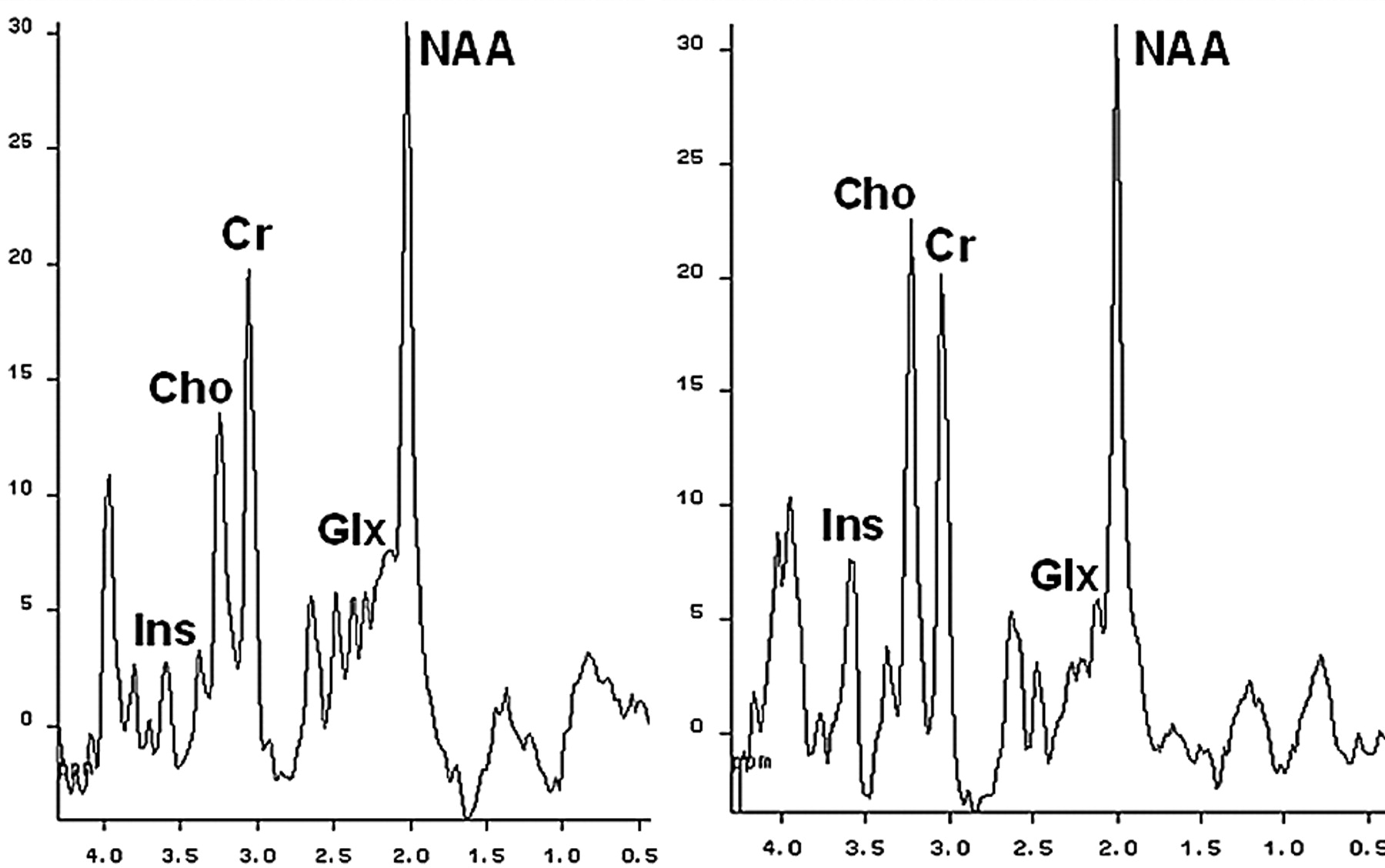
  • Fig 2.
    • Download figure
    • Open in new tab
    • Download powerpoint
    Fig 2.

    1H-MR-spectroscopy water-suppressed proton spectra of an 8-mL voxel located in the parietal region including predominantly normal-appearing white matter in a patient with cirrhosis before (left) and after (right) liver transplantation, recorded with a stimulated echo acquisition mode pulse sequence (TR/TE, 1600/20 ms; acquisitions, 256). The main resonances correspond to N-acetylaspartate (NAA, 2.0 ppm), glutamine/glutamate (Glx, 2.1–2.5 ppm), creatine/phosphocreatine (Cr, 3.02 ppm), choline-containing compounds (Cho, 3.2 ppm), and myo-inositol (Ins, 3.55 ppm). The initial spectrum shows an increase in the glutamate/glutamine region and a decrease in the myo-inositol and choline resonances. These abnormalities normalized after liver transplantation. Normal NAA indices are seen in both examinations.

  • Fig 3.
    • Download figure
    • Open in new tab
    • Download powerpoint
    Fig 3.

    A, Transverse T2-weighted fast FLAIR images obtained in a patient with liver cirrhosis during an episode of hepatic encephalopathy. Observe the symmetric areas of increased signal intensity along the corticospinal tract in both cerebral hemispheres. B, This signal-intensity abnormality almost completely reverses on a follow-up study obtained few months later, when the patient showed no signs of overt hepatic encephalopathy.

  • Fig 4.
    • Download figure
    • Open in new tab
    • Download powerpoint
    Fig 4.

    A, Baseline MR imaging study (transverse fast FLAIR T2-weighted image) of a 56-year-old patient with hepatitis C cirrhosis without overt hepatic encephalopathy. Multiple focal WMLs in both cerebral hemispheres are attributed to small-vessel disease. B, A new scan obtained 2 years later during an episode of hepatic encephalopathy shows marked increase in the size of these focal WMLs. C, A new follow-up scan after complete resolution of neurologic symptoms shows a decrease in the size of the WMLs. This last scan was almost identical to the first study. A lacunar infarct is seen in the deep right frontal white matter.

Tables

  • Figures
    • View popup
    Table 1:

    Classification of hepatic encephalopathy (HE)

    HELiver DiseaseExtrahepatic Portal-Systemic ShuntingNeurologic ManifestationsSpecific Features
    Acute episode
        In cirrhosisCirrhosisVariableAcute confusional state to comaUsually precipitated
        In acute liver failureAcute liver failureAbsentAcute confusional state to comaFrequently complicated by brain edema and intracranial hypertension
    Chronic
        RelapsingCirrhosisSevereRelapsing episodes of encephalopathyUsually without precipitating factors
        PersistentCirrhosisSeverePersistent cognitive or motor abnormalitiesGenerally related to surgically induced shunts
    Minimal HECirrhosisVariableAsymptomaticAbnormalities revealed by neuropsychological or neurophysiologic tests
    In patients with portal-systemic bypass with no intrinsic hepatocellular diseaseAbsentLarge shuntsRelapsing episodes and persistent abnormalitiesRare disorder, secondary to congenital abnormalities, surgical shunts, or portal vein thrombosis
    • View popup
    Table 2:

    West Haven criteria for semiquantitative grading of HE

    GradeCriteria
    1Trivial lack of awareness, euphoria or anxiety, shortened attention span, impaired performance of addition (Sixty-seven percent of patients with cirrhosis may have minimal HE.)
    2Lethargy or apathy, minimal disorientation for time or place, subtle personality change, inappropriate behavior, impaired performance of subtraction
    3Somnolence to semistupor, but responsive to verbal stimuli; confusion; gross disorientation
    4Coma (unresponsive to verbal or noxious stimuli)
    • View popup
    Table 3:

    MR imaging features in patients with cirrhosis with or without overt HE

    T1WI Sequence1H-MR spectroscopyMTRT2/FLAIR SequenceDWI
    MR imaging abnormalitiesBilateral, symmetric high signal intensity of the globus pallidus and substantia nigraIncrease in glutamine/glutamate signal; depletion of myo-inositol signal; decrease in choline signal; normal NAA signalMild (10%) and diffuse decrease in normal-appearing white matterDiffuse white matter high signal intensities involving predominantly the hemispheric corticospinal tract; focal high-signal T2 lesions in subcortical hemispheric white matterIncrease mean diffusivity in hemispheric white matter; normal fractional anisotropy
    PathogenesisIncreased brain tissue concentration of manganeseOsmolar adaptation of intra-astrocytic accumulation of glutamineMild and diffuse brain edemaMild and diffuse brain edemaInterstitial brain edema
    Functional consequencesParkinsonism (particularly if substantia nigra is involved)Overt hepatic encephalopathy (particularly with increase in glutamine/glutamate)Functional abnormalities of the corticospinal tract on TMSFunctional abnormalities of the corticospinal tract on TMS; cognitive impairmentCognitive impairment
    • Note:—T1WI Indicates T1-weighted imaging; NAA, N-acetylaspartate; TMS, transcranial magnetic stimulation; FLAIR, fluid-attenuated inversion recovery; DWI, diffusion-weighted imaging; MTR, magnetization transfer ratio.

PreviousNext
Back to top

In this issue

American Journal of Neuroradiology: 29 (9)
American Journal of Neuroradiology
Vol. 29, Issue 9
October 2008
  • Table of Contents
  • Index by author
Advertisement
Print
Download PDF
Email Article

Thank you for your interest in spreading the word on American Journal of Neuroradiology.

NOTE: We only request your email address so that the person you are recommending the page to knows that you wanted them to see it, and that it is not junk mail. We do not capture any email address.

Enter multiple addresses on separate lines or separate them with commas.
MR Imaging Findings in Hepatic Encephalopathy
(Your Name) has sent you a message from American Journal of Neuroradiology
(Your Name) thought you would like to see the American Journal of Neuroradiology web site.
CAPTCHA
This question is for testing whether or not you are a human visitor and to prevent automated spam submissions.
Cite this article
A. Rovira, J. Alonso, J. Córdoba
MR Imaging Findings in Hepatic Encephalopathy
American Journal of Neuroradiology Oct 2008, 29 (9) 1612-1621; DOI: 10.3174/ajnr.A1139

Citation Manager Formats

  • BibTeX
  • Bookends
  • EasyBib
  • EndNote (tagged)
  • EndNote 8 (xml)
  • Medlars
  • Mendeley
  • Papers
  • RefWorks Tagged
  • Ref Manager
  • RIS
  • Zotero
0 Responses
Respond to this article
Share
Bookmark this article
MR Imaging Findings in Hepatic Encephalopathy
A. Rovira, J. Alonso, J. Córdoba
American Journal of Neuroradiology Oct 2008, 29 (9) 1612-1621; DOI: 10.3174/ajnr.A1139
del.icio.us logo Twitter logo Facebook logo Mendeley logo
  • Tweet Widget
  • Facebook Like
  • Google Plus One
Purchase

Jump to section

  • Article
    • Abstract
    • Clinical Features
    • Pathogenesis of HE
    • Manganese
    • MR Imaging Marker of Manganese Accumulation within the CNS
    • Ammonia
    • Diffuse Brain Edema in Chronic Liver Failure
    • Conclusions
    • Acknowledgments
    • References
  • Figures & Data
  • Info & Metrics
  • Responses
  • References
  • PDF

Related Articles

  • No related articles found.
  • PubMed
  • Google Scholar

Cited By...

  • Meta-analysis of magnetic resonance spectroscopy in the diagnosis of hepatic encephalopathy
  • What to see when you are looking at confusion: a review of the neuroimaging of acute encephalopathy
  • Neuroimaging in Patients with Abnormal Blood Glucose Levels
  • Acute Hyperammonemic Encephalopathy in Adults: Imaging Findings
  • Acute Hepatic Encephalopathy: Diffusion-Weighted and Fluid-Attenuated Inversion Recovery Findings, and Correlation with Plasma Ammonia Level and Clinical Outcome
  • Crossref
  • Google Scholar

This article has not yet been cited by articles in journals that are participating in Crossref Cited-by Linking.

More in this TOC Section

  • An Atlas of Neonatal Neurovascular Imaging Anatomy as Depicted with Microvascular Imaging: The Intracranial Arteries
  • An Atlas of Neonatal Neurovascular Imaging Anatomy as Depicted with Microvascular Imaging: The Intracranial Veins
  • Clinical Translation of Hyperpolarized 13C Metabolic Probes for Glioma Imaging
Show more Review Articles

Similar Articles

Advertisement

Indexed Content

  • Current Issue
  • Accepted Manuscripts
  • Article Preview
  • Past Issues
  • Editorials
  • Editor's Choice
  • Fellows' Journal Club
  • Letters to the Editor
  • Video Articles

Cases

  • Case Collection
  • Archive - Case of the Week
  • Archive - Case of the Month
  • Archive - Classic Case

More from AJNR

  • Trainee Corner
  • Imaging Protocols
  • MRI Safety Corner
  • Book Reviews

Multimedia

  • AJNR Podcasts
  • AJNR Scantastics

Resources

  • Turnaround Time
  • Submit a Manuscript
  • Submit a Video Article
  • Submit an eLetter to the Editor/Response
  • Manuscript Submission Guidelines
  • Statistical Tips
  • Fast Publishing of Accepted Manuscripts
  • Graphical Abstract Preparation
  • Imaging Protocol Submission
  • Evidence-Based Medicine Level Guide
  • Publishing Checklists
  • Author Policies
  • Become a Reviewer/Academy of Reviewers
  • News and Updates

About Us

  • About AJNR
  • Editorial Board
  • Editorial Board Alumni
  • Alerts
  • Permissions
  • Not an AJNR Subscriber? Join Now
  • Advertise with Us
  • Librarian Resources
  • Feedback
  • Terms and Conditions
  • AJNR Editorial Board Alumni

American Society of Neuroradiology

  • Not an ASNR Member? Join Now

© 2025 by the American Society of Neuroradiology All rights, including for text and data mining, AI training, and similar technologies, are reserved.
Print ISSN: 0195-6108 Online ISSN: 1936-959X

Powered by HighWire