Advertisement

Main menu

  • Home
  • Content
    • Current Issue
    • Accepted Manuscripts
    • Article Preview
    • Past Issue Archive
    • Video Articles
    • AJNR Case Collection
    • Case of the Week Archive
    • Case of the Month Archive
    • Classic Case Archive
  • Special Collections
    • AJNR Awards
    • Low-Field MRI
    • Alzheimer Disease
    • ASNR Foundation Special Collection
    • Photon-Counting CT
    • View All
  • Multimedia
    • AJNR Podcasts
    • AJNR SCANtastic
    • Trainee Corner
    • MRI Safety Corner
    • Imaging Protocols
  • For Authors
    • Submit a Manuscript
    • Submit a Video Article
    • Submit an eLetter to the Editor/Response
    • Manuscript Submission Guidelines
    • Statistical Tips
    • Fast Publishing of Accepted Manuscripts
    • Graphical Abstract Preparation
    • Imaging Protocol Submission
    • Author Policies
  • About Us
    • About AJNR
    • Editorial Board
    • Editorial Board Alumni
  • More
    • Become a Reviewer/Academy of Reviewers
    • Subscribers
    • Permissions
    • Alerts
    • Feedback
    • Advertisers
    • ASNR Home

User menu

  • Alerts
  • Log in

Search

  • Advanced search
American Journal of Neuroradiology

American Journal of Neuroradiology

ASHNR American Society of Functional Neuroradiology ASHNR American Society of Pediatric Neuroradiology ASSR
  • Alerts
  • Log in

Advanced Search

  • Home
  • Content
    • Current Issue
    • Accepted Manuscripts
    • Article Preview
    • Past Issue Archive
    • Video Articles
    • AJNR Case Collection
    • Case of the Week Archive
    • Case of the Month Archive
    • Classic Case Archive
  • Special Collections
    • AJNR Awards
    • Low-Field MRI
    • Alzheimer Disease
    • ASNR Foundation Special Collection
    • Photon-Counting CT
    • View All
  • Multimedia
    • AJNR Podcasts
    • AJNR SCANtastic
    • Trainee Corner
    • MRI Safety Corner
    • Imaging Protocols
  • For Authors
    • Submit a Manuscript
    • Submit a Video Article
    • Submit an eLetter to the Editor/Response
    • Manuscript Submission Guidelines
    • Statistical Tips
    • Fast Publishing of Accepted Manuscripts
    • Graphical Abstract Preparation
    • Imaging Protocol Submission
    • Author Policies
  • About Us
    • About AJNR
    • Editorial Board
    • Editorial Board Alumni
  • More
    • Become a Reviewer/Academy of Reviewers
    • Subscribers
    • Permissions
    • Alerts
    • Feedback
    • Advertisers
    • ASNR Home
  • Follow AJNR on Twitter
  • Visit AJNR on Facebook
  • Follow AJNR on Instagram
  • Join AJNR on LinkedIn
  • RSS Feeds

AJNR Awards, New Junior Editors, and more. Read the latest AJNR updates

OtherSpine Imaging and Spine Image-Guided Interventions

Significance and Targeting of Small, Central Clefts in Severe Fractures Treated With Vertebroplasty

A. Ehteshami Rad, L.A. Gray and D.F. Kallmes
American Journal of Neuroradiology August 2008, 29 (7) 1285-1287; DOI: https://doi.org/10.3174/ajnr.A1066
A. Ehteshami Rad
  • Find this author on Google Scholar
  • Find this author on PubMed
  • Search for this author on this site
L.A. Gray
  • Find this author on Google Scholar
  • Find this author on PubMed
  • Search for this author on this site
D.F. Kallmes
  • Find this author on Google Scholar
  • Find this author on PubMed
  • Search for this author on this site
  • Article
  • Figures & Data
  • Info & Metrics
  • Responses
  • References
  • PDF
Loading

Article Figures & Data

Figures

  • Tables
  • Fig 1.
    • Download figure
    • Open in new tab
    • Download powerpoint
    Fig 1.

    Anteroposterior and lateral plain radiographs during vertebroplasty at L3 show the tip of the 11-gauge cannula in the ventral aspect of the midline of the vertebral body. Barium-opacified cement fills a small ventral cleft and a small portion of the right lateral aspect of the vertebral body, with a small amount of extravasation through the superior endplate.
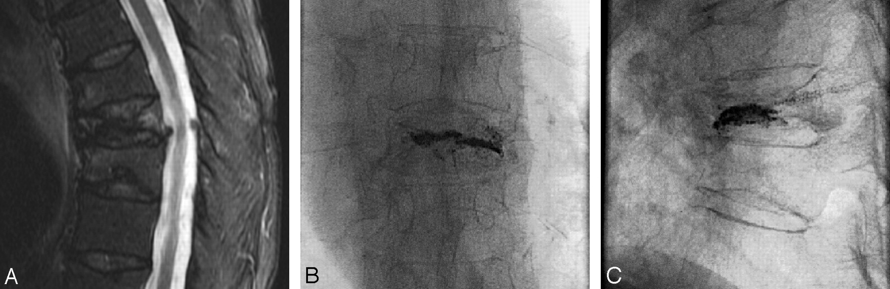
  • Fig 2.
    • Download figure
    • Open in new tab
    • Download powerpoint
    Fig 2.

    A, Sagittal T2-weighted MR imaging of the lumbar spine shows severe fractures at T12 and L2 with fluid-filled clefts. B, lateral plain radiographs after vertebroplasty show cement-filled clefts corresponding with clefts seen on preprocedure MR imaging shown in A.

  • Fig 3.
    • Download figure
    • Open in new tab
    • Download powerpoint
    Fig 3.

    A, Sagittal T2-weighted MR imaging shows severe fracture at T8 without evidence of a cleft. B and C, Anteroposterior and lateral plain radiographs after vertebroplasty show cement filling a linear cleft.
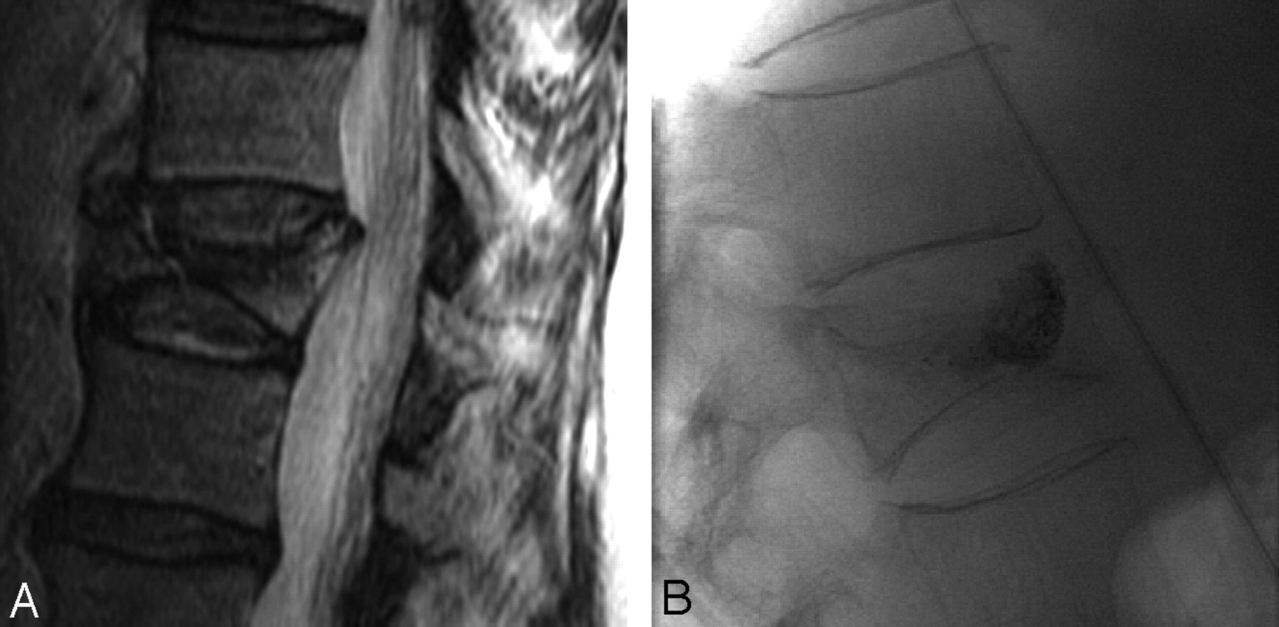
  • Fig 4.
    • Download figure
    • Open in new tab
    • Download powerpoint
    Fig 4.

    A, Sagittal T2-weighted image shows small cleft within the severe fracture at T12. B, Lateral plain radiograph after vertebroplasty shows filling of a large cleft.

Tables

  • Figures
  • Pain relief after vertebroplasty procedure

    WorseNo ChangeDecreasedCompletely Improved
    Pain at rest
        Postoperative (11)0164
        1 week (11)1154
        1 month (9)0135
        6 months (6)0024
    Pain with activity
        Postoperative (11)0074
        1 week (11)0083
        1 month (9)0063
        6 months (6)0051
    • Note:—Numbers in parentheses indicate number of patients available at each follow-up time point, and numbers in each column indicate severity of pain on the basis of a semiquantitative pain scale (0–10).

PreviousNext
Back to top

In this issue

American Journal of Neuroradiology: 29 (7)
American Journal of Neuroradiology
Vol. 29, Issue 7
August 2008
  • Table of Contents
  • Index by author
Advertisement
Print
Download PDF
Email Article

Thank you for your interest in spreading the word on American Journal of Neuroradiology.

NOTE: We only request your email address so that the person you are recommending the page to knows that you wanted them to see it, and that it is not junk mail. We do not capture any email address.

Enter multiple addresses on separate lines or separate them with commas.
Significance and Targeting of Small, Central Clefts in Severe Fractures Treated With Vertebroplasty
(Your Name) has sent you a message from American Journal of Neuroradiology
(Your Name) thought you would like to see the American Journal of Neuroradiology web site.
CAPTCHA
This question is for testing whether or not you are a human visitor and to prevent automated spam submissions.
Cite this article
A. Ehteshami Rad, L.A. Gray, D.F. Kallmes
Significance and Targeting of Small, Central Clefts in Severe Fractures Treated With Vertebroplasty
American Journal of Neuroradiology Aug 2008, 29 (7) 1285-1287; DOI: 10.3174/ajnr.A1066

Citation Manager Formats

  • BibTeX
  • Bookends
  • EasyBib
  • EndNote (tagged)
  • EndNote 8 (xml)
  • Medlars
  • Mendeley
  • Papers
  • RefWorks Tagged
  • Ref Manager
  • RIS
  • Zotero
0 Responses
Respond to this article
Share
Bookmark this article
Significance and Targeting of Small, Central Clefts in Severe Fractures Treated With Vertebroplasty
A. Ehteshami Rad, L.A. Gray, D.F. Kallmes
American Journal of Neuroradiology Aug 2008, 29 (7) 1285-1287; DOI: 10.3174/ajnr.A1066
del.icio.us logo Twitter logo Facebook logo Mendeley logo
  • Tweet Widget
  • Facebook Like
  • Google Plus One
Purchase

Jump to section

  • Article
    • Abstract
    • Patients and Technique
    • Results
    • Discussion
    • References
  • Figures & Data
  • Info & Metrics
  • Responses
  • References
  • PDF

Related Articles

  • No related articles found.
  • PubMed
  • Google Scholar

Cited By...

  • Asymptomatic and Unrecognized Cement Pulmonary Embolism Commonly Occurs with Vertebroplasty
  • Injury to the Vertebral Endplate-Disk Complex Associated with Osteoporotic Vertebral Compression Fractures
  • Clinical Outcomes with Hemivertebral Filling during Percutaneous Vertebroplasty
  • Efficacy of Percutaneous Vertebroplasty for Multiple Synchronous and Metachronous Vertebral Compression Fractures
  • Crossref
  • Google Scholar

This article has not yet been cited by articles in journals that are participating in Crossref Cited-by Linking.

More in this TOC Section

  • Dynamic CT Myelography: Patient Positioning
  • Evaluation of SIH MR Scoring Systems in Normals
  • Management Outcomes For VO Spine Biopsy
Show more SPINE IMAGING AND SPINE IMAGE-GUIDED INTERVENTIONS

Similar Articles

Advertisement

Indexed Content

  • Current Issue
  • Accepted Manuscripts
  • Article Preview
  • Past Issues
  • Editorials
  • Editor's Choice
  • Fellows' Journal Club
  • Letters to the Editor
  • Video Articles

Cases

  • Case Collection
  • Archive - Case of the Week
  • Archive - Case of the Month
  • Archive - Classic Case

More from AJNR

  • Trainee Corner
  • Imaging Protocols
  • MRI Safety Corner
  • Book Reviews

Multimedia

  • AJNR Podcasts
  • AJNR Scantastics

Resources

  • Turnaround Time
  • Submit a Manuscript
  • Submit a Video Article
  • Submit an eLetter to the Editor/Response
  • Manuscript Submission Guidelines
  • Statistical Tips
  • Fast Publishing of Accepted Manuscripts
  • Graphical Abstract Preparation
  • Imaging Protocol Submission
  • Evidence-Based Medicine Level Guide
  • Publishing Checklists
  • Author Policies
  • Become a Reviewer/Academy of Reviewers
  • News and Updates

About Us

  • About AJNR
  • Editorial Board
  • Editorial Board Alumni
  • Alerts
  • Permissions
  • Not an AJNR Subscriber? Join Now
  • Advertise with Us
  • Librarian Resources
  • Feedback
  • Terms and Conditions
  • AJNR Editorial Board Alumni

American Society of Neuroradiology

  • Not an ASNR Member? Join Now

© 2025 by the American Society of Neuroradiology All rights, including for text and data mining, AI training, and similar technologies, are reserved.
Print ISSN: 0195-6108 Online ISSN: 1936-959X

Powered by HighWire