Advertisement

Main menu

  • Home
  • Content
    • Current Issue
    • Accepted Manuscripts
    • Article Preview
    • Past Issue Archive
    • Video Articles
    • AJNR Case Collection
    • Case of the Week Archive
    • Case of the Month Archive
    • Classic Case Archive
  • Special Collections
    • AJNR Awards
    • Low-Field MRI
    • Alzheimer Disease
    • ASNR Foundation Special Collection
    • Photon-Counting CT
    • View All
  • Multimedia
    • AJNR Podcasts
    • AJNR SCANtastic
    • Trainee Corner
    • MRI Safety Corner
    • Imaging Protocols
  • For Authors
    • Submit a Manuscript
    • Submit a Video Article
    • Submit an eLetter to the Editor/Response
    • Manuscript Submission Guidelines
    • Statistical Tips
    • Fast Publishing of Accepted Manuscripts
    • Graphical Abstract Preparation
    • Imaging Protocol Submission
    • Author Policies
  • About Us
    • About AJNR
    • Editorial Board
    • Editorial Board Alumni
  • More
    • Become a Reviewer/Academy of Reviewers
    • Subscribers
    • Permissions
    • Alerts
    • Feedback
    • Advertisers
    • ASNR Home

User menu

  • Alerts
  • Log in

Search

  • Advanced search
American Journal of Neuroradiology

American Journal of Neuroradiology

ASHNR American Society of Functional Neuroradiology ASHNR American Society of Pediatric Neuroradiology ASSR
  • Alerts
  • Log in

Advanced Search

  • Home
  • Content
    • Current Issue
    • Accepted Manuscripts
    • Article Preview
    • Past Issue Archive
    • Video Articles
    • AJNR Case Collection
    • Case of the Week Archive
    • Case of the Month Archive
    • Classic Case Archive
  • Special Collections
    • AJNR Awards
    • Low-Field MRI
    • Alzheimer Disease
    • ASNR Foundation Special Collection
    • Photon-Counting CT
    • View All
  • Multimedia
    • AJNR Podcasts
    • AJNR SCANtastic
    • Trainee Corner
    • MRI Safety Corner
    • Imaging Protocols
  • For Authors
    • Submit a Manuscript
    • Submit a Video Article
    • Submit an eLetter to the Editor/Response
    • Manuscript Submission Guidelines
    • Statistical Tips
    • Fast Publishing of Accepted Manuscripts
    • Graphical Abstract Preparation
    • Imaging Protocol Submission
    • Author Policies
  • About Us
    • About AJNR
    • Editorial Board
    • Editorial Board Alumni
  • More
    • Become a Reviewer/Academy of Reviewers
    • Subscribers
    • Permissions
    • Alerts
    • Feedback
    • Advertisers
    • ASNR Home
  • Follow AJNR on Twitter
  • Visit AJNR on Facebook
  • Follow AJNR on Instagram
  • Join AJNR on LinkedIn
  • RSS Feeds

AJNR Awards, New Junior Editors, and more. Read the latest AJNR updates

Research ArticleNeurointervention

Posterior Inferior Cerebellar Artery Aneurysms: Incidence, Clinical Presentation, and Outcome of Endovascular Treatment

J.P. Peluso, W.J. van Rooij, M. Sluzewski, G.N. Beute and C.B. Majoie
American Journal of Neuroradiology January 2008, 29 (1) 86-90; DOI: https://doi.org/10.3174/ajnr.A0758
J.P. Peluso
  • Find this author on Google Scholar
  • Find this author on PubMed
  • Search for this author on this site
W.J. van Rooij
  • Find this author on Google Scholar
  • Find this author on PubMed
  • Search for this author on this site
M. Sluzewski
  • Find this author on Google Scholar
  • Find this author on PubMed
  • Search for this author on this site
G.N. Beute
  • Find this author on Google Scholar
  • Find this author on PubMed
  • Search for this author on this site
C.B. Majoie
  • Find this author on Google Scholar
  • Find this author on PubMed
  • Search for this author on this site
  • Article
  • Figures & Data
  • Supplemental
  • Info & Metrics
  • Responses
  • References
  • PDF
Loading

Article Figures & Data

Figures

  • Fig 1.
    • Download figure
    • Open in new tab
    • Download powerpoint
    Fig 1.

    A 49-year-old man presenting with left abducens and right facial nerve palsies. A, MR imaging demonstrates an aneurysm compressing the medulla and pons with surrounding edema. B, Left vertebral angiogram shows a PICA aneurysm. C, After coiling, complete aneurysm occlusion is seen. D, MR imaging 6 months later shows regression of edema; cranial nerve palsies are cured.

  • Fig 2.
    • Download figure
    • Open in new tab
    • Download powerpoint
    Fig 2.

    A 52-year-old woman with an incidentally found PICA aneurysm. A, 3D left vertebral angiogram demonstrates PICA aneurysm with the PICA originating from the sac. B, Six-month follow-up angiogram shows complete occlusion with preservation of flow in the PICA.
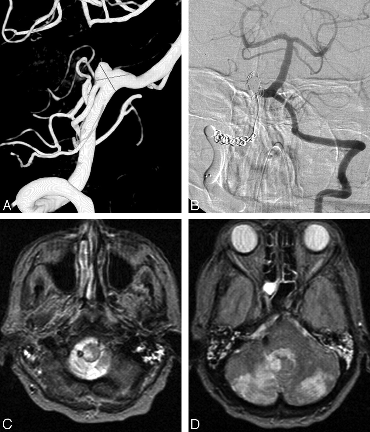
  • Fig 3.
    • Download figure
    • Open in new tab
    • Download powerpoint
    Fig 3.

    Ruptured PICA aneurysm in a 62-year-old man presenting in poor clinical condition. A, 3D right vertebral angiogram shows a wide-necked PICA aneurysm. This aneurysm was treated with coil occlusion, including the parent PICA (internal trapping). B, CT scan 6 months later demonstrates infarction in the PICA territory; however, the patient was asymptomatic.

  • Fig 4.
    • Download figure
    • Open in new tab
    • Download powerpoint
    Fig 4.

    Acutely ruptured PICA aneurysm in a 46-year-old woman. A, 3D left vertebral angiogram shows PICA aneurysm broad-based on the VA and PICA, originating from the sac. B, Angiography in the process of coiling, with occlusion of the PICA including the VA and PICA origin. MR imaging 3 months later (not shown) revealed small bilateral peripheral asymptomatic cerebellar infarctions, probably as a result of vasospasm.

  • Fig 5.
    • Download figure
    • Open in new tab
    • Download powerpoint
    Fig 5.

    A 68-year-old woman presenting 18 days after SAH from PICA aneurysm. A, Right vertebral angiogram demonstrates PICA aneurysm with PICA originating from the sac. B, Left vertebral angiogram after coil occlusion of the right VA proximal to the PICA (arrow) demonstrates persistent opacification of the aneurysm. MR imaging 6 months later (not shown) was equivocal for aneurysm thrombosis; there was no recurrent hemorrhage during 7 years of clinical follow-up.

  • Fig 6.
    • Download figure
    • Open in new tab
    • Download powerpoint
    Fig 6.

    A 71-year-old woman presenting with acute SAH in poor clinical condition. A, 3D right vertebral angiogram shows broad-based PICA aneurysm with PICA originating from the sac. B, Right vertebral angiogram after coil occlusion of the aneurysm and coil and balloon occlusion of the right VA. C and D, MR imaging 2 weeks later shows right lateral medullary infarct and bilateral PICA infarctions. The patient was right hemiparetic.

PreviousNext
Back to top

In this issue

American Journal of Neuroradiology: 29 (1)
American Journal of Neuroradiology
Vol. 29, Issue 1
January 2008
  • Table of Contents
  • Index by author
Advertisement
Print
Download PDF
Email Article

Thank you for your interest in spreading the word on American Journal of Neuroradiology.

NOTE: We only request your email address so that the person you are recommending the page to knows that you wanted them to see it, and that it is not junk mail. We do not capture any email address.

Enter multiple addresses on separate lines or separate them with commas.
Posterior Inferior Cerebellar Artery Aneurysms: Incidence, Clinical Presentation, and Outcome of Endovascular Treatment
(Your Name) has sent you a message from American Journal of Neuroradiology
(Your Name) thought you would like to see the American Journal of Neuroradiology web site.
CAPTCHA
This question is for testing whether or not you are a human visitor and to prevent automated spam submissions.
Cite this article
J.P. Peluso, W.J. van Rooij, M. Sluzewski, G.N. Beute, C.B. Majoie
Posterior Inferior Cerebellar Artery Aneurysms: Incidence, Clinical Presentation, and Outcome of Endovascular Treatment
American Journal of Neuroradiology Jan 2008, 29 (1) 86-90; DOI: 10.3174/ajnr.A0758

Citation Manager Formats

  • BibTeX
  • Bookends
  • EasyBib
  • EndNote (tagged)
  • EndNote 8 (xml)
  • Medlars
  • Mendeley
  • Papers
  • RefWorks Tagged
  • Ref Manager
  • RIS
  • Zotero
0 Responses
Respond to this article
Share
Bookmark this article
Posterior Inferior Cerebellar Artery Aneurysms: Incidence, Clinical Presentation, and Outcome of Endovascular Treatment
J.P. Peluso, W.J. van Rooij, M. Sluzewski, G.N. Beute, C.B. Majoie
American Journal of Neuroradiology Jan 2008, 29 (1) 86-90; DOI: 10.3174/ajnr.A0758
del.icio.us logo Twitter logo Facebook logo Mendeley logo
  • Tweet Widget
  • Facebook Like
  • Google Plus One
Purchase

Jump to section

  • Article
    • Abstract
    • Patients and Methods
    • Results
    • Discussion
    • Conclusions
    • Footnotes
    • References
  • Figures & Data
  • Supplemental
  • Info & Metrics
  • Responses
  • References
  • PDF

Related Articles

  • PubMed
  • Google Scholar

Cited By...

  • Flow diversion for the treatment of posterior inferior cerebellar artery aneurysms: a novel classification and strategies
  • Endovascular treatment of posterior inferior cerebellar artery aneurysms: a 7-year single-center experience
  • Endovascular treatment of PICA aneurysms with a Low-profile Visualized Intraluminal Support (LVIS Jr) device
  • Retrograde access to the posterior inferior cerebellar artery in balloon-assisted coiling of posterior inferior cerebellar artery aneurysms
  • Endovascular treatment of intracranial aneurysms with detachable coils: correlation between aneurysm volume, packing, and angiographic recurrence
  • Review of 2 Decades of Aneurysm-Recurrence Literature, Part 1: Reducing Recurrence after Endovascular Coiling
  • Results of Screening for Intracranial Aneurysms in Patients with Coarctation of the Aorta
  • Endovascular Treatment of Ruptured Vertebral Artery Dissecting Aneurysms Involving the Posterior Inferior Cerebellar Artery
  • Incidence and Risk Factors of Recurrence After Endovascular Treatment of Intracranial Vertebrobasilar Dissecting Aneurysms
  • Clinical and Angiographic Follow-Up of Stent-Only Therapy for Acute Intracranial Vertebrobasilar Dissecting Aneurysms
  • Crossref
  • Google Scholar

This article has not yet been cited by articles in journals that are participating in Crossref Cited-by Linking.

More in this TOC Section

  • Connectivity for remote neurovascular procedures
  • Cavernous dural AVF treated by transfacial route
  • A Retrospective Study in Tentorial DAVFs
Show more NEUROINTERVENTION

Similar Articles

Advertisement

Indexed Content

  • Current Issue
  • Accepted Manuscripts
  • Article Preview
  • Past Issues
  • Editorials
  • Editor's Choice
  • Fellows' Journal Club
  • Letters to the Editor
  • Video Articles

Cases

  • Case Collection
  • Archive - Case of the Week
  • Archive - Case of the Month
  • Archive - Classic Case

More from AJNR

  • Trainee Corner
  • Imaging Protocols
  • MRI Safety Corner
  • Book Reviews

Multimedia

  • AJNR Podcasts
  • AJNR Scantastics

Resources

  • Turnaround Time
  • Submit a Manuscript
  • Submit a Video Article
  • Submit an eLetter to the Editor/Response
  • Manuscript Submission Guidelines
  • Statistical Tips
  • Fast Publishing of Accepted Manuscripts
  • Graphical Abstract Preparation
  • Imaging Protocol Submission
  • Evidence-Based Medicine Level Guide
  • Publishing Checklists
  • Author Policies
  • Become a Reviewer/Academy of Reviewers
  • News and Updates

About Us

  • About AJNR
  • Editorial Board
  • Editorial Board Alumni
  • Alerts
  • Permissions
  • Not an AJNR Subscriber? Join Now
  • Advertise with Us
  • Librarian Resources
  • Feedback
  • Terms and Conditions
  • AJNR Editorial Board Alumni

American Society of Neuroradiology

  • Not an ASNR Member? Join Now

© 2025 by the American Society of Neuroradiology All rights, including for text and data mining, AI training, and similar technologies, are reserved.
Print ISSN: 0195-6108 Online ISSN: 1936-959X

Powered by HighWire