Advertisement

Main menu

  • Home
  • Content
    • Current Issue
    • Accepted Manuscripts
    • Article Preview
    • Past Issue Archive
    • Video Articles
    • AJNR Case Collection
    • Case of the Week Archive
    • Case of the Month Archive
    • Classic Case Archive
  • Special Collections
    • AJNR Awards
    • Low-Field MRI
    • Alzheimer Disease
    • ASNR Foundation Special Collection
    • Photon-Counting CT
    • View All
  • Multimedia
    • AJNR Podcasts
    • AJNR SCANtastic
    • Trainee Corner
    • MRI Safety Corner
    • Imaging Protocols
  • For Authors
    • Submit a Manuscript
    • Submit a Video Article
    • Submit an eLetter to the Editor/Response
    • Manuscript Submission Guidelines
    • Statistical Tips
    • Fast Publishing of Accepted Manuscripts
    • Graphical Abstract Preparation
    • Imaging Protocol Submission
    • Author Policies
  • About Us
    • About AJNR
    • Editorial Board
    • Editorial Board Alumni
  • More
    • Become a Reviewer/Academy of Reviewers
    • Subscribers
    • Permissions
    • Alerts
    • Feedback
    • Advertisers
    • ASNR Home

User menu

  • Alerts
  • Log in

Search

  • Advanced search
American Journal of Neuroradiology

American Journal of Neuroradiology

ASHNR American Society of Functional Neuroradiology ASHNR American Society of Pediatric Neuroradiology ASSR
  • Alerts
  • Log in

Advanced Search

  • Home
  • Content
    • Current Issue
    • Accepted Manuscripts
    • Article Preview
    • Past Issue Archive
    • Video Articles
    • AJNR Case Collection
    • Case of the Week Archive
    • Case of the Month Archive
    • Classic Case Archive
  • Special Collections
    • AJNR Awards
    • Low-Field MRI
    • Alzheimer Disease
    • ASNR Foundation Special Collection
    • Photon-Counting CT
    • View All
  • Multimedia
    • AJNR Podcasts
    • AJNR SCANtastic
    • Trainee Corner
    • MRI Safety Corner
    • Imaging Protocols
  • For Authors
    • Submit a Manuscript
    • Submit a Video Article
    • Submit an eLetter to the Editor/Response
    • Manuscript Submission Guidelines
    • Statistical Tips
    • Fast Publishing of Accepted Manuscripts
    • Graphical Abstract Preparation
    • Imaging Protocol Submission
    • Author Policies
  • About Us
    • About AJNR
    • Editorial Board
    • Editorial Board Alumni
  • More
    • Become a Reviewer/Academy of Reviewers
    • Subscribers
    • Permissions
    • Alerts
    • Feedback
    • Advertisers
    • ASNR Home
  • Follow AJNR on Twitter
  • Visit AJNR on Facebook
  • Follow AJNR on Instagram
  • Join AJNR on LinkedIn
  • RSS Feeds

AJNR Awards, New Junior Editors, and more. Read the latest AJNR updates

Research ArticleHead and Neck Imaging

Comparison of 45° Oblique Reformats with Axial Reformats in CT Evaluation of the Vestibular Aqueduct

B. Ozgen, M.E. Cunnane, P.A. Caruso and H.D. Curtin
American Journal of Neuroradiology January 2008, 29 (1) 30-34; DOI: https://doi.org/10.3174/ajnr.A0735
B. Ozgen
  • Find this author on Google Scholar
  • Find this author on PubMed
  • Search for this author on this site
M.E. Cunnane
  • Find this author on Google Scholar
  • Find this author on PubMed
  • Search for this author on this site
P.A. Caruso
  • Find this author on Google Scholar
  • Find this author on PubMed
  • Search for this author on this site
H.D. Curtin
  • Find this author on Google Scholar
  • Find this author on PubMed
  • Search for this author on this site
  • Article
  • Figures & Data
  • Info & Metrics
  • Responses
  • References
  • PDF
Loading

Article Figures & Data

Figures

  • Tables
  • Fig 1.
    • Download figure
    • Open in new tab
    • Download powerpoint
    Fig 1.

    Reformatted image of a CT of the temporal bone in the 45° oblique plane, demonstrating the position of the vestibular aqueduct.

  • Fig 2.
    • Download figure
    • Open in new tab
    • Download powerpoint
    Fig 2.

    Planning of the axial reformats based on the sagittal scout image. Axial reformats were obtained parallel to the axis of the cochlea (A). The obtained axial reformat demonstrates the vestibular aqueduct (arrow) (B).

  • Fig 3.
    • Download figure
    • Open in new tab
    • Download powerpoint
    Fig 3.

    Planning of the 45° oblique reformats based on the axial scout image. The 45° oblique reformats were obtained parallel to the plane of the SSC (A). The obtained reformat demonstrates the vestibular aqueduct (arrow) (B).
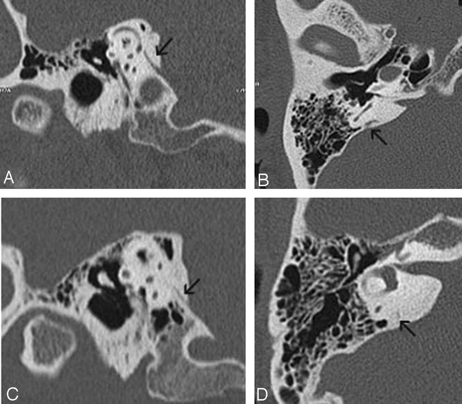
  • Fig 4.
    • Download figure
    • Open in new tab
    • Download powerpoint
    Fig 4.

    Grading of the aqueductal visibility. In 1 patient, there is a grade 3 well-visualized aqueduct in both 45° oblique (A) and axial (B) reformats. In another patient, the vestibular aqueduct is judged to be grade 2 (thin but visible) in 45° oblique formats (C), and grade 1 (difficult to see/very thin) on axial reformats (D).
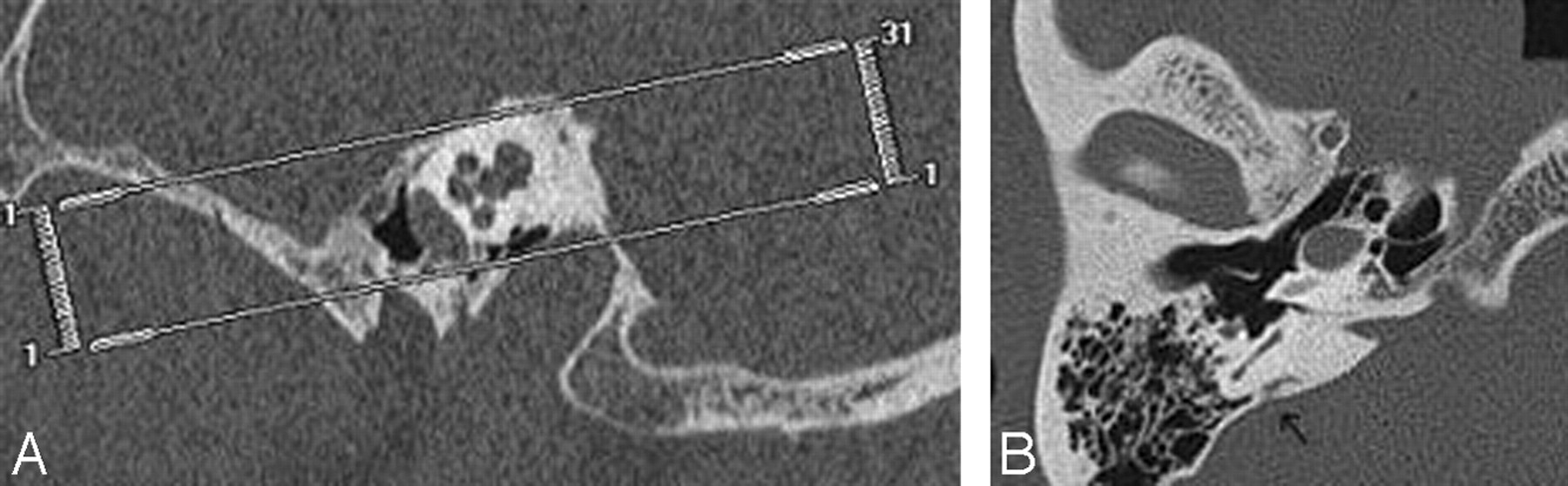
  • Fig 5.
    • Download figure
    • Open in new tab
    • Download powerpoint
    Fig 5.

    Measurement of the aqueducts: positioning of the measurement bars at the 2 described levels (midportion of the postisthmic segment and external aperture) in (A) and in axial (B, C) reformats.

  • Fig 6.
    • Download figure
    • Open in new tab
    • Download powerpoint
    Fig 6.

    Diagram demonstrating the plane of the axial image across the vestibular aqueduct on the 45° oblique reformat. A line is drawn connecting 2 points of the lateral semicircular canal. This line demonstrates the obliquity of the axial reformat to the axis of the vestibular aqueduct.

Tables

  • Figures
    • View popup
    Table 1:

    Patient demographics and indications for temporal bone CT studies

    SubjectsAge (y)/GenderIndication for CT
    Patient 111/MMastoiditis
    Patient 216/MCholesteatoma
    Patient 39/MCholesteatoma
    Patient 445/MCholesteatoma
    Patient 533/FCholesteatoma
    Patient 619 months/MCholesteatoma
    Patient 77/FCholesteatoma
    Patient 810/FCholesteatoma
    Patient 950/MConductive hearing loss
    Patient 1053/FRule out semicircular canal dehiscence
    Patient 1149/FRule out semicircular canal dehiscence
    Patient 1273/MMalignant otitis externa
    Patient 1373/MExternal auditory canal osteoma
    Patient 1445/FConductive hearing loss
    Patient 1520 months/FCholesteatoma
    • View popup
    Table 2:

    Visibility grade of 30 vestibular aqueducts viewed by 2 readers on 45° oblique and axial reformats

    Visibility GradeObserver 1Observer 1Observer 2Observer 2
    45° ObliqueAxial45° ObliqueAxial
    11 (3%)3 (10%)1 (3%)4 (13%)
    23 (10%)9 (30%)6 (20%)11 (37%)
    326 (87%)18 (60%)23 (77%)15 (15%)
    • View popup
    Table 3:

    Vestibular aqueduct measurements in 45° oblique plane

    Measurement (mm)Mean ± SDMinimumMaximumMedian
    Width at midportion of the postisthmic segment0.482 ± 0.0980.30.730.455
    Width at the external aperture0.616 ± 0.1330.40.850.6
    • View popup
    Table 4:

    Comparison of measurements obtained from vestibular aqueducts in axial views (paired Student t test) for both ears

    Measurement (mm)Mean ± SDAxialP
    External aperture0.616 ± 0.1330.741 ± 0.200.006
PreviousNext
Back to top

In this issue

American Journal of Neuroradiology: 29 (1)
American Journal of Neuroradiology
Vol. 29, Issue 1
January 2008
  • Table of Contents
  • Index by author
Advertisement
Print
Download PDF
Email Article

Thank you for your interest in spreading the word on American Journal of Neuroradiology.

NOTE: We only request your email address so that the person you are recommending the page to knows that you wanted them to see it, and that it is not junk mail. We do not capture any email address.

Enter multiple addresses on separate lines or separate them with commas.
Comparison of 45° Oblique Reformats with Axial Reformats in CT Evaluation of the Vestibular Aqueduct
(Your Name) has sent you a message from American Journal of Neuroradiology
(Your Name) thought you would like to see the American Journal of Neuroradiology web site.
CAPTCHA
This question is for testing whether or not you are a human visitor and to prevent automated spam submissions.
Cite this article
B. Ozgen, M.E. Cunnane, P.A. Caruso, H.D. Curtin
Comparison of 45° Oblique Reformats with Axial Reformats in CT Evaluation of the Vestibular Aqueduct
American Journal of Neuroradiology Jan 2008, 29 (1) 30-34; DOI: 10.3174/ajnr.A0735

Citation Manager Formats

  • BibTeX
  • Bookends
  • EasyBib
  • EndNote (tagged)
  • EndNote 8 (xml)
  • Medlars
  • Mendeley
  • Papers
  • RefWorks Tagged
  • Ref Manager
  • RIS
  • Zotero
0 Responses
Respond to this article
Share
Bookmark this article
Comparison of 45° Oblique Reformats with Axial Reformats in CT Evaluation of the Vestibular Aqueduct
B. Ozgen, M.E. Cunnane, P.A. Caruso, H.D. Curtin
American Journal of Neuroradiology Jan 2008, 29 (1) 30-34; DOI: 10.3174/ajnr.A0735
del.icio.us logo Twitter logo Facebook logo Mendeley logo
  • Tweet Widget
  • Facebook Like
  • Google Plus One
Purchase

Jump to section

  • Article
    • Abstract
    • Patients and Methods
    • Discussion
    • Conclusion
    • References
  • Figures & Data
  • Info & Metrics
  • Responses
  • References
  • PDF

Related Articles

  • No related articles found.
  • PubMed
  • Google Scholar

Cited By...

  • Vestibular Aqueduct Measurements in the 45{degrees} Oblique (Pöschl) Plane
  • Crossref
  • Google Scholar

This article has not yet been cited by articles in journals that are participating in Crossref Cited-by Linking.

More in this TOC Section

  • Diagnostic Performance of US in Neck Node NIRADS 2
  • Hydrops Herniation into the Semicircular Canals
  • MRI of the Parasellar Ligaments
Show more HEAD AND NECK IMAGING

Similar Articles

Advertisement

Indexed Content

  • Current Issue
  • Accepted Manuscripts
  • Article Preview
  • Past Issues
  • Editorials
  • Editor's Choice
  • Fellows' Journal Club
  • Letters to the Editor
  • Video Articles

Cases

  • Case Collection
  • Archive - Case of the Week
  • Archive - Case of the Month
  • Archive - Classic Case

More from AJNR

  • Trainee Corner
  • Imaging Protocols
  • MRI Safety Corner
  • Book Reviews

Multimedia

  • AJNR Podcasts
  • AJNR Scantastics

Resources

  • Turnaround Time
  • Submit a Manuscript
  • Submit a Video Article
  • Submit an eLetter to the Editor/Response
  • Manuscript Submission Guidelines
  • Statistical Tips
  • Fast Publishing of Accepted Manuscripts
  • Graphical Abstract Preparation
  • Imaging Protocol Submission
  • Evidence-Based Medicine Level Guide
  • Publishing Checklists
  • Author Policies
  • Become a Reviewer/Academy of Reviewers
  • News and Updates

About Us

  • About AJNR
  • Editorial Board
  • Editorial Board Alumni
  • Alerts
  • Permissions
  • Not an AJNR Subscriber? Join Now
  • Advertise with Us
  • Librarian Resources
  • Feedback
  • Terms and Conditions
  • AJNR Editorial Board Alumni

American Society of Neuroradiology

  • Not an ASNR Member? Join Now

© 2025 by the American Society of Neuroradiology All rights, including for text and data mining, AI training, and similar technologies, are reserved.
Print ISSN: 0195-6108 Online ISSN: 1936-959X

Powered by HighWire