Advertisement

Main menu

  • Home
  • Content
    • Current Issue
    • Accepted Manuscripts
    • Article Preview
    • Past Issue Archive
    • Video Articles
    • AJNR Case Collection
    • Case of the Week Archive
    • Case of the Month Archive
    • Classic Case Archive
  • Special Collections
    • AJNR Awards
    • Low-Field MRI
    • Alzheimer Disease
    • ASNR Foundation Special Collection
    • Photon-Counting CT
    • View All
  • Multimedia
    • AJNR Podcasts
    • AJNR SCANtastic
    • Trainee Corner
    • MRI Safety Corner
    • Imaging Protocols
  • For Authors
    • Submit a Manuscript
    • Submit a Video Article
    • Submit an eLetter to the Editor/Response
    • Manuscript Submission Guidelines
    • Statistical Tips
    • Fast Publishing of Accepted Manuscripts
    • Graphical Abstract Preparation
    • Imaging Protocol Submission
    • Author Policies
  • About Us
    • About AJNR
    • Editorial Board
    • Editorial Board Alumni
  • More
    • Become a Reviewer/Academy of Reviewers
    • Subscribers
    • Permissions
    • Alerts
    • Feedback
    • Advertisers
    • ASNR Home

User menu

  • Alerts
  • Log in

Search

  • Advanced search
American Journal of Neuroradiology

American Journal of Neuroradiology

ASHNR American Society of Functional Neuroradiology ASHNR American Society of Pediatric Neuroradiology ASSR
  • Alerts
  • Log in

Advanced Search

  • Home
  • Content
    • Current Issue
    • Accepted Manuscripts
    • Article Preview
    • Past Issue Archive
    • Video Articles
    • AJNR Case Collection
    • Case of the Week Archive
    • Case of the Month Archive
    • Classic Case Archive
  • Special Collections
    • AJNR Awards
    • Low-Field MRI
    • Alzheimer Disease
    • ASNR Foundation Special Collection
    • Photon-Counting CT
    • View All
  • Multimedia
    • AJNR Podcasts
    • AJNR SCANtastic
    • Trainee Corner
    • MRI Safety Corner
    • Imaging Protocols
  • For Authors
    • Submit a Manuscript
    • Submit a Video Article
    • Submit an eLetter to the Editor/Response
    • Manuscript Submission Guidelines
    • Statistical Tips
    • Fast Publishing of Accepted Manuscripts
    • Graphical Abstract Preparation
    • Imaging Protocol Submission
    • Author Policies
  • About Us
    • About AJNR
    • Editorial Board
    • Editorial Board Alumni
  • More
    • Become a Reviewer/Academy of Reviewers
    • Subscribers
    • Permissions
    • Alerts
    • Feedback
    • Advertisers
    • ASNR Home
  • Follow AJNR on Twitter
  • Visit AJNR on Facebook
  • Follow AJNR on Instagram
  • Join AJNR on LinkedIn
  • RSS Feeds

AJNR Awards, New Junior Editors, and more. Read the latest AJNR updates

Research ArticleSpine Imaging and Spine Image-Guided Interventions

Pain Improvement after Intradiskal Lidocaine Administration in Provocation Lumbar Diskography: Association with Diskographic Contrast Leakage

W.S. Bartynski and W.E. Rothfus
American Journal of Neuroradiology August 2007, 28 (7) 1259-1265; DOI: https://doi.org/10.3174/ajnr.A0559
W.S. Bartynski
  • Find this author on Google Scholar
  • Find this author on PubMed
  • Search for this author on this site
W.E. Rothfus
  • Find this author on Google Scholar
  • Find this author on PubMed
  • Search for this author on this site
  • Article
  • Figures & Data
  • Info & Metrics
  • Responses
  • References
  • PDF
Loading

Article Figures & Data

Figures

  • Tables
  • Fig 1.
    • Download figure
    • Open in new tab
    • Download powerpoint
    Fig 1.

    Patient is a 39-year-old man with an injury 20 years ago related to an overturned military vehicle. He has long-standing lower back pain that has recently worsened (VAS score, 8/10). Significant degenerative disk changes are present at L4-5 and L5-S1 on MR imaging, and surgery is now being considered. After 3-mL contrast injection at L5-S1, severe concordant lower back pain was provoked (VAS score, 8/10), with no improvement after 1 mL of intradiskal lidocaine administration.

    A, Frontal diskographic fluoroscopic image obtained after contrast injection at L5-S1 demonstrates degenerative changes with nuclear and annular fragmentation (arrow) but no contrast leakage.

    B. Axial postdiskographic CT image obtained at L5-S1 demonstrates significant degenerative disk changes with central and peripheral annular tears (arrows) without leakage.

  • Fig 2.
    • Download figure
    • Open in new tab
    • Download powerpoint
    Fig 2.

    Patient is a 36-year-old man with long-standing back pain and left leg pain (VAS score, 8/10), who had prior diskectomy at L4-5, with only limited improvement. Diskography was requested for follow-up assessment. After 2-mL contrast injection at L5-S1, severe nonconcordant pain was provoked (VAS score, 8/10) with no improvement after 1 mL of intradiskal lidocaine administration. His pain did reproduce concordantly at the diskectomy level.

    A, Outside MR sagittal T2-weighted image demonstrates significant degenerative disk changes at L4-5 and L5-S1 (arrowheads) with a high-intensity zone noted along the posterior L5-S1 disk margin (arrow).

    B, Lateral diskographic fluoroscopic image obtained after contrast injection at L5-S1 demonstrates degenerative changes (arrow), without evidence of contrast leakage. Contrast dilution was noted throughout the disk after lidocaine administration (not shown), but provoked pain did not improve.

    C, Axial postdiskographic CT image obtained at L5-S1 demonstrates a contained radial tear with focal disk protrusion and focal contrast accumulation without epidural leakage (arrow).
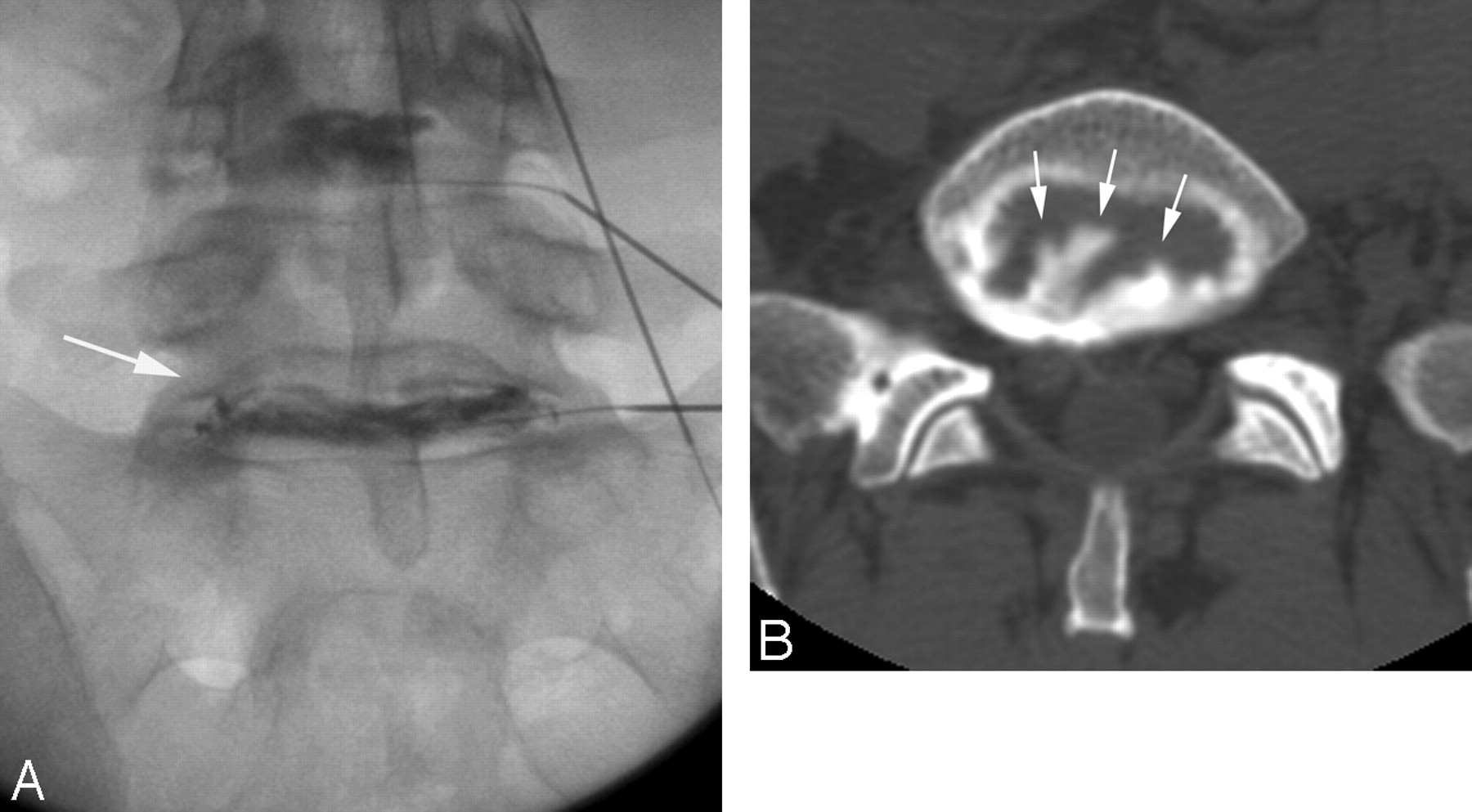
  • Fig 3.
    • Download figure
    • Open in new tab
    • Download powerpoint
    Fig 3.

    Patient is a 50-year-old woman with long-standing lower back pain, not associated with injury (VAS score, 10/10), with an outside MR imaging study demonstrating mild degenerative disk changes at L4-5 and significant degenerative disk changes at L5-S1 but no evidence of disk protrusion. The patient is currently being evaluated further by provocation diskography for potential surgical treatment. After 2.5-mL contrast injection at L5-S1, her severe and concordant lower back pain reproduced (VAS score, 9–9.5/10). Partial improvement in her concordant pain occurred after intradiskal administration of 1-mL lidocaine.

    A, Frontal diskographic fluoroscopic image obtained after contrast injection at L5-S1 demonstrates fragmentation and degenerative changes of both nuclear and annular components (arrow), without evidence of contrast leakage.

    B, Axial postdiskographic CT image at L5-S1 demonstrates degenerative disk changes with fragmentation and tears of the annulus (arrows) but without any epidural contrast leakage.

  • Fig 4.
    • Download figure
    • Open in new tab
    • Download powerpoint
    Fig 4.

    Patient is a 40-year-old man with long-standing severe lower back pain (VAS score, 10/10) and some leg diskomfort, without specific injury or trauma, with an outside MR imaging study that demonstrated degenerative disk changes at L4-5 and L5-S1. He was being considered for surgical intervention, and diskography was requested. After 3-mL contrast injection at L4-5, the patient developed severe and concordant pain (VAS score, 10/10). Injection of 1.5-mL lidocaine into this painful disk resulted in complete or near-complete elimination of the provoked pain.

    A, Lateral diskographic fluoroscopic image at L4-5 demonstrates degenerative disk changes (arrow) with clear contrast leakage extending into the epidural space (arrowheads).

    B, Axial postdiskographic CT image at L4-5 demonstrates complex degenerative changes with both nuclear fragmentation and annular tears (arrow) and a small disk protrusion and focal leakage of contrast into the adjacent epidural space on the left (arrowhead).
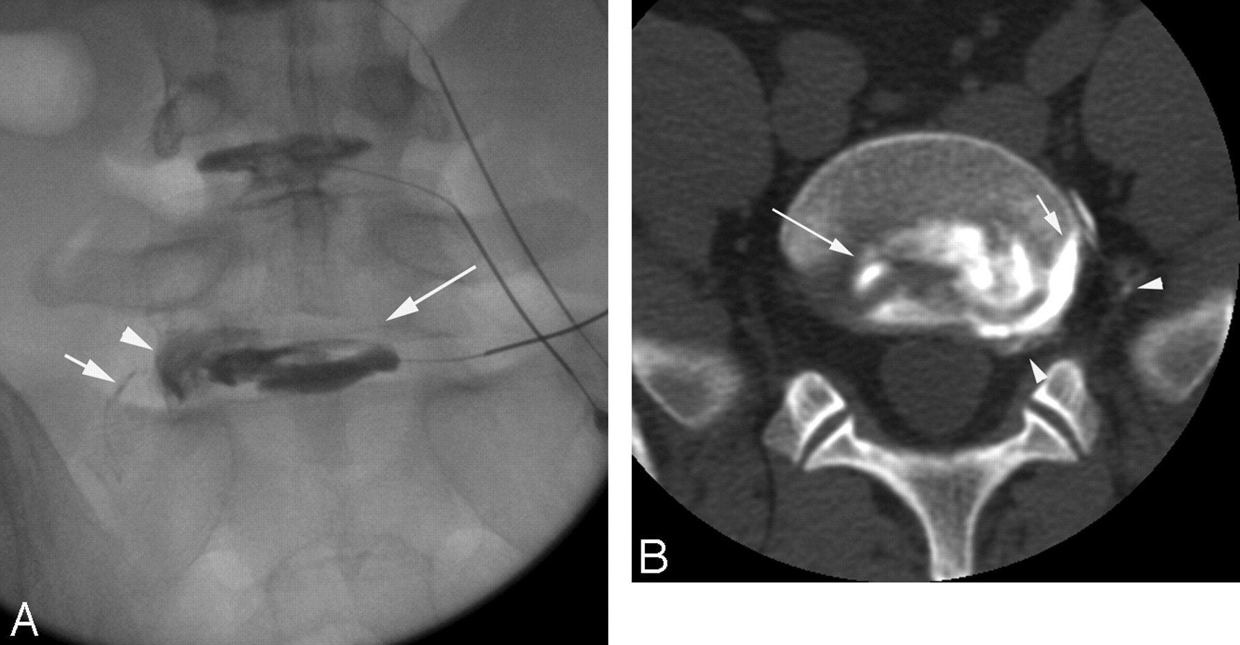
  • Fig 5.
    • Download figure
    • Open in new tab
    • Download powerpoint
    Fig 5.

    Patient is a 35-year-old man with long-standing lower back pain along with minor leg diskomfort, experiencing increasing pain during the past year. Outside MR imaging demonstrated significant degenerative changes at L4-5 and L5-S1, with possible spondylolysis of L5. Currently, he claimed the back pain would “bring him to his knees” (VAS score, 10/10), and diskography was requested in consideration of surgical intervention. After 3-mL contrast was injected at L5-S1, severe and concordant LBP was provoked without leg pain (VAS score, 10/10). Injection of 1-mL lidocaine resulted in complete elimination of his provoked back pain.

    A, Frontal diskographic image at L5-S1 demonstrates degenerative disk changes with annular tears and fragmentation (arrow), along with a peripheral annular tear projecting over the left foraminal disk margin (arrowhead) and focal leakage into the foramen and far lateral region (short arrow).

    B, Axial postdiskographic CT image demonstrates internal annular tears and fragmentation (arrow) and a peripheral annular tear on the left (short arrow), with leakage into the epidural space, L5-S1 foramen, and far lateral region (arrowheads).

Tables

  • Figures
    • View popup
    Table 1:

    Disk-space response to lidocaine administration: 182 studied lumbar levels

    Disk FeaturePain Response to Lidocaine AdministrationTotal
    Complete ReliefPartial ReliefNo Relief
    Leaking741511100
    Contained1795682
    Total912467182
    • View popup
    Table 2:

    Disk-space response to lidocaine administration: 33 postoperative levels

    Disk FeaturePain Response to Lidocaine AdministrationTotal
    Complete ReliefPartial ReliefNo Relief
    Leaking102416
    Contained131317
    Total1151733
    • View popup
    Table 3:

    Disk-space response to lidocaine administration: 149 nonoperative levels

    Disk FeaturePain Response to Lidocaine AdministrationTotal
    Complete ReliefPartial ReliefNo Relief
    Leaking6413784
    Contained1664365
    Total801950149
PreviousNext
Back to top

In this issue

American Journal of Neuroradiology: 28 (7)
American Journal of Neuroradiology
Vol. 28, Issue 7
August 2007
  • Table of Contents
  • Index by author
Advertisement
Print
Download PDF
Email Article

Thank you for your interest in spreading the word on American Journal of Neuroradiology.

NOTE: We only request your email address so that the person you are recommending the page to knows that you wanted them to see it, and that it is not junk mail. We do not capture any email address.

Enter multiple addresses on separate lines or separate them with commas.
Pain Improvement after Intradiskal Lidocaine Administration in Provocation Lumbar Diskography: Association with Diskographic Contrast Leakage
(Your Name) has sent you a message from American Journal of Neuroradiology
(Your Name) thought you would like to see the American Journal of Neuroradiology web site.
CAPTCHA
This question is for testing whether or not you are a human visitor and to prevent automated spam submissions.
Cite this article
W.S. Bartynski, W.E. Rothfus
Pain Improvement after Intradiskal Lidocaine Administration in Provocation Lumbar Diskography: Association with Diskographic Contrast Leakage
American Journal of Neuroradiology Aug 2007, 28 (7) 1259-1265; DOI: 10.3174/ajnr.A0559

Citation Manager Formats

  • BibTeX
  • Bookends
  • EasyBib
  • EndNote (tagged)
  • EndNote 8 (xml)
  • Medlars
  • Mendeley
  • Papers
  • RefWorks Tagged
  • Ref Manager
  • RIS
  • Zotero
0 Responses
Respond to this article
Share
Bookmark this article
Pain Improvement after Intradiskal Lidocaine Administration in Provocation Lumbar Diskography: Association with Diskographic Contrast Leakage
W.S. Bartynski, W.E. Rothfus
American Journal of Neuroradiology Aug 2007, 28 (7) 1259-1265; DOI: 10.3174/ajnr.A0559
del.icio.us logo Twitter logo Facebook logo Mendeley logo
  • Tweet Widget
  • Facebook Like
  • Google Plus One
Purchase

Jump to section

  • Article
    • Abstract
    • Materials and Methods
    • Results
    • Discussion
    • Conclusion
    • Acknowledgments
    • References
  • Figures & Data
  • Info & Metrics
  • Responses
  • References
  • PDF

Related Articles

  • No related articles found.
  • PubMed
  • Google Scholar

Cited By...

  • Enhancing Annular Fissures and High-Intensity Zones: Pain, Internal Derangement, and Anesthetic Response at Provocation Lumbar Discography
  • Crossref
  • Google Scholar

This article has not yet been cited by articles in journals that are participating in Crossref Cited-by Linking.

More in this TOC Section

  • Dynamic CT Myelography: Patient Positioning
  • Evaluation of SIH MR Scoring Systems in Normals
  • Management Outcomes For VO Spine Biopsy
Show more SPINE IMAGING AND SPINE IMAGE-GUIDED INTERVENTIONS

Similar Articles

Advertisement

Indexed Content

  • Current Issue
  • Accepted Manuscripts
  • Article Preview
  • Past Issues
  • Editorials
  • Editor's Choice
  • Fellows' Journal Club
  • Letters to the Editor
  • Video Articles

Cases

  • Case Collection
  • Archive - Case of the Week
  • Archive - Case of the Month
  • Archive - Classic Case

More from AJNR

  • Trainee Corner
  • Imaging Protocols
  • MRI Safety Corner
  • Book Reviews

Multimedia

  • AJNR Podcasts
  • AJNR Scantastics

Resources

  • Turnaround Time
  • Submit a Manuscript
  • Submit a Video Article
  • Submit an eLetter to the Editor/Response
  • Manuscript Submission Guidelines
  • Statistical Tips
  • Fast Publishing of Accepted Manuscripts
  • Graphical Abstract Preparation
  • Imaging Protocol Submission
  • Evidence-Based Medicine Level Guide
  • Publishing Checklists
  • Author Policies
  • Become a Reviewer/Academy of Reviewers
  • News and Updates

About Us

  • About AJNR
  • Editorial Board
  • Editorial Board Alumni
  • Alerts
  • Permissions
  • Not an AJNR Subscriber? Join Now
  • Advertise with Us
  • Librarian Resources
  • Feedback
  • Terms and Conditions
  • AJNR Editorial Board Alumni

American Society of Neuroradiology

  • Not an ASNR Member? Join Now

© 2025 by the American Society of Neuroradiology All rights, including for text and data mining, AI training, and similar technologies, are reserved.
Print ISSN: 0195-6108 Online ISSN: 1936-959X

Powered by HighWire