
Article Figures & Data
Figures
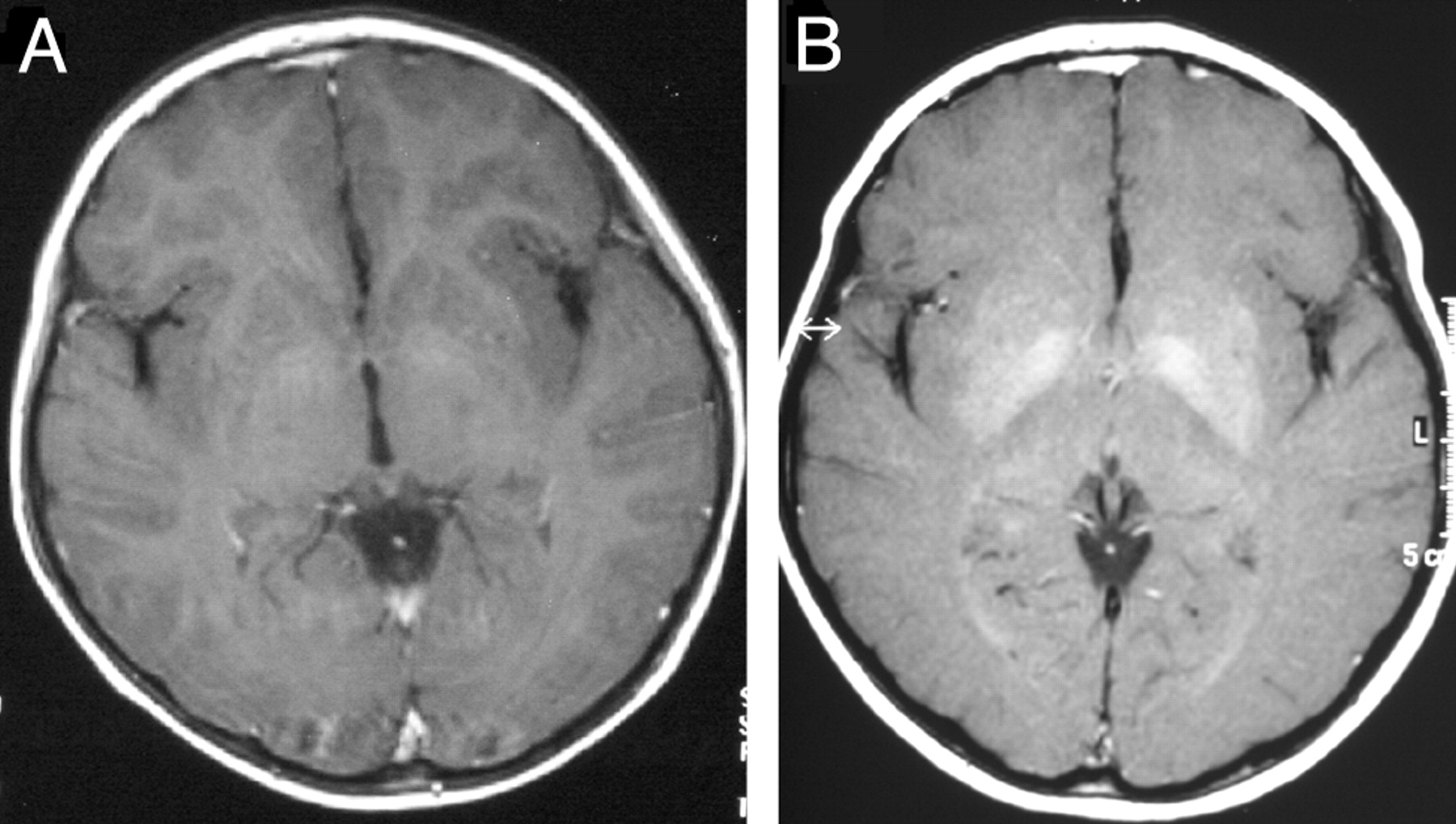
- Fig 1.
Course of signal intensity alteration in the basal ganglia in patient 6.
A, Axial T1-weighted images show mild hyperintense signal intensity alterations limited to the pallidum.
B, Three years later, prominent hyperintense signal intensity alterations involve the pallidum and the putamen.
- Fig 2.
Course of cerebellar atrophy in patient 4. Sagittal T1-weighted images show mild cerebellar atrophy at the time of the diagnosis of ND-LCH (A). Seven years later the atrophy is more pronounced (B).
- Fig 3.
Course of cerebellar MR imaging findings in patient 8. Left column, T1-weighted images (T1WI); right column, T2-weighted images (T2WI). The first signal intensity abnormalities in the cerebellum were detected at the age of 4 years (1994) and were composed of subtle increased signal intensities on T2WI limited to the region of the dentate nucleus. In 1997, the signal intensity abnormalities on T1- and T2WI involved the dentate nucleus, surrounding white matter, middle cerebellar peduncle, and pons. In 2001, the patient developed an obstructive hydrocephalus, a thinning of the corpus callosum, and progressive cerebellar neurologic symptoms. At the last follow-up (2005), the dentate nucleus appeared atrophic.
- Fig 4.
(A) Axial T2-weighted image obtained 2 months after a biopsy of the left cerebellar cortex (arrow) shows extensive hyperintensities in the cerebellar white matter extending to the middle cerebellar peduncle and the dorsal pons.
B and C, Cerebellar biopsy: the cerebellar cortex shows diminished thickness of the molecular layer. Immunocytochemistry for microtubule associated protein II (MAP II) reveals a massive loss of Purkinje cells and their dendrites. (B; ×60), the adjacent sections, stained for glial fibrillary acidic protein, show massive astrocytic gliosis, reflected by thick radial glial cell processes in the molecular layer (C; ×60). (Courtesy of Prof Hans Lassmann, Brain Research Institute, Medical University of Vienna, Vienna, Austria).


Tables
Patient 1 2 3 4 5 6 7 8 9 Sex Male Male Female Male Male Male Male Female Female Age at diagnosis of LCH 1 y 10 mo 2 y 9 mo 1 y 10 mo 3 y 4 mo 2 y 2 y 8 mo 16 y 10 mo 2 y 10 mo 6 mo Organs involved at diagnosis of LCH Bone, skin, liver, lung, spleen, hematopoietic system Bone, skin Bone, skin, liver Bone, skin, DI Bone, skin Bone, skin, liver, spleen Bone, skin, DI Bone, skin, hematopoietic system, DI Skin, liver Course of LCH (outside the CNS) Chronic active 3 RA 2 RA no RA 4 RA 1 RA 2 RA 2 RA Chronic active Age at diagnosis of ND-LCH 6 y 6 mo 7 y 3 mo 5 y 7 mo 4 y 5 mo 2 y 6 mo 8 y 2 mo 17 y 11 mo 4 y 3 mo 4 y 3 mo Interval diagnosis LCH to the diagnosis of ND 4 y 7 mo 4 y 7 mo 3 y 10 mo 1 y 1 mo 5 mo 5 y 7 mo 1y 1 y 5 mo 3 y 9 mo MR imaging follow-up period 7 y 5 mo 10 y 3 mo 5 y 2 mo 7 y 6 mo 11 y 7 y 3 mo 5 y 11 y 8 mo 6 y 8 mo No. of evaluated MR imaging studies 4 4 3 3 5 4 3 5 3 Age at last MR imaging 13 y 10 mo 17 y 6 mo 10 y 10 mo 11 y 8 mo 13 y 1 mo 15 y 5 mo 22 y 11 mo 15 y 10 mo 10 y 10 mo Additional intracranial abnormalities HPR abnormalities** −/+ +/+ +/+ +/+ −/+ +/+ +/+ +/+ +/+ Space-occupying meningeal lesions** −/− −/+ −/− −/− −/+ −/− −/− +/+ −/− Craniofacial bone lesions** +/+ +/+ +/+ +/− +/+ +/− +/+ +/+ −/− Space-occupying intraparenchymal lesions** −/− −/− −/− −/− −/− −/− +/+ −/− −/− DI* −/+ +/+ −/+ +/+ −/+ +/+ +/+ +/+ +/+ Growth hormone deficiency* −/+ +/+ −/+ −/− −/− −/− +/+ −/+ −/− Neurologic symptoms* −/− −/− −/− Ataxia/severe cerebellar symptoms −/− −/− −/− −/ataxia, spastic diplegia −/− Note:—ND indicates neurodegeneration; LCH, Langerhans cell histiocytosis; y, year; mo, month; DI, diabetes insipidus; RA, reactivation; HPR, hypothalamic pituitary region; −, absent; +, present.
* At diagnosis/at the last follow-up.
** before or at the diagnosis of ND-LCH/after the diagnosis of ND-LCH.
- Table 2:
Course of radiologic ND-LCH in the cerebellum, anterior pons, and basal ganglia in 9 patients
Patient Follow-Up At Diagnosis 3 years 6 years 9 years 12 years 1 Cerebellum ++ ++ ++ ++ Basal ganglia − + ++ ++ Anterior pons − − − − 2 Cerebellum ++ ++ ++ ++ Basal ganglia + + ++ ++ Anterior pons − − − − 3 Cerebellum + + + Basal ganglia + + ++ Anterior pons − − − 4 Cerebellum +++* +++* +++* Basal ganglia +* ++* ++* Anterior pons +* +* +* 5 Cerebellum + ++ +++ +++ +++ Basal ganglia − + + + + Anterior pons − − + + + 6 Cerebellum +++ +++ +++ +++ Basal ganglia + +++ +++ +++ Anterior pons − − − − 7 Cerebellum + + + Basal ganglia − + + Anterior pons − − − 8 Cerebellum + +++ +++* +++* +++* Basal ganglia − + ++* ++* ++* Anterior pons − + ++* ++* ++* 9 Cerebellum ++ ++ +++ Basal ganglia − − ++ Anterior pons − − − Note:—ND indicates neurodegeneration; LCH, Langerhans cell histiocytosis; Cerebellum: +, mild signal alterations limited to the dentate nucleus; ++, moderate signal alterations in dentate nucleus; +++, extensive signal alterations in the dentate nucleus, cerebellar white matter, middle cerebellar peduncles, and dorsal part of the pons. Anterior pons: −, normal; +, subtle signal alterations; ++, prominent signal alterations. Basal ganglia: −, normal; +, mild signal alterations limited to the pallidum; ++, moderate signal alterations limited to the pallidum; +++, extensive alterations involving the pallidum and the putamen.
* Radiologic ND-LCH associated with neurologic symptoms.
In this issue


Jump to section
Related Articles
Cited By...
- Mechanism of neurodegeneration mediated by clonal inflammatory microglia
- Erdheim-Chester Disease
- Erdheim-Chester Disease
- Neuroimaging in Pediatric Patients with Juvenile Xanthogranuloma of the CNS
- Waxing and Waning Neuroimaging Abnormalities in Langerhans Cell Histiocytosis
- Erdheim-Chester disease presenting with chorea and mimicking IgG4-related disorder
- The spectrum of immune-mediated and inflammatory lesions of the brainstem: Clues to diagnosis
- T1 Signal Measurements in Pediatric Brain: Findings after Multiple Exposures to Gadobenate Dimeglumine for Imaging of Nonneurologic Disease
- Cerebellar leukoencephalopathy: Most likely histiocytosis-related