
Article Figures & Data
Figures
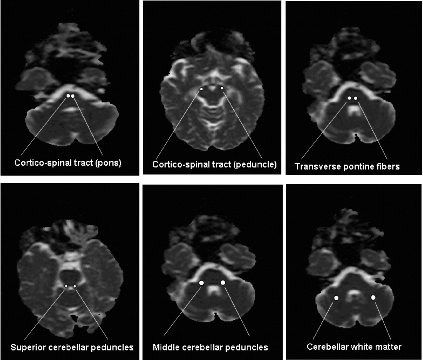
- Fig 1.
Positioning of ROI for ADC and FA measurements.
- Fig 2.
Scatterplot of ADC (A) and FA (B) measurements. For each ROI, the columns are presented in the following order: controls, patients with SCA1, and patients with SCA2.


Tables
Features SCA1 (n = 14) SCA2 (n = 11) CAG repeats (range) 40–58 35–47 Age at onset (years) 40.3 ± 10 35.9 ± 10 Disease duration (years) 8.2 ± 4.9 7.4 ± 4.4 Dysarthria (%) 100 87 Nystagmus (%) 21 0 Vertical gaze palsy (%) 43 73 Horizontal gaze palsy (%) 14 33 Slow saccades (%) 36 87 Increased bicep reflexes (%) 93 20 Increased patellar reflexes (%) 100 20 Lower limb spasticity (%) 71 13 Babinski sign (%) 79 60 Decreased vibration sense (%) 83 87 Dysphagia (%) 50 80 Amyotrophy (%) 7 0 Cognitive impairment (%) 7 13 Sphincteric disturbances (%) 29 47 Note:—CAG indicates cytosine adenine guanine.
- Table 2:
Results from post hoc comparisons for the apparent diffusion coefficient (ADC) between controls and patients with SCA1 and SCA2
ADC Controls SCA1 SCA2 SCA1-controls SCA2-controls SCA1-SCA2 CST (cerebral peduncle) 0.70 ± 0.04 0.75 ± 0.08 0.82 ± 0.08 n.s. *** ** CST (pons) 0.68 ± 0.05 0.71 ± 0.05 0.78 ± 0.06 n.s. *** *** TPF 0.69 ± 0.04 0.72 ± 0.06 0.78 ± 0.09 n.s. *** *** SCP 0.86 ± 0.05 0.88 ± 0.05 0.92 ± 0.06 n.s. ** * MCP 0.68 ± 0.04 0.81 ± 0.08 0.88 ± 0.12 *** *** * CWM 0.66 ± 0.04 0.78 ± 0.04 0.89 ± 0.11 *** *** *** Note:—CST indicates corticospinal tract, TPF, transverse pontine fibers; SCP, superior cerebellar peduncles; MCP, middle cerebellar peduncles; CWM, cerebellar white matter; n.s., not significant.
Values are expressed as mean ± SD.
* P < .05,
** P < .01,
*** P < .001.
- Table 3:
Results from post hoc comparisons for fractional anisotropy (FA) between controls and patients with SCA1 and SCA2
FA Controls SCA1 SCA2 SCA1-Controls SCA2-Controls SCA1-SCA2 CST (cerebral peduncle) 0.63 ± 0.06 0.53 ± 0.09 0.48 ± 0.07 ** *** * CST (pons) 0.47 ± 0.05 0.39 ± 0.09 0.38 ± 0.07 ** ** n.s. TPF 0.47 ± 0.06 0.42 ± 0.09 0.34 ± 0.11 * *** *** SCP 0.48 ± 0.04 0.42 ± 0.05 0.43 ± 0.07 **. * n.s. MCP 0.54 ± 0.07 0.44 ± 0.08 0.40 ± 0.12 ** *** n.s. CWM 0.45 ± 0.05 0.30 ± 0.11 0.34 ± 0.10 *** *** n.s. Note:—CST indicates corticospinal tract; TPF, transverse pontine fibers; SCP, superior cerebellar peduncles; MCP, middle cerebellar peduncles; CWM, cerebellar white matter; n.s., not significant.
Values are expressed as mean ± SD.
* P < .05,
** P < .01,
*** P < .001.
In this issue


Jump to section
Related Articles
Cited By...
- Association of the Level of Neurofilament Light With Disease Severity in Patients With Spinocerebellar Ataxia Type 2
- Longitudinal single-cell transcriptional dynamics throughout neurodegeneration in SCA1
- Ataxia Severity Correlates with White Matter Degeneration in Spinocerebellar Ataxia Type 7
- Progression of Microstructural Damage in Spinocerebellar Ataxia Type 2: A Longitudinal DTI Study
- Macro- and Microstructural Changes in Patients with Spinocerebellar Ataxia Type 6: Assessment of Phylogenetic Subdivisions of the Cerebellum and the Brain Stem
- Partial deletion of AFG3L2 causing spinocerebellar ataxia type 28
- Comparison of 3D FLAIR, 2D FLAIR, and 2D T2-Weighted MR Imaging of Brain Stem Anatomy
- Efficiency of Fractional Anisotropy and Apparent Diffusion Coefficient on Diffusion Tensor Imaging in Prognosis of Neonates with Hypoxic-Ischemic Encephalopathy: A Methodologic Prospective Pilot Study