
Article Figures & Data
Figures
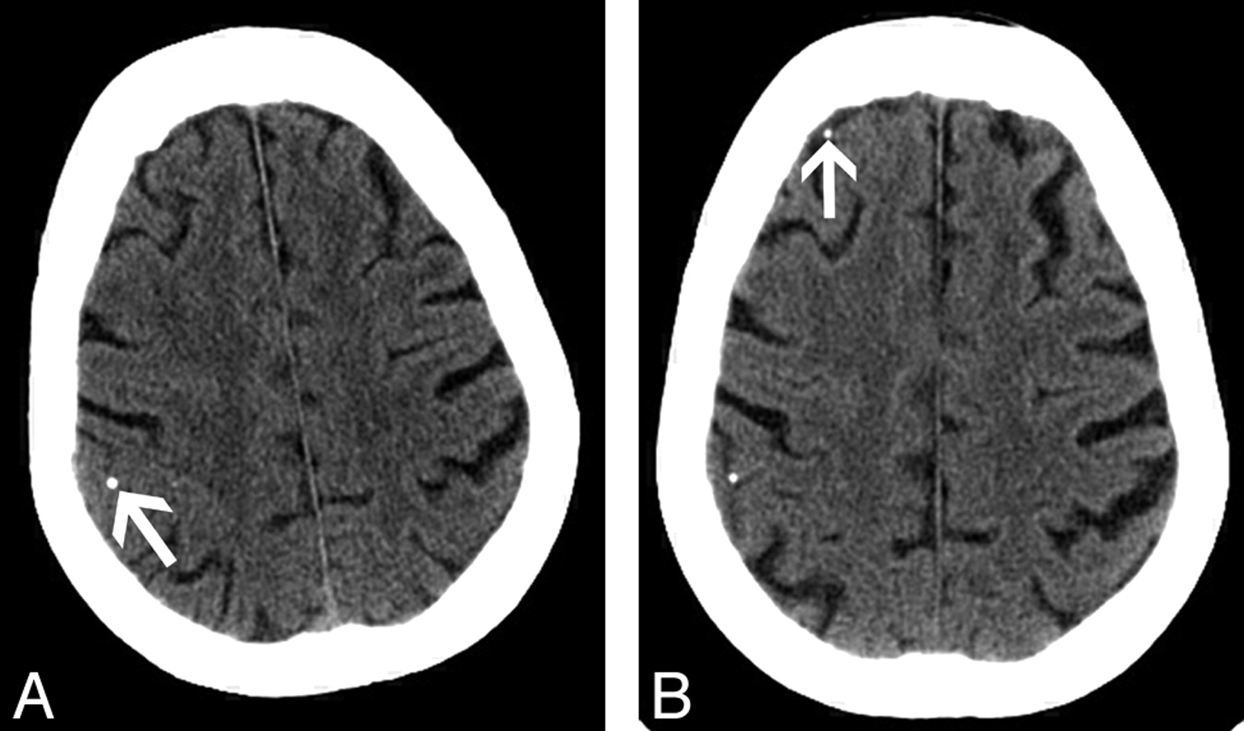
- Fig 1.
A 66-year-old man with a calcified cerebral embolus to the left middle cerebral artery.
A, Axial 2.5-mm image from a noncontrast brain CT scan shows a calcified embolus in the distal M1 segment of the left middle cerebral artery (arrow).
B, Axial 2.5-mm image obtained at a higher level shows a normal left parietal lobe, with normal gray-white matter differentiation.
C, Coronal image from a CT angiogram from the level of the aortic arch to the circle of Willis shows calcified embolus in the distal M1 segment of the left middle cerebral artery (arrow).
D, Coronal oblique image from the same examination as C shows high-grade stenosis of the left internal carotid artery with extensive calcified plaque.
E, Follow-up noncontrast brain CT scan, obtained 1 day after A and B, shows an acute infarct in the left middle cerebral artery territory with loss of normal gray-white matter differentiation and local mass effect.
- Fig 2.
A 70-year-old woman with a left middle cerebral artery territory infarct after a coronary angiogram.
A, Axial 2.5-mm image from noncontrast brain CT shows a calcified embolus in a Sylvian branch of the left middle cerebral artery (arrow).
B, Axial 2.5-mm image obtained from the same brain CT as A shows a calcific gyral attenuation in the right frontal lobe (arrow), very likely representing a second calcified cerebral embolus.
C, Axial 2.5-mm image (obtained at the same axial level as A), from noncontrast brain CT performed 1 day after A and B, shows that the previously identified calcified cerebral embolus in a Sylvian branch of the left middle cerebral artery is no longer visualized.
D, Axial 2.5-mm image (obtained at same axial level as B) from the same examination as C shows unchanged cortical attenuation in the right frontal lobe. Two new calcific densities are identified in the left precentral gyrus (arrows), consistent with downstream migration and fragmentation of the previously identified calcified cerebral embolus.
- Fig 3.
A 61-year-old woman with a calcified cerebral embolus to the left posterior cerebral artery.
A, Axial 2.5-mm image from noncontrast brain CT shows a calcified cerebral embolus in the left posterior cerebral artery (arrow).
B, Axial 2.5-mm image obtained at a higher level shows a long-standing area of infarction in the periventricular deep white matter of the left frontal lobe.
C. Axial 2.5-mm image (obtained at the same axial level as A), from noncontrast brain CT performed 5 months after A and B, shows that the previously identified calcified embolus is no longer seen in the region of the proximal left posterior cerebral artery. Note the new area of infarction in the left temporal lobe.
D, Axial 2.5-mm image (obtained at the same axial level as B), from the same examination as C, shows downstream migration of the previously identified calcified cerebral embolus into the left occipital lobe (arrow).
- Fig 4.
A 86-year-old woman with new onset of confusion after a recent myocardial infarct.
A, Axial 2.5-mm image from noncontrast brain CT shows a gyral calcification in the posterior right frontal lobe (arrow).
B, Axial 2.5-mm image (obtained at the same axial level as A) from noncontrast brain CT performed after cardiac arrest (5 days after A). The previously identified gyral calcification in the posterior right frontal lobe appears unchanged. Note the presence of a new gyral calcification in the anterior right frontal lobe (arrow), consistent with a calcified cerebral embolus.


