
Article Figures & Data
Figures
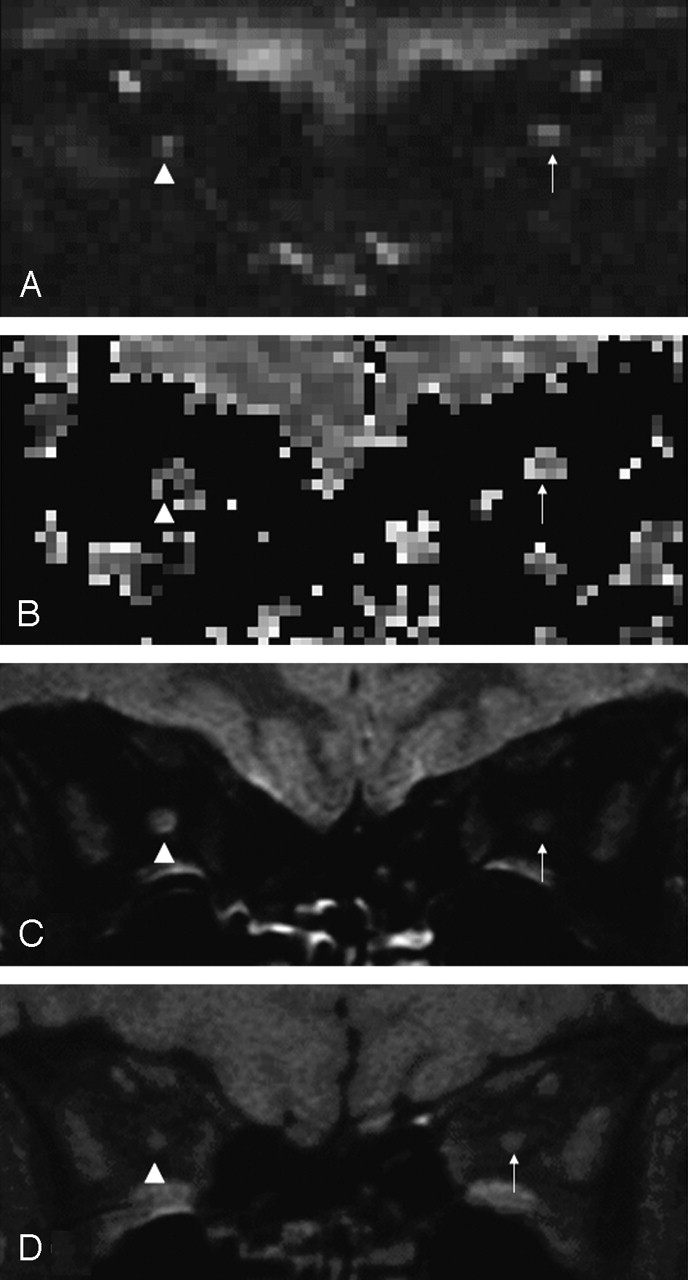
- Fig 1.
Orbital images from a 36-year-old woman 1 year after right-sided optic neuritis. Diseased optic nerve indicated by arrowhead, and contralateral healthy optic nerve indicated by arrow.
A, Nondiffusion weighted (b = 0) image.
B, ADC map.
C, FSE image demonstrating high signal intensity lesion in the right optic nerve.
D, sTE fFLAIR image demonstrating right optic nerve atrophy.
- Fig 2.
Scatter graph showing ADCs for diseased optic nerves 1 year after acute optic neuritis, healthy contralateral optic nerves, and control optic nerves.
- Fig 3.
Association between logMAR visual acuity and diseased optic nerve ADC (rS = 0.73; P = .001).
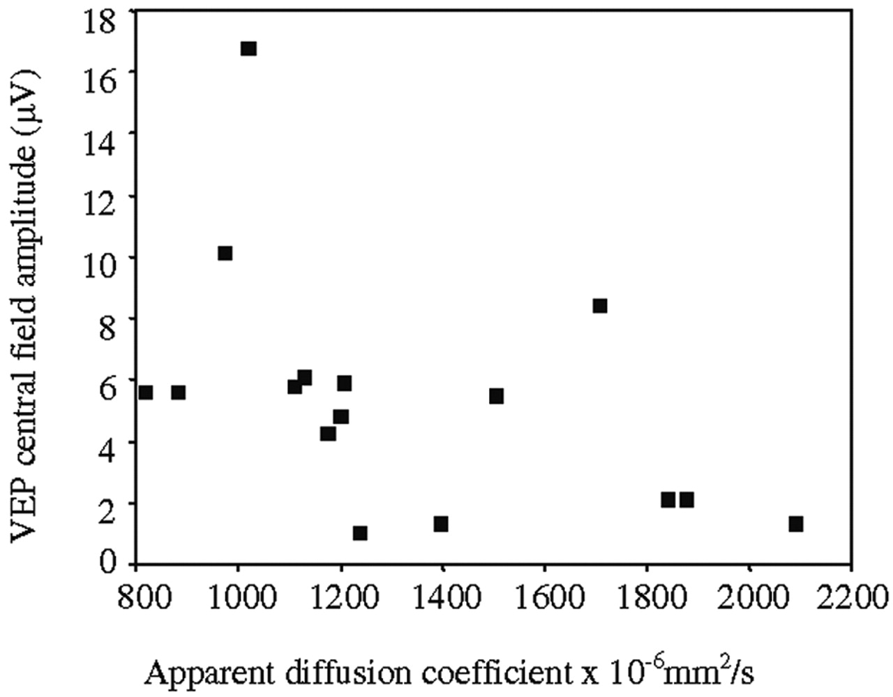
- Fig 4.
Association between 1-year VEP central field amplitude and diseased optic nerve ADC (rS = −0.57, P = .021).


Tables
Variable Mean (×10−6 mm2/s) Within-Subject SD 95% Reference Rangea CV (%)b Reliability Coefficientc Analysis/re-analysis: Control mean (n = 10) 928 23 ±45 2.5 0.99 Diseased optic nerves (n = 16) 1324 97 ±189 7.3 0.94 Healthy contralateral optic nerves (n = 14) 990 31 ±61 3.2 0.96 Image/reimage: Control mean (n = 7) 946 95 ±186 10.0 0.58 a 1.96 × within-subject SD; 95% of measurements are expected to lie within this departure from the true value.
b CV, coefficient of variation.
c The proportion of total variance due to between-subject variation. Under assumptions that are plausible here, 1 minus this value is the proportion of variation due to measurement error.
- TABLE 2:
Association between diseased optic nerve ADC and clinical and electrophysiologic measurements
SRCC P Value logMAR visual acuity 0.73 0.001 30-2 Humphrey mean deviation (dB) −0.80 <0.001 FM 100 Hue square root of error score 0.83 <0.001 VEP whole-field amplitude (μV) −0.64 0.008 VEP whole-field latency (ms) 0.58 0.019 VEP central-field amplitude (μV) −0.57 0.021 VEP central-field latency (ms) 0.58 0.019 Note.—FM 100 Hue, Farnsworth Munsell 100 Hue test; SRCC, Spearman’s rank correlation coefficient; VEP = visual evoked potentials.
In this issue


Jump to section
Related Articles
Cited By...
- MRI signs helpful in the differentiation of patients with anterior ischaemic optic neuropathy and optic neuritis
- Reduced Field-of-View Diffusion Tensor Imaging of the Optic Nerve in Retinitis Pigmentosa at 3T
- Imaging outcomes for trials of remyelination in multiple sclerosis
- Diffusion Tensor Imaging of the Optic Nerve in Multiple Sclerosis: Association with Retinal Damage and Visual Disability
- Diffusion Changes in the Vitreous Humor of the Eye during Aging
- Diffusion Tensor Imaging of the Optic Nerve in Subacute Anterior Ischemic Optic Neuropathy at 3T
- Directional diffusivity changes in the optic nerve and optic radiation in optic neuritis
- Radial diffusivity in remote optic neuritis discriminates visual outcomes
- Disability in optic neuritis correlates with diffusion tensor-derived directional diffusivities
- Retinal Peripapillary Nerve Fiber Layer Thickness in Neuromyelitis Optica
- Diffusion MRI in multiple sclerosis